-
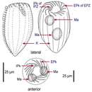
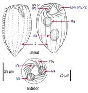
Fig 1: Strombidinopsis acuminatum Line drawing of protargol stained cells, showing kineties, oral structures and nuclei
-
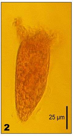
Fig 2: Strombidinopsis acuminatum Lugol's fixed cell
-
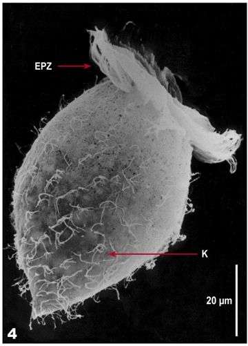
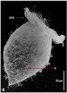
Fig 4: Strombidinopsis acuminatum SEM of Lugol's fixed cell
-
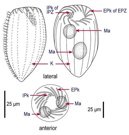
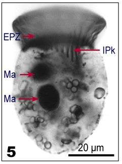
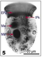
Fig 5: Strombidinopsis acuminatum protargol stained cell, lateral view: Nuclei and internal polykinetids
-
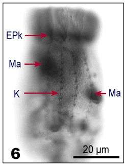
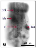
Fig 6: Strombidinopsis acuminatum protargol stained cell: Cell surface, showing the kineties
-
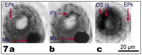
Fig 7a-c: Strombidinopsis acuminatum Apical view of protargol-stained cells, showing details of the oral structures: a. Above the cell (showing the cilia of the EPk); b. At the base of the cilia, showing the EPk; c. Within the cell (showing the IPk), OS - outer segment, IS - inner segment
-
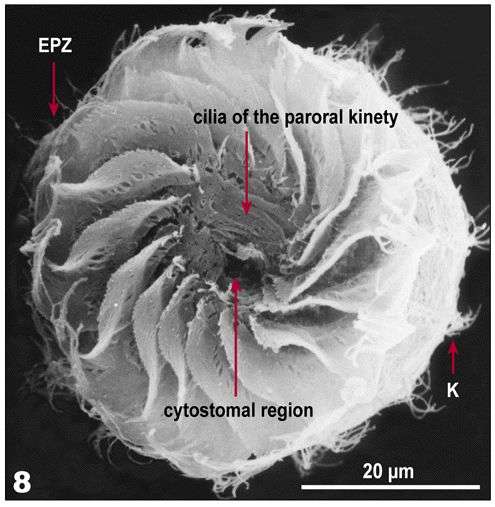
Fig 8: Strombidinopsis acuminatum SEM image, apical view of the oral region