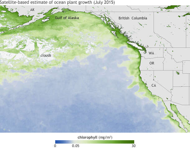
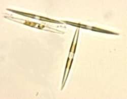
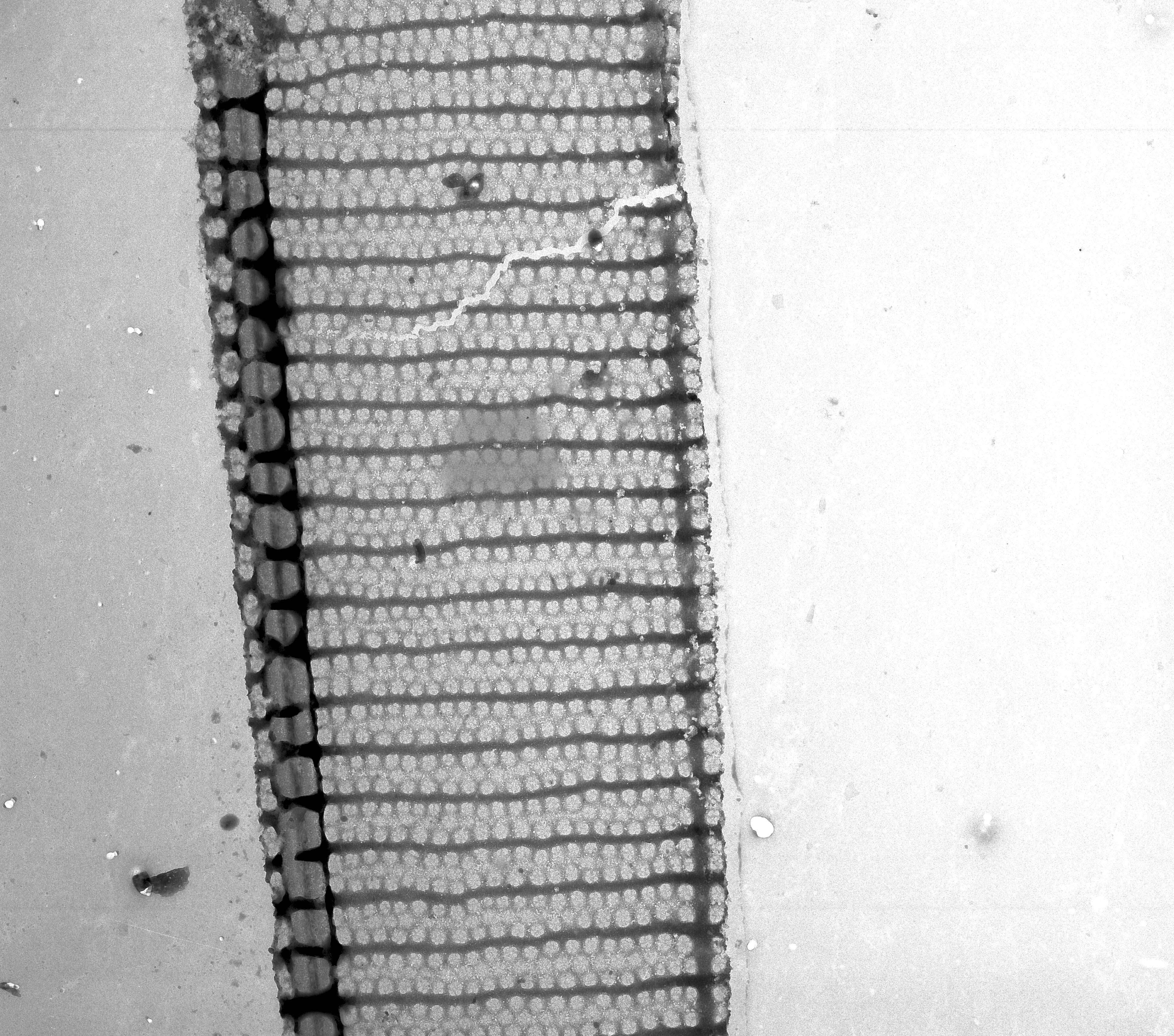
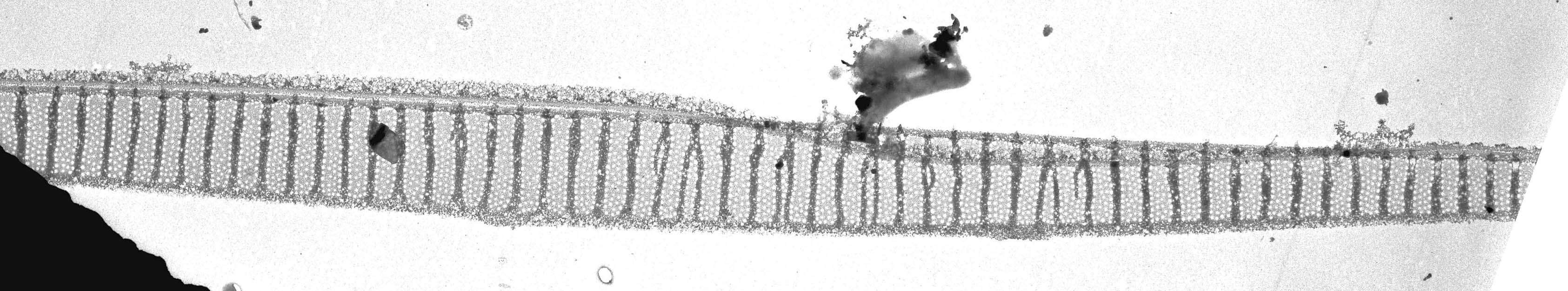
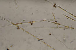
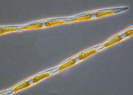
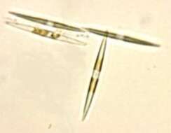
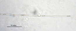
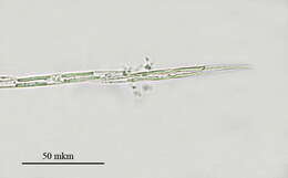
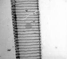
Description: English: Pseudo-nitzschia multiseries strain "CCMP 2708" : diatoms viewed under a light microscope Français : Diatomées de l'espèce Pseudo-nitzschia multiseries souche "CCMP 2708" prise en microscopie photonique. Date: 4 June 2007. Source: Own work. Author:
Guillmot44. Photo prise au centre Ifremer de Nantes (France) durant mes travaux. La diatomée Pseudo-nitzschia multiseries est productrice d'acide domoïque, une neurotoxine amnésiante. Licensing[
edit] I, the copyright holder of this work, hereby publish it under the following licenses: : Permission is granted to copy, distribute and/or modify this document under the terms of the
GNU Free Documentation License, Version 1.2 or any later version published by the
Free Software Foundation; with no Invariant Sections, no Front-Cover Texts, and no Back-Cover Texts. A copy of the license is included in the section entitled
GNU Free Documentation License.http://www.gnu.org/copyleft/fdl.htmlGFDLGNU Free Documentation Licensetruetrue. : This file is licensed under the
Creative Commons Attribution-Share Alike
3.0 Unported,
2.5 Generic,
2.0 Generic and
1.0 Generic license. :. https://creativecommons.org/licenses/by-sa/3.0 CC BY-SA 3.0 Creative Commons Attribution-Share Alike 3.0 truetrue. You may select the license of your choice.