-
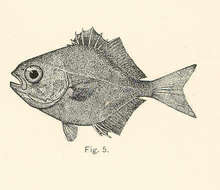
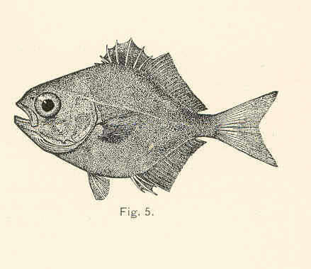
Trachynotus rhomboides.
-
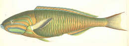
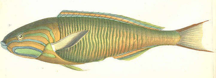
Iulus Lutescens.
-
Nesrine Akkari, Pavel Stoev, Henrik Enghoff
Zookeys
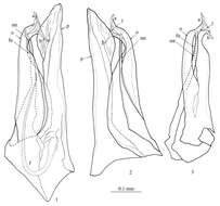
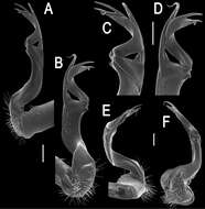
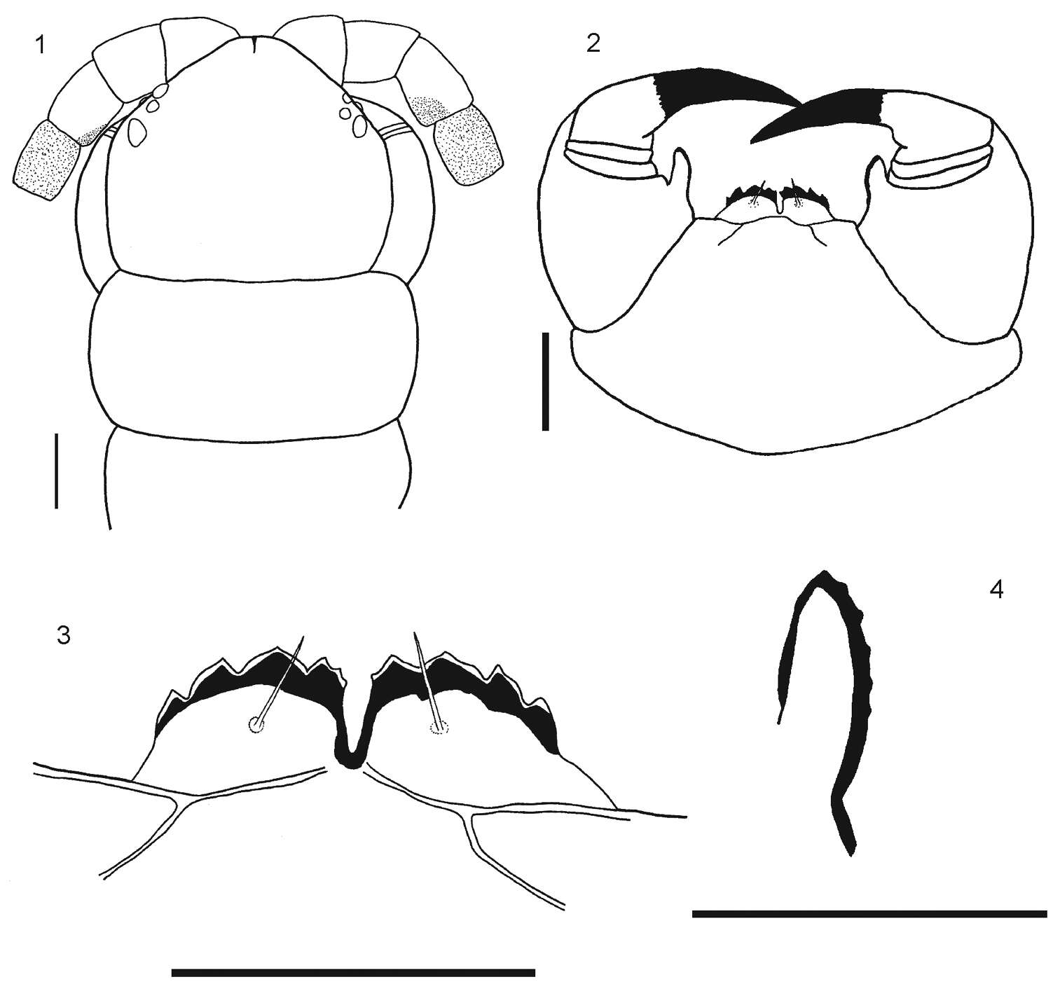
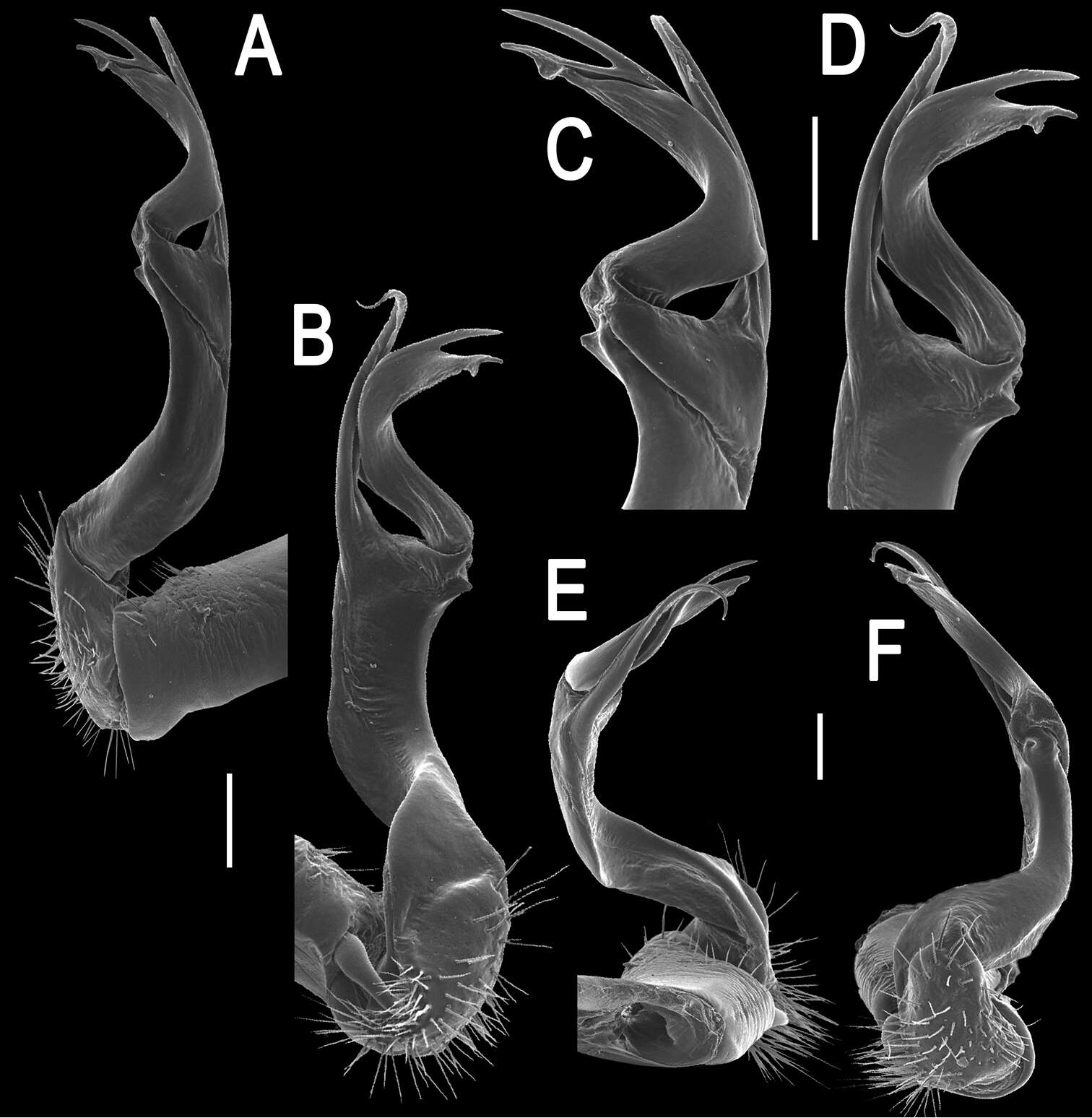
Figures 1−3.Mammamia profuga gen. n., sp. n., gonopods: 1 right gonopod, mesal view 2 right gonopod, lateral view 3 left posterior gonopod, anterior view. Abbreviations: a, b: opisthomerital processes a and b, f: flagellum, fu: furrow, l: lamella, mt: mesomerital process, o: opisthomerite, p: promerite.
-
Sergei I. Golovatch, Youbang Li, Weixin Liu, Jean-Jacques Geoffroy
Zookeys
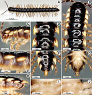
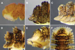
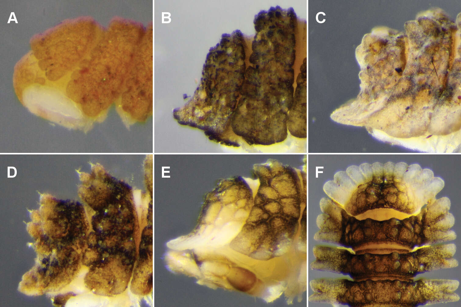
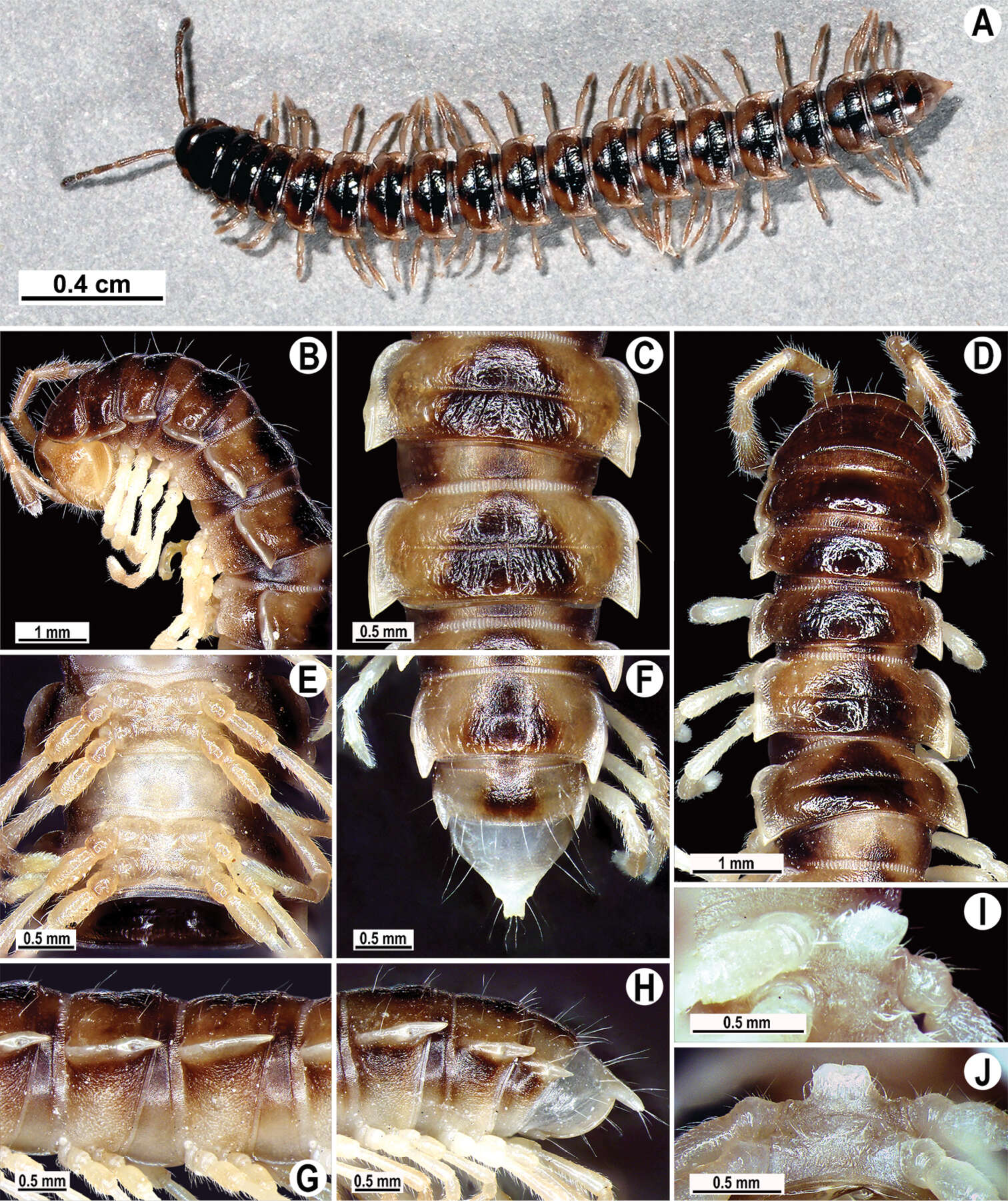
Figure 1.Desmoxytes eupterygota sp. n., ♂ paratype from near Tianhe. A–C anterior part of body, dorsal, ventral and lateral views, respectively D midbody segments, dorsal view E telson, ventral view F midbody leg, front view.Photographed not to scale.
-
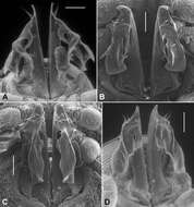
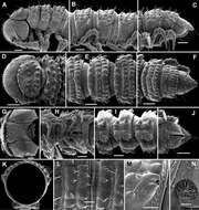
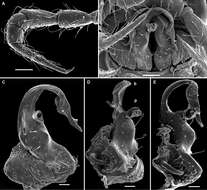
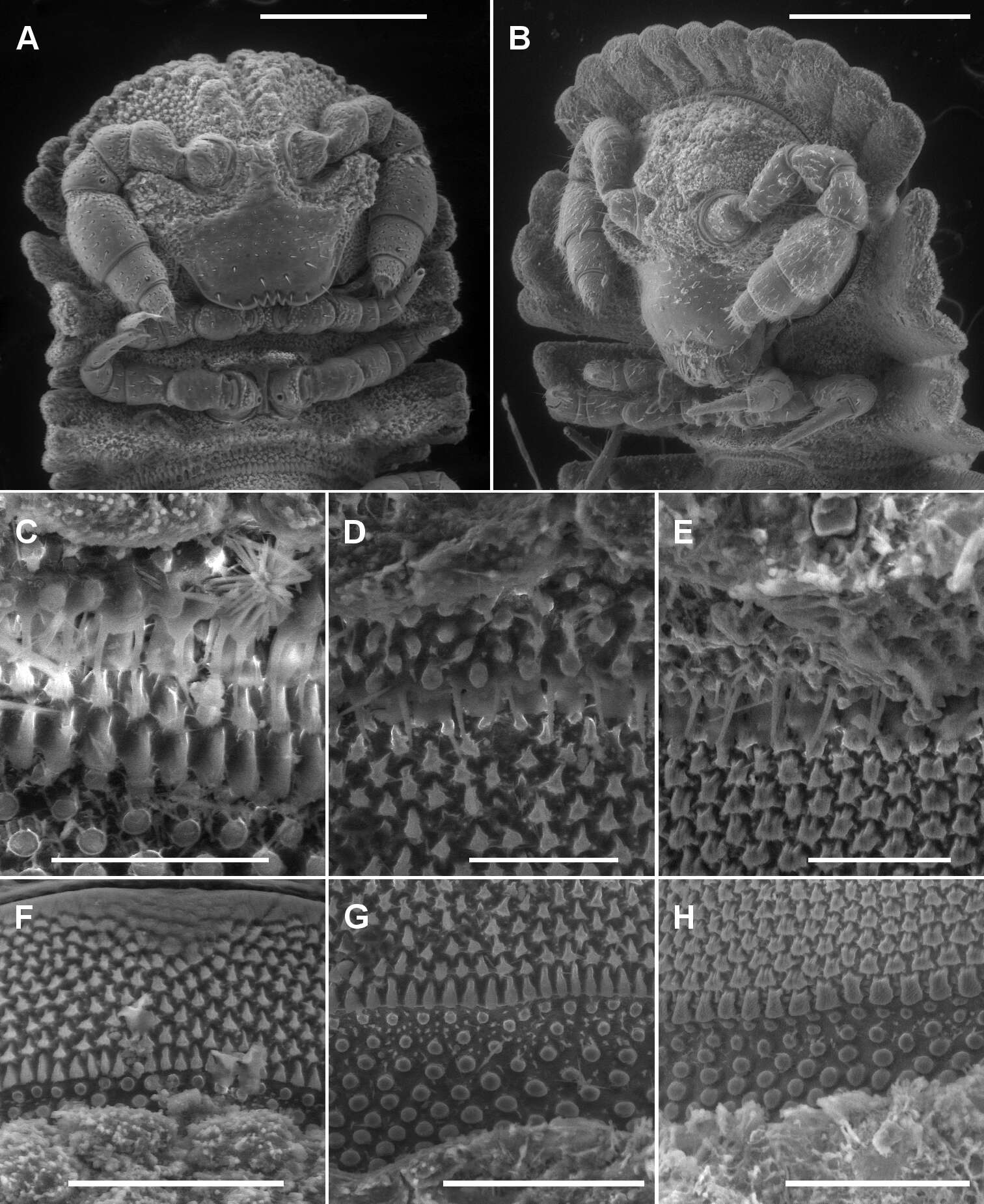
Figure 2.A, B Ventral views of head of Asticopyrgodesmus maiala sp. n., male paratype ex ANIC 64-000220 (A) and Notopyrgodesmus weiri sp. n., male paratype ANIC 64-000249 C, D, E Views of lobe-and-spike limbus on midbody rings (anterior at top) of Asticopyrgodesmus maiala sp. n., male paratype ex ANIC 64-000220 (C), Nephopyrgodesmus eungella sp. n., male paratype ex ANIC 64-000231 (D) and Notopyrgodesmus kulla sp. n., male paratype ex ANIC 64-000243 (E) F, G, H Views of prozonite sculpture on midbody rings (anterior at top) of Asticopyrgodesmus lamingtonensis sp. n., male paratype ex ANIC 64-000217 (F), Notopyrgodesmus eungella sp. n., male paratype ex ANIC 64-000231 (G) and Notopyrgodesmus kulla sp. n., male paratype ex ANIC 64-000243 (H). Scanning electron micrographs of uncoated specimens; scale bars: A = 0.5 mm, B = 0.2 mm, C, D, E, H = 0.05 mm, F, G = 0.1 mm.
-
Figure 1–4.1 Rhysida celeris from Ecuador. Cephalic plate 2 Forcipular Coxosternum 3 Tooth plates 4 Forcipular trochanteroprefemur process. Scale bars 1 mm.
-
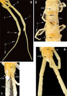
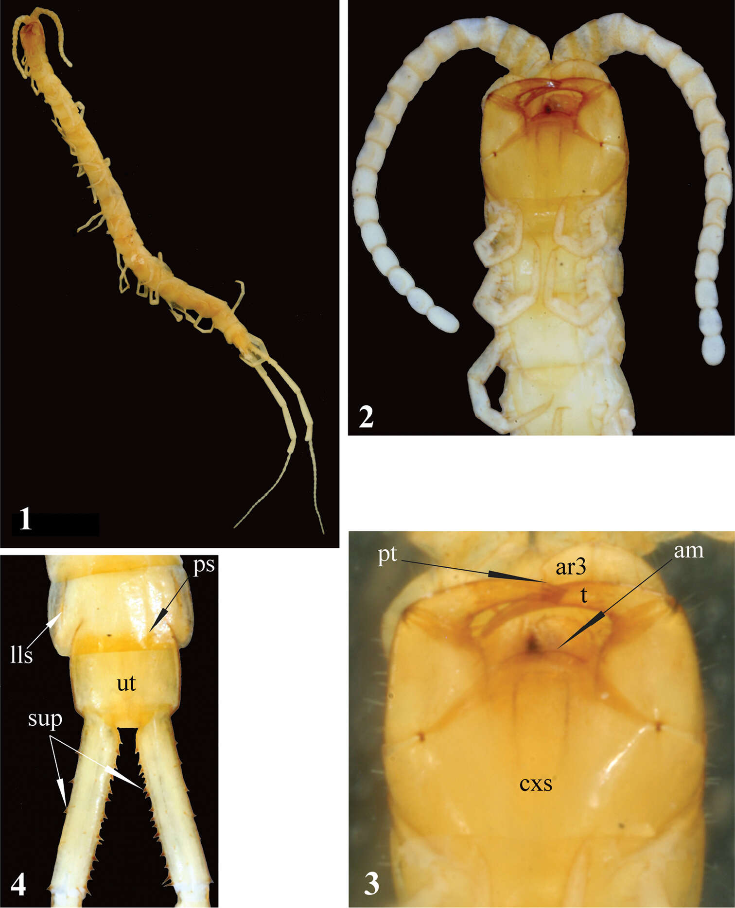
Figures 1–4.Newportia stoevi,sp. n. 1 Habitus 2 Head and anterior segments, ventral view 3 Forcipular segment, ventral view 4 Tergites 22 and 23 and prefemora of ultimate legs, dorsal view; (pt) – pretarsus of second maxilla, (ar3) – article 3 of telopodite of second maxilla, (cxs) – forcipular coxosternite, (am) – anterior margin of coxosternite, (t) – tarsungulum, (ps) – paramedian sutures, (lls) – lateral longitudinal sutures, (ut) – tergite of ultimate leg-bearing segment, (sup) – spurs of ultimate prefemur.
-
Sergei I. Golovatch, Jean-Jacques Geoffroy, Pavel Stoev, Didier Vanden Spiegel
Zookeys
Figure 12. Retrodesmus dammermani Chamberlin, 1945, ♂ holotype from Java, Indonesia; A–C habitus, dorsal, lateral and ventral views, respectively.
-
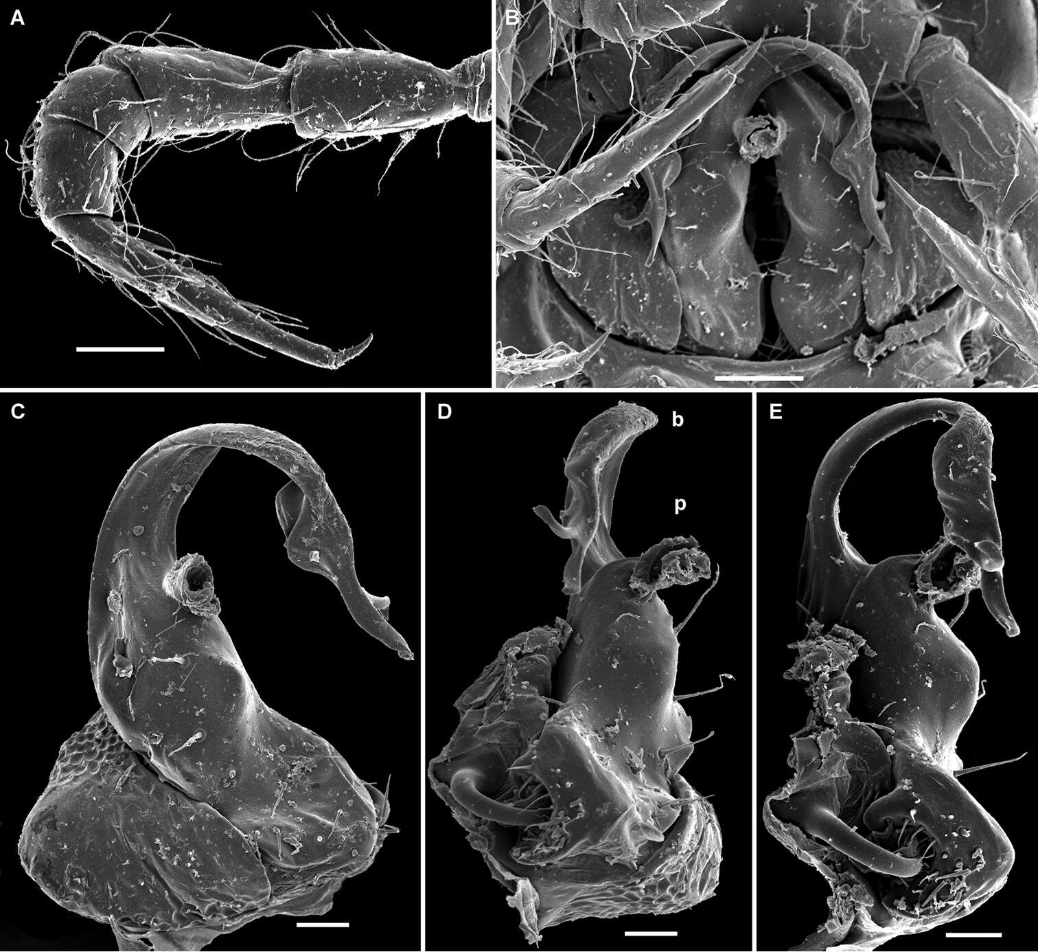
Figure 6.Posterior gonopod views. A Agathodesmus adelphus sp. n., holotype, QM S96015 B Agathodesmus aenigmaticus sp. n., paratype ex QM S96018 C Agathodesmus agnus sp. n., paratype ex QM S96021 D Agathodesmus anici sp. n., holotype, ANIC 64-000327. A and D are uncoated specimens; scale bars = 0.1 mm. Note two mites on Agathodesmus adelphus sp. n. telopodites.
-
Natdanai Likhitrakarn, Sergei I. Golovatch, Somsak Panha
Zookeys
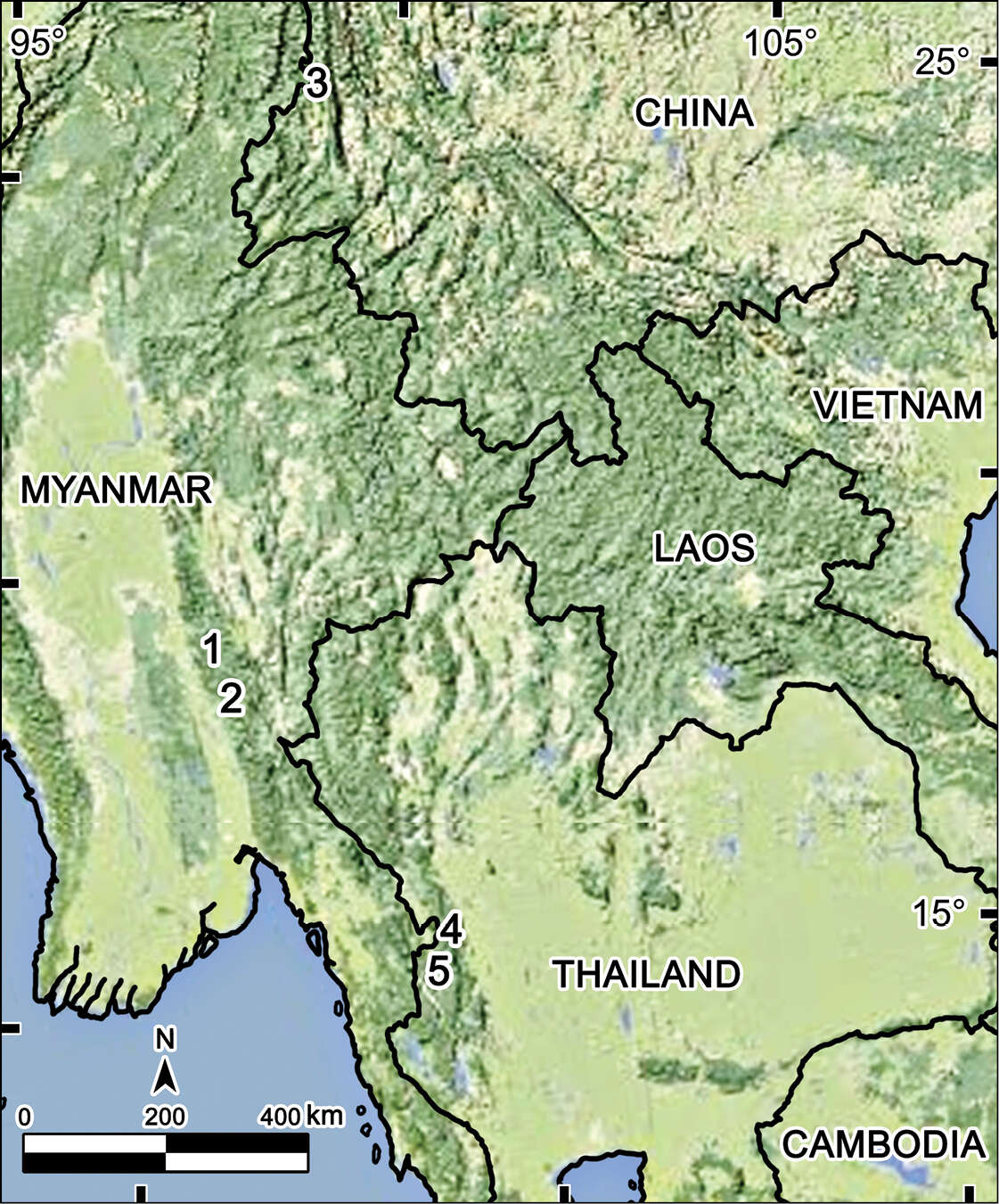
Figure 4.Distribution of Tetracentrosternus species. 1,2 Tetracentrosternus subspinosus (1 Carin Cheba (= Bia-po) 2 Puepoli) 3 Tetracentrosternus hoffmani (Mt Gaoligong Shan) 4,5 Tetracentrosternus theelorsuensis sp. n. (4 Thee Lor Sue Waterfall 5 Pa Wai Waterfall).
-
Natdanai Likhitrakarn, Sergei I. Golovatch, Somsak Panha
Zookeys
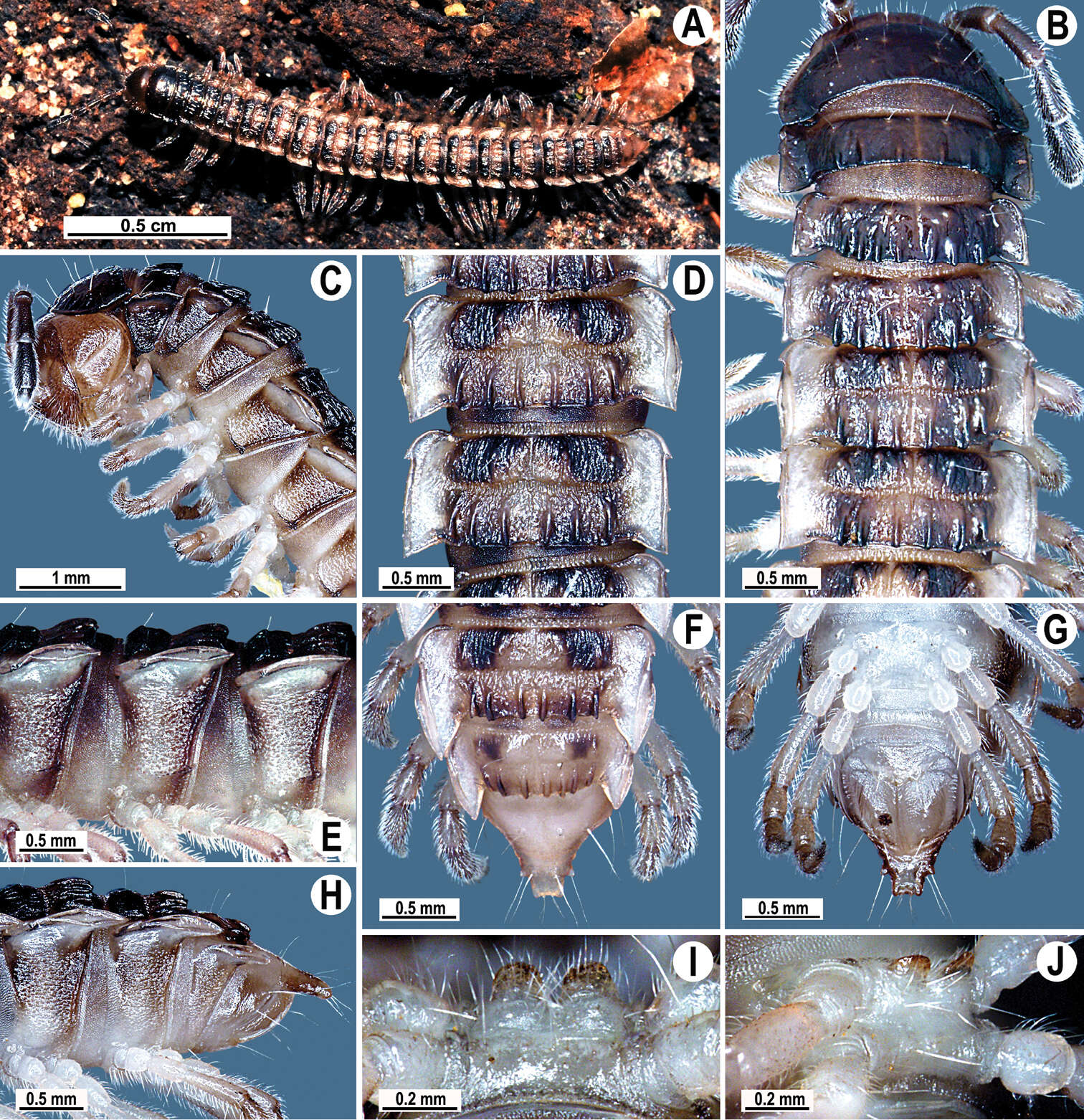
Figure 1.Orthomorpha paviei Brölemann, 1896, ♂ from Laos. A habitus, live coloration; B, C anterior part of body, dorsal and lateral views, respectivelyD, E segments 10 and 11, dorsal and lateral views, respectively F–H posterior part of body, dorsal, ventral and lateral views, respectivelyI, J sternal cones between coxae 4, subcaudal and sublateral views, respectively.
-
Sergei I. Golovatch, Jean-Jacques Geoffroy, Didier VandenSpiegel
Zookeys
Figure 1.Cocacolaria hauseri Hoffman, 1987, ♂ from Natawa forest, Espirito Santo Island, Vanuatu; A, D anterior part of body, lateral and dorsal views, respectively B, E, I midbody segments, lateral, dorsal and ventral views, respectively C, F, J posterior part of body, lateral, dorsal and ventral views, respectively G head, ventral view H body segments 6 and 7, ventral view K cross-section of a midbody segment, caudal view L midbody tergal sculpture and setae, dorsal view M caudal part of a midbody pore-bearing paratergite, lateral view N midbody spiracle. Scale bars: A–F, H–K 0.1 mm; G, L, M 0.05 mm; N 0.02 mm.
-
Natdanai Likhitrakarn, Sergei I. Golovatch, Somsak Panha
Zookeys
Figure 1.Tylopus corrugatus sp. n., ♂ holotype; A habitus, live coloration B, C anterior part of body, dorsal and lateral views, respectively D, E segments 10 and 11, dorsal and lateral views, respectively F–H posterior part of body, dorsal, ventral and lateral views, respectively I, J sternal cones between coxae 4, subcaudal and sublateral views, respectively.
-
Chao-Chun Chen, Sergei I. Golovatch, Hsueh-Wen Chang, Shyh-Hwang Chen
Zookeys
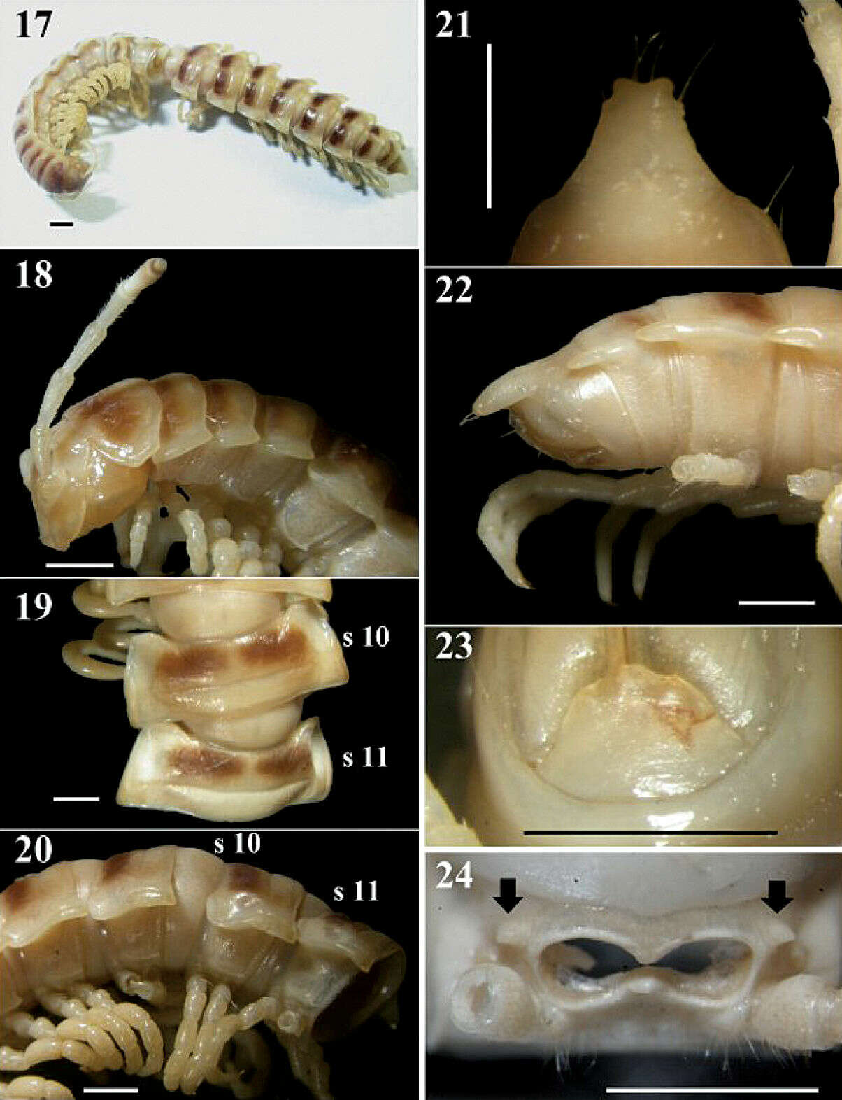
Figures 17–24.Chamberlinius pessior sp. n., holotype. 17 Entire body, dorsal view 18 Anterior body portion, lateral 19, 20 segments 10 and 11, dorsal and lateral views, respectively 21, 22 Epiproct, dorsal and lateral views, respectively 23 Hypoproct, ventral view 24 Spiracle-bearing cones lateral to gonopod aperture (arrows). Scale bar: 1.0 mm. s10 and s11: segments 10 and 11 separately.
-
Nesrine Akkari, Pavel Stoev, Henrik Enghoff
Zookeys
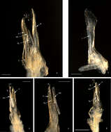
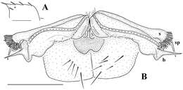
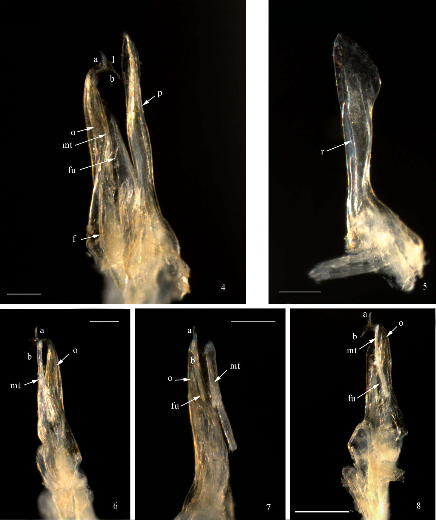
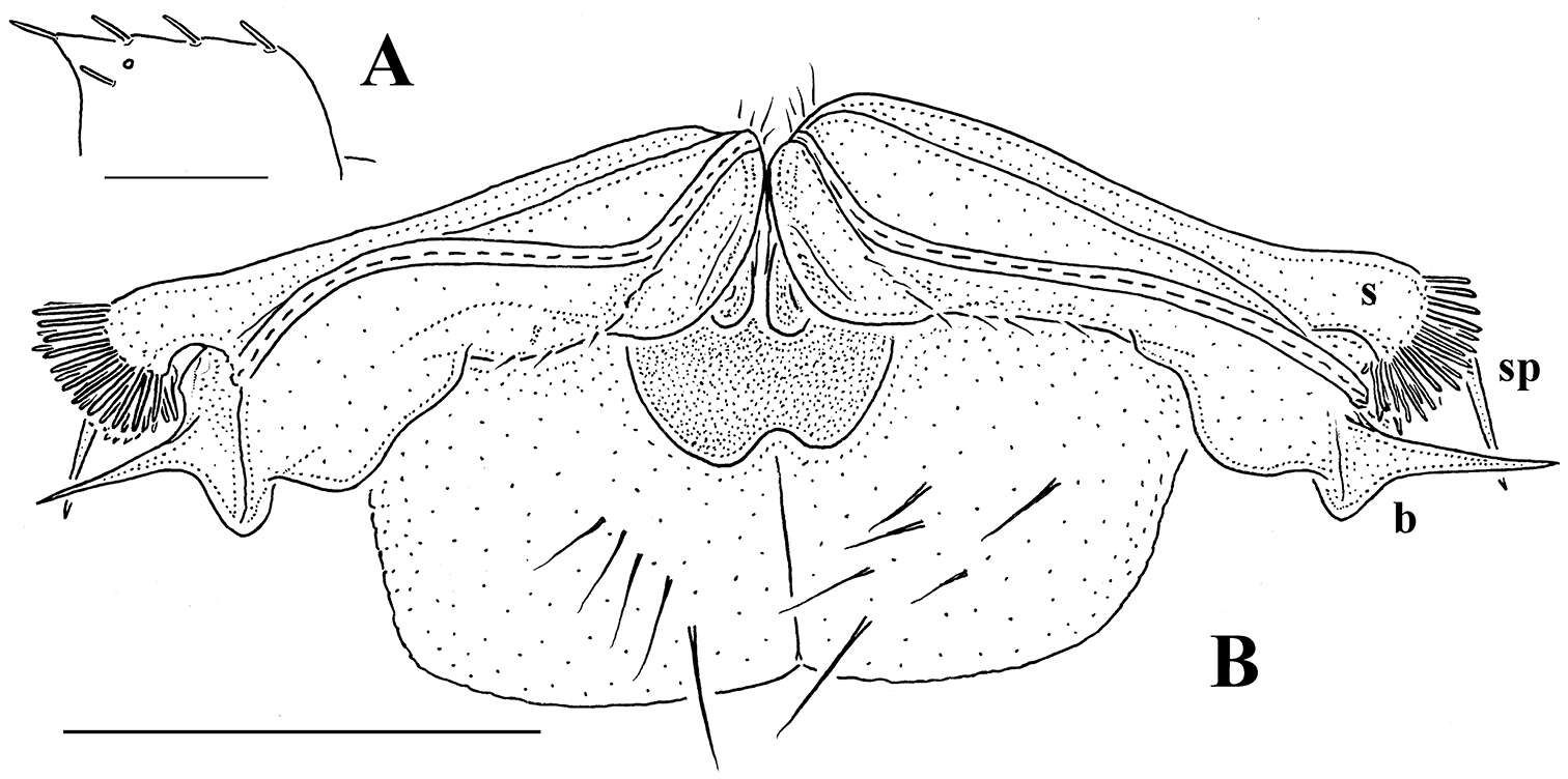
Figures 4−8.Mammamia profuga gen. n., sp. n., gonopods: 4 right gonopod, mesal view 5 left promerite, posterior view 6 left posterior gonopod, lateral view 7 left posterior gonopod, postero-lateral view 8 left posterior gonopod, posterior view. Abbreviations: a, b: opisthomerital processes a and b, f: flagellum, fu: furrow, l: lamella, mt: mesomerital process, o: opisthomerite, p: promerite, r: ridge. Scale bar: 0.1 mm.
-
Sergei I. Golovatch, Youbang Li, Weixin Liu, Jean-Jacques Geoffroy
Zookeys
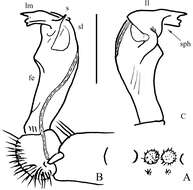
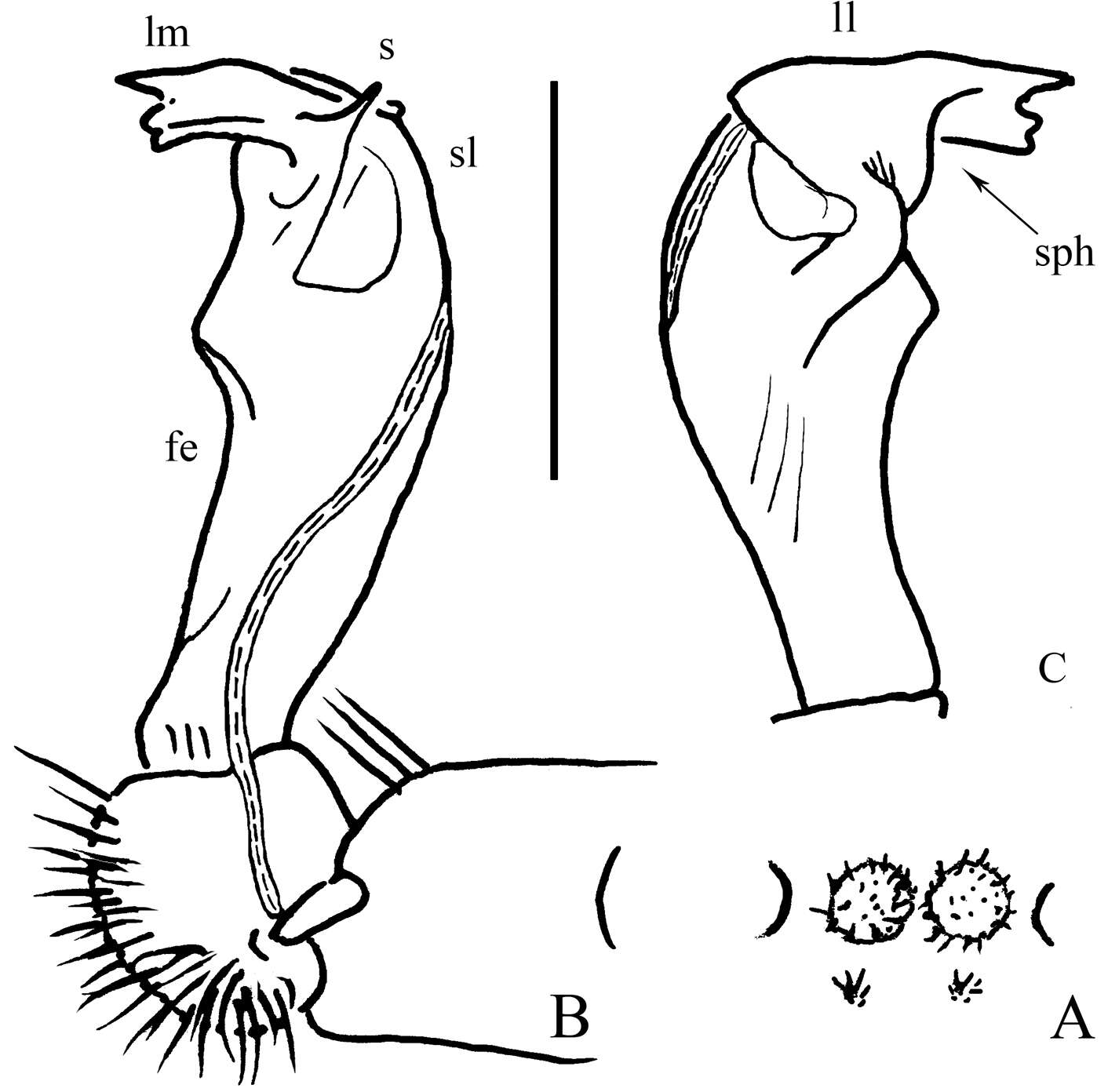
Figure 2.Desmoxytes eupterygota sp. n., ♂ paratype from near Tianhe. A sternal cones between coxae 4, ventral view B and C right gonopod, mesal and lateral views, respectively. Scale bar: 1.0 (A) and 0.5 mm (B, C). Designations: fe femorite sph solenophore sl solenomere ll lamina lateralis lm lamina medialis s spinule on lm
-
Figure 3.A–E Left lateral views of anterior end. A Asticopyrgodesmus maiala sp. n., holotype B Nephopyrgodesmus eungella sp. n., male paratype ex ANIC 64-000231 C Notopyrgodesmus kulla sp. n., male paratype ex ANIC 64-000242 D Nephopyrgodesmus lanosus sp. n., holotype E Nephopyrgodesmus weiri sp. n., holotype F Nephopyrgodesmus weiri sp. n., male paratype ANIC 64-000251, dorsal view of anterior end. Images not to same scale.
-
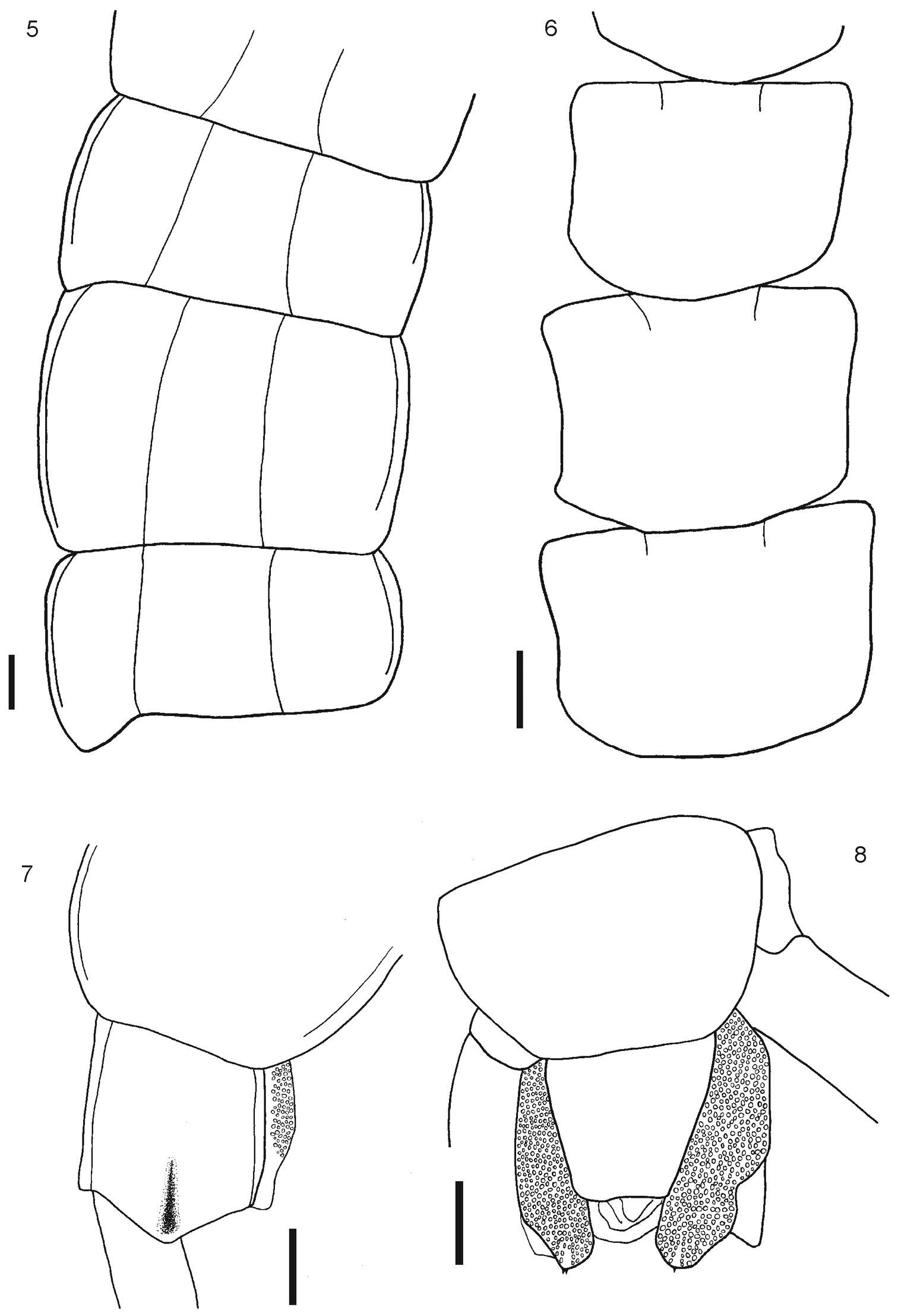
Figure 5–8. 5 Tergites 11, 12 and 13 6 Sternites 4, 5 and 6 7 Tergite 21 8 Segment 21 showing sternite 21 and coxopleuron. Scale bars 1 mm.
-
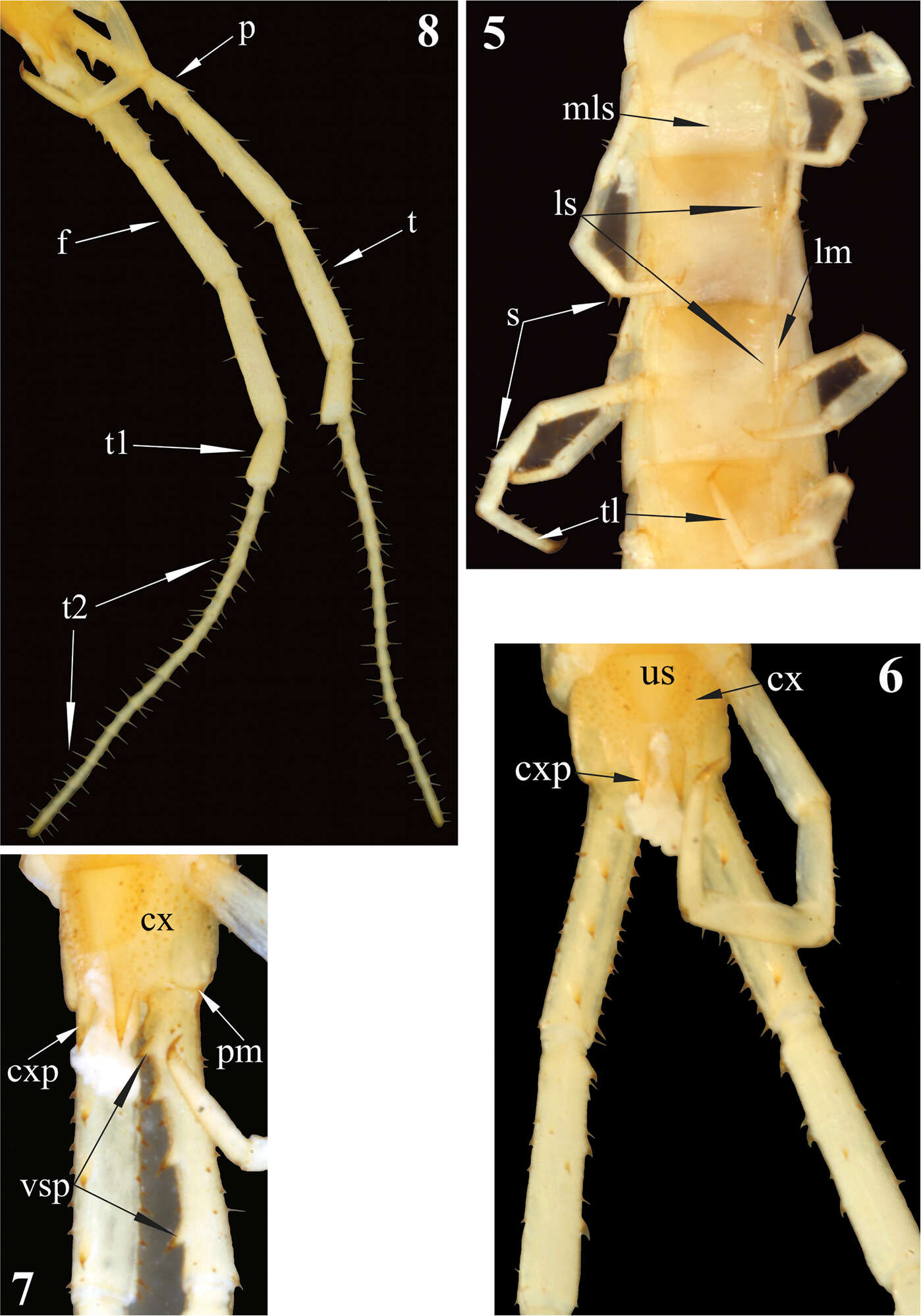
Figures 5–8.Newportia stoevi,sp. n. 5 Segments and midbody legs, ventral view 6 Posterior body end, ventral view 7 Left side of ultimate leg-bearing segment and prefemora of ultimate legs, ventro-lateral view 8 Ultimate legs, ventro-lateral view; (mls) – median longitudinal sulcus, (ls) – lateral sutures, (lm) – lateral margination, (s) – setae, (tl) – monoarticulated tarsus of locomotory leg, (us) – sternite of ultimate leg-bearing segment, (cx) – coxopleuron, (cxp) – coxopleural process, (pm) – posterior margin of pleuron of ultimate leg-bearing segment, (vsp) – ventral spinous processes of ultimate prefemur, (p) – prefemur, (f) – femur, (t) – tibia, (t1) – tarsus 1, (t2) – tarsus 2.
-
Sergei I. Golovatch, Jean-Jacques Geoffroy, Pavel Stoev, Didier Vanden Spiegel
Zookeys
Figure 13. Retrodesmus dammermani Chamberlin, 1945, ♂ holotype from Java, Indonesia A left paratergite 10, dorsal view B both gonopods in situ, ventral view. – Scale bars: 0.2 mm.
-
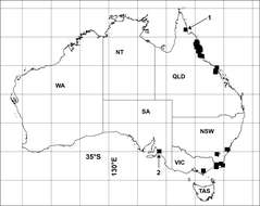
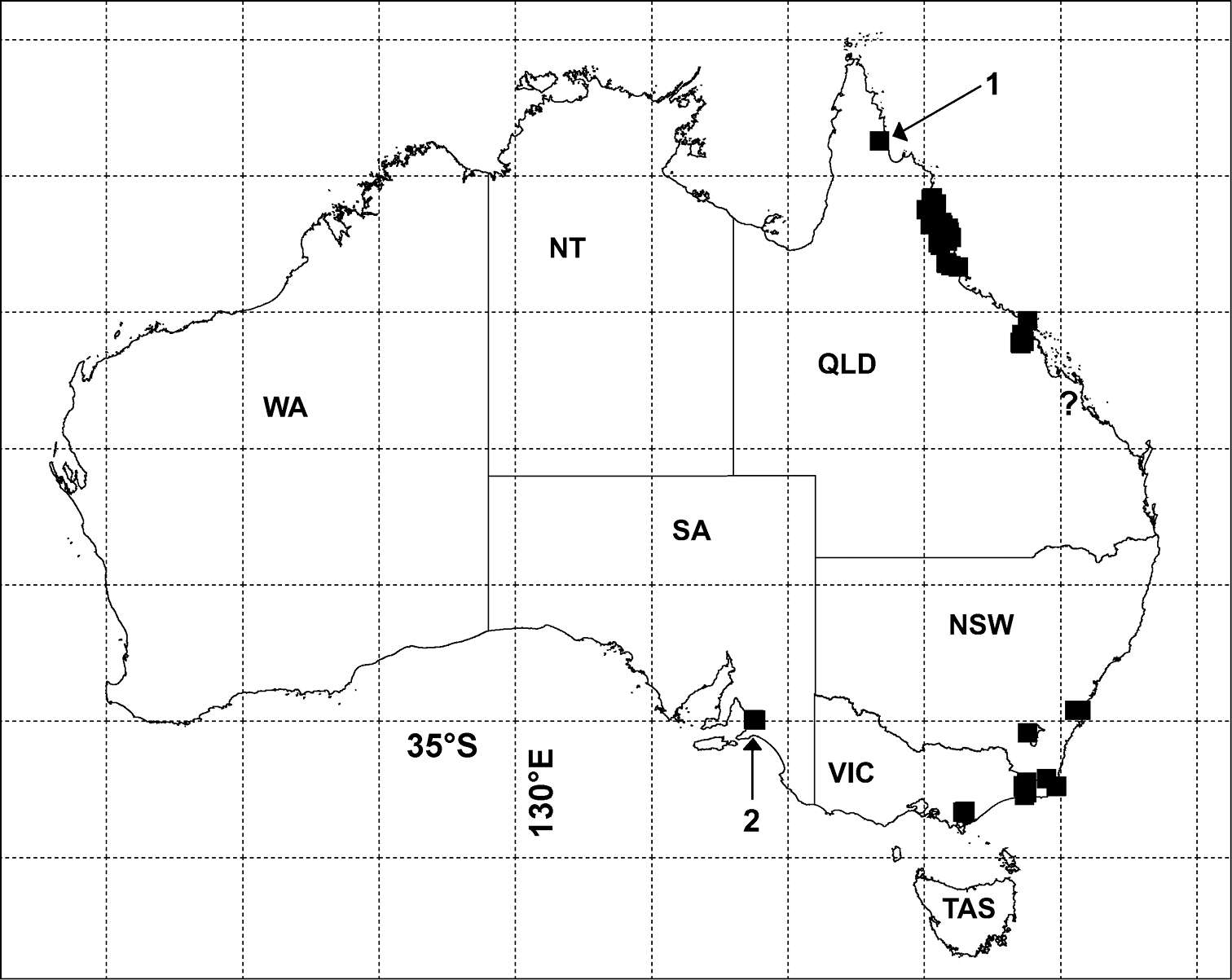
Figure 1.Localities for Agathodesmus spp. in Australia (filled black squares) as of July 2012. 1 = only known locality for Agathodesmus anici sp. n. 2 = cluster of 4 localities for Agathodesmus chandleri sp. n., ? = questionable Cammoo Caves locality for Agathodesmus agnus sp. n.; see Figs 11-13 for other species. Geographic projection, 5° latitude-longitude grid. NSW = New South Wales, NT = Northern Territory, QLD = Queensland, SA = South Australia, TAS = Tasmania, VIC = Victoria, WA = Western Australia.
-
Natdanai Likhitrakarn, Sergei I. Golovatch, Somsak Panha
Zookeys
Figure 1.Tetracentrosternus theelorsuensis sp. n., ♂ holotype. A habitus, live coloration B, D anterior part of body, lateral and dorsal views, respectively C, E, G segments 10 and 11, dorsal, ventral and lateral views, respectively F, H posterior part of body, dorsal and lateral views, respectively I, J sternal lobe between coxae 4, sublateral and subcaudal views, respectively.
-
Natdanai Likhitrakarn, Sergei I. Golovatch, Somsak Panha
Zookeys
Figure 2.Orthomorpha paviei Brölemann, 1896, ♂ from Laos, left gonopod. A, B lateral and mesal views, respectively C, D telopodite, lateral and mesal views, respectively E, F distal part, subcaudal and suboral views, respectively. Scale bar: 0.2 mm.
-
Sergei I. Golovatch, Jean-Jacques Geoffroy, Didier VandenSpiegel
Zookeys
Figure 2.Cocacolaria hauseri Hoffman, 1987, ♂ from Natawa forest, Espirito Santo Island, Vanuatu; A midbody leg, lateral view B gonopod aperture with both gonopods in situ, ventral view C right gonopod, lateral view D, E left gonopod, caudomesal and mesal views, respectively. Scale bars: A, B 0.05 mm; C–E 0.02 mm. Designations of gonopod structures in text.