-
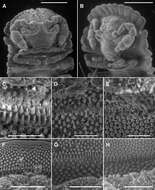
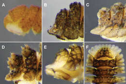
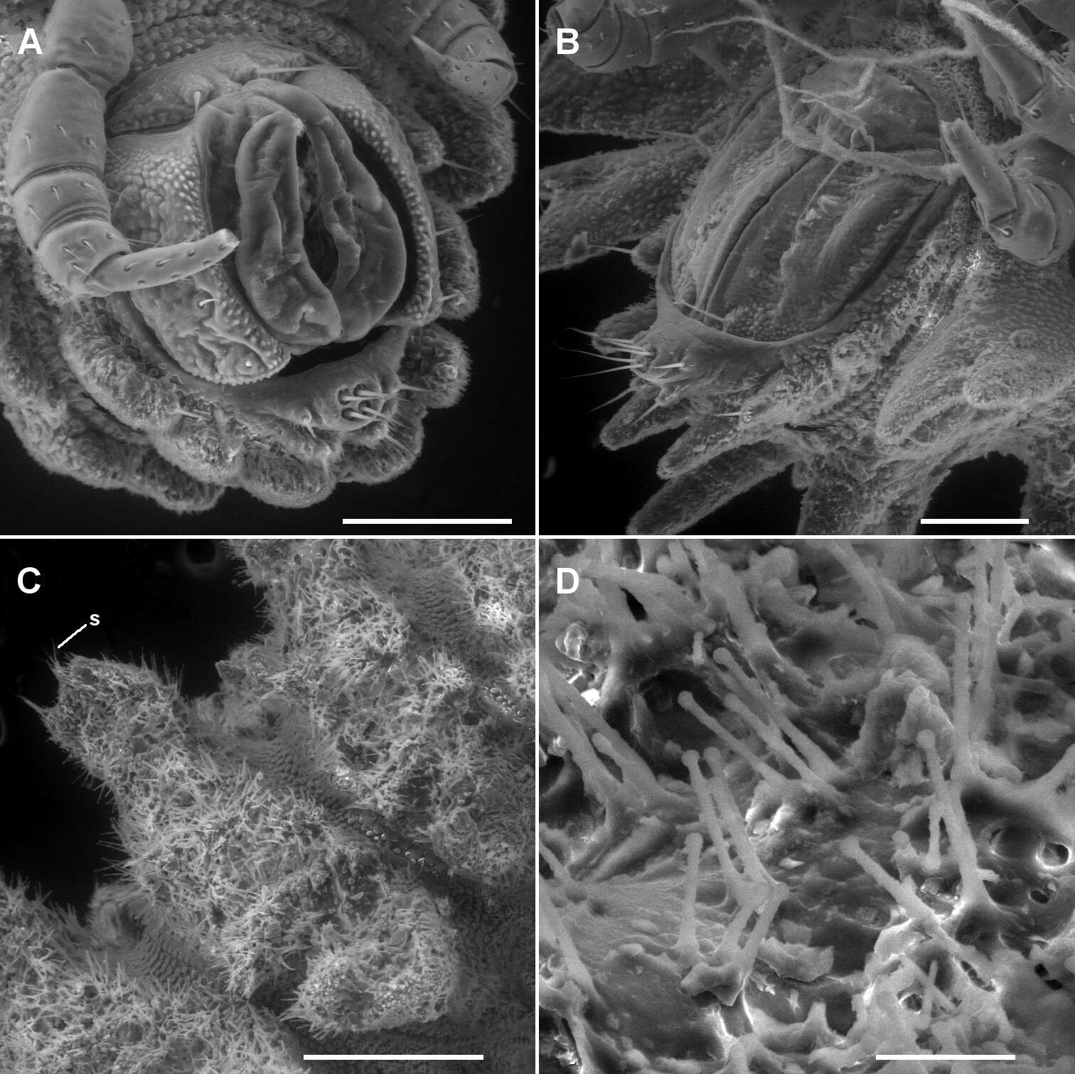
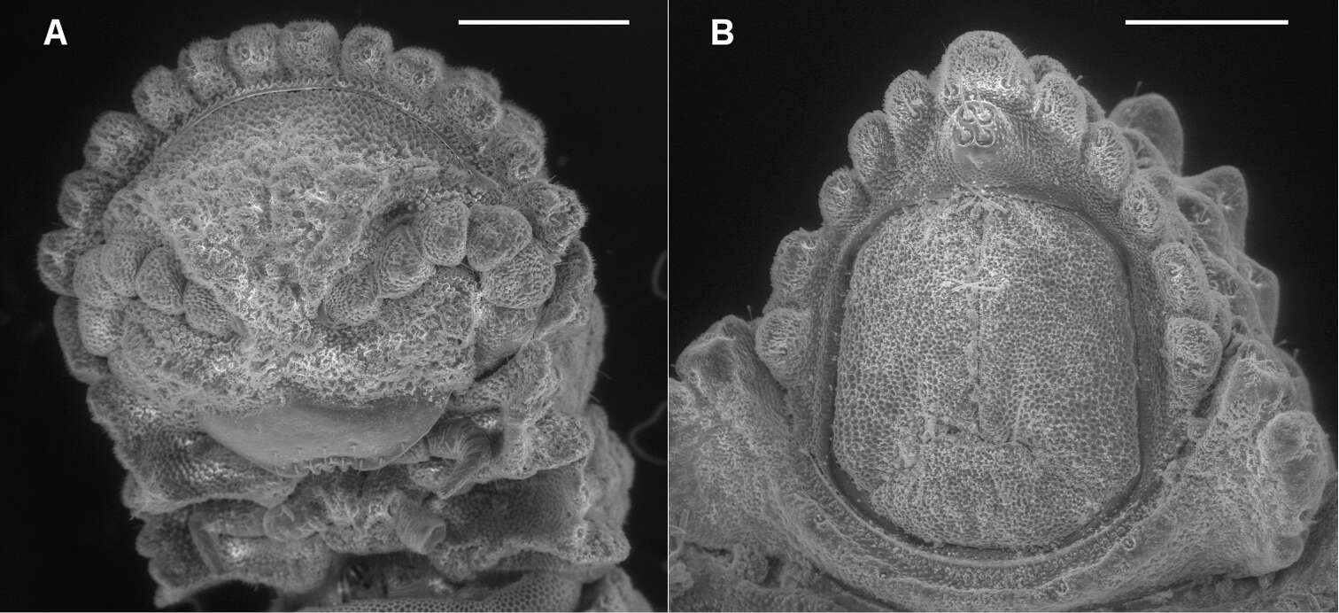
Figure 6.A, B Ventrolateral views of telson of Asticopyrgodesmus maiala sp. n., male paratype ex ANIC 64-000220 (A) and Notopyrgodesmus weiri sp. n., male paratype ANIC 64-000249 (B) C Right lateral view of midbody ring of Notopyrgodesmus kulla sp. n., male paratype ex ANIC 64-000243, anterior to upper right; s = ‘spine’ on tip of paramedian tubercle D Close-up of hair-like cuticular outgrowths on specimen in C Scanning electron micrographs of uncoated specimens; scale bars: A, B = 0.1 mm, C = 0.25 mm, D = 0.025 mm.
-
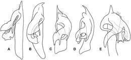
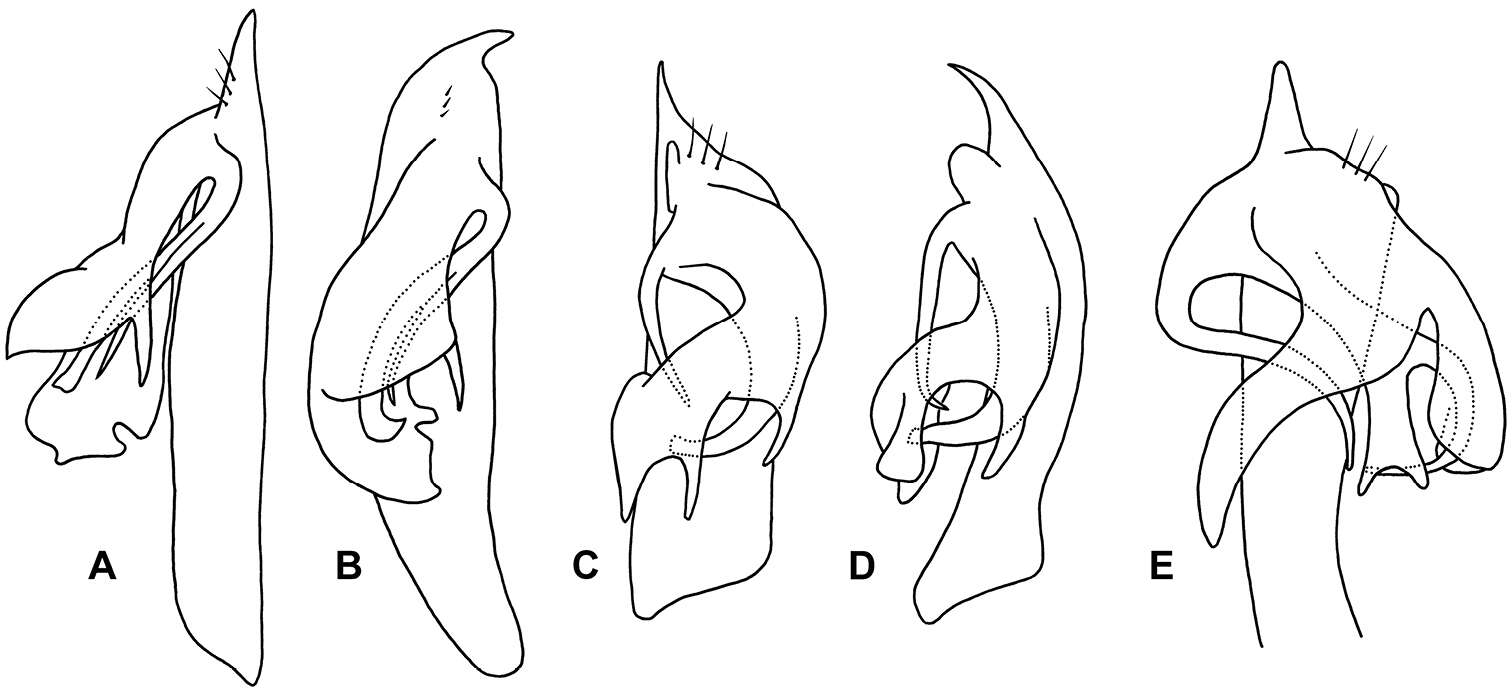
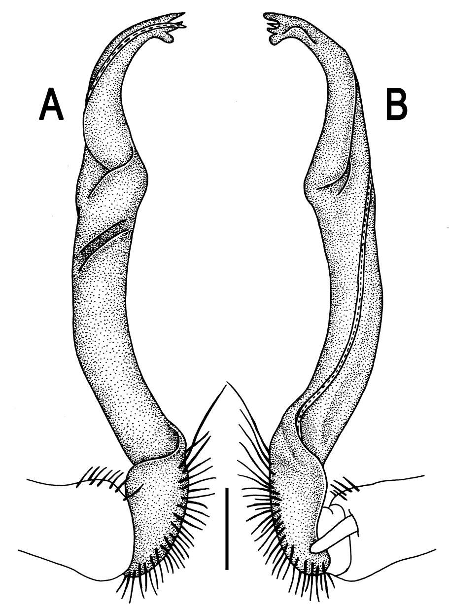
Figure 8.Agathodesmus spp. gonopod telopodites, not to same scale. A, B Agathodesmus gayundah sp. n., paratype ex QM S96039, right gonopod, posterior (A) and lateral (B) views C, D Agathodesmus millaa sp. n., paratype ex QM S96055, left gonopod, posterior (C) and lateral (D) views, as not shown in lateral view E Agathodesmus quintanus sp. n., paratype ex QM S96074, left gonopod, posterior and slightly basal view.
-
Sergei I. Golovatch, Jean-Jacques Geoffroy, Didier VandenSpiegel
Zookeys
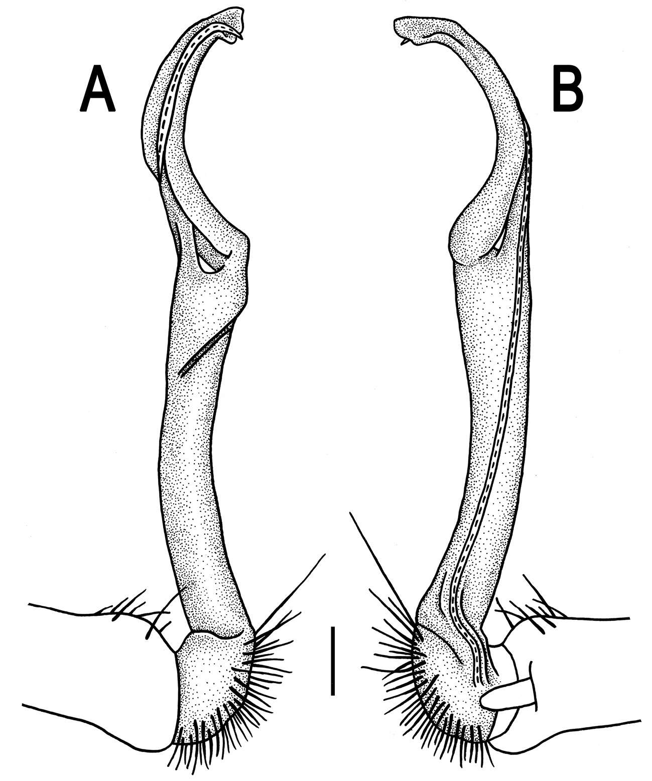
Figure 14.Helicodesmus anichkini sp. n., ♂ paratype; A both gonopods in situ, ventral view B–D right gonopod, anteroventral, ventrolateral and lateral views, respectively. Scale bars: A, B, D 0.05 mm; C 0.02 mm. Designations of gonopod structures in text.
-
Natdanai Likhitrakarn, Sergei I. Golovatch, Somsak Panha
Zookeys
Figure 10.Tylopus hilaris (Attems, 1937), ♂ holotype; A, B anterior part of body, dorsal and lateral views, respectively C segments 10 and 11, dorsal view D segments 9–11, lateral view E–G posterior part of body, lateral, dorsal and ventral views, respectively H, I sternal cones between coxae 4, subcaudal and sublateral views, respectively.
-
Natdanai Likhitrakarn, Sergei I. Golovatch, Somsak Panha
Zookeys
Figure 108.Orthomorpha similanensis sp. n., ♂ holotype. A, B right gonopod, lateral and mesal views, respectively.Scale bar: 0.2 mm.
-
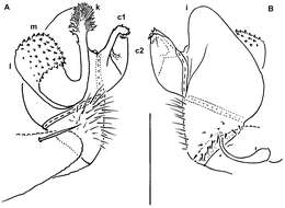
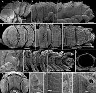
Figure 3.Left ventrolateral views of gonopods of Prosopodesmus species. A Prosopodesmus crater sp. n., paratype, ANIC 64–000212. B Prosopodesmus kirrama sp. n., paratype, QM S91627. Tip of left gonopod telopodite of Prosopodesmus crater is broken. Scale bars = 0.2 mm. Image contrast is low because specimens are uncoated.
-
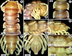
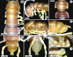
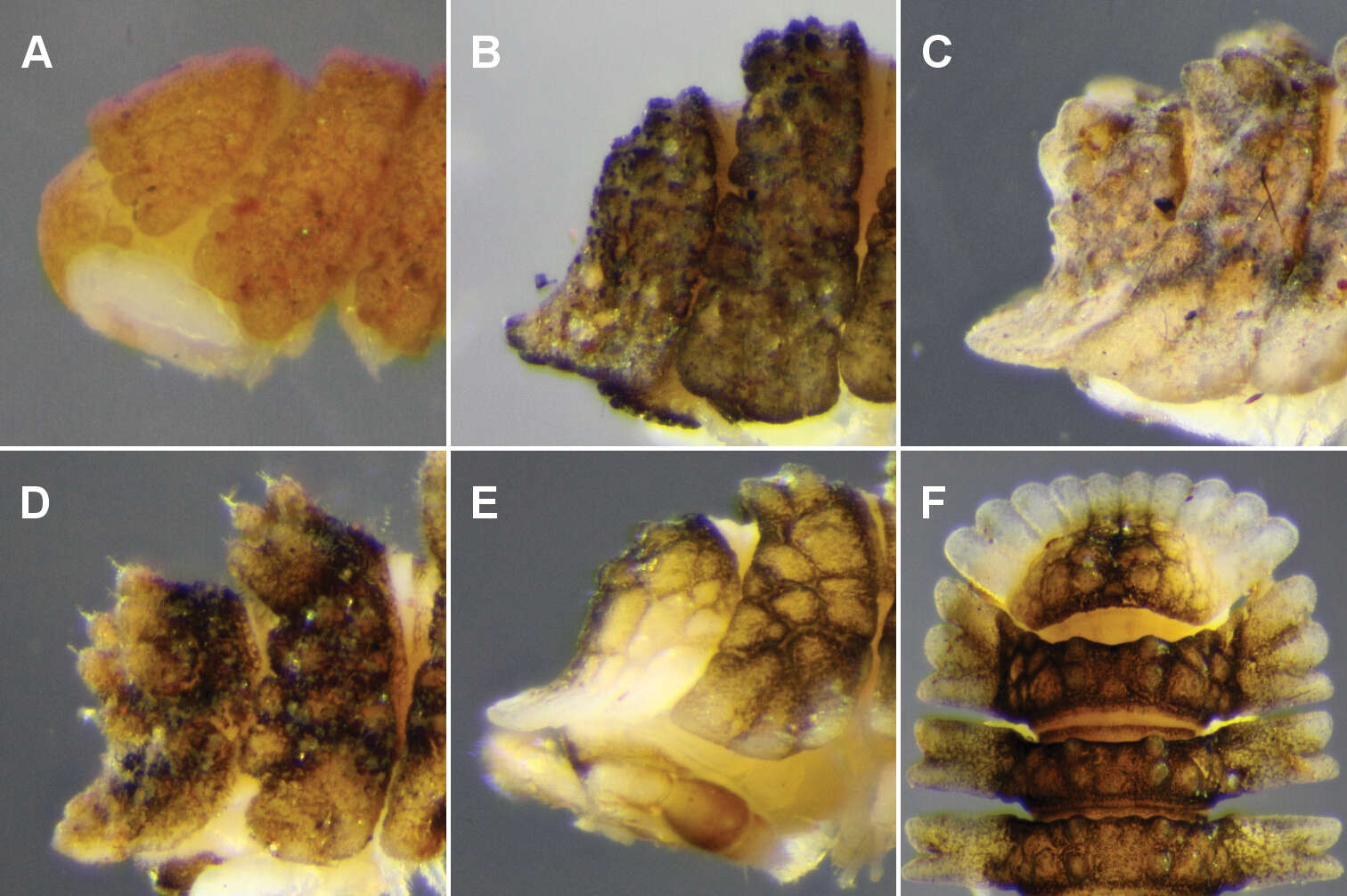
Figure 10.A Notopyrgodesmus kulla sp. n., male (left) and female (right) paratypes ex QM S92793. Scale bar = 5 mm B–D Dorsal views of midbody rings of males, not to same scale B Notopyrgodesmus kulla sp. n., paratype ex QM S92793 C Notopyrgodesmus lanosus sp. n., holotype D Notopyrgodesmus weiri sp. n., paratype ANIC 64-000251.
-
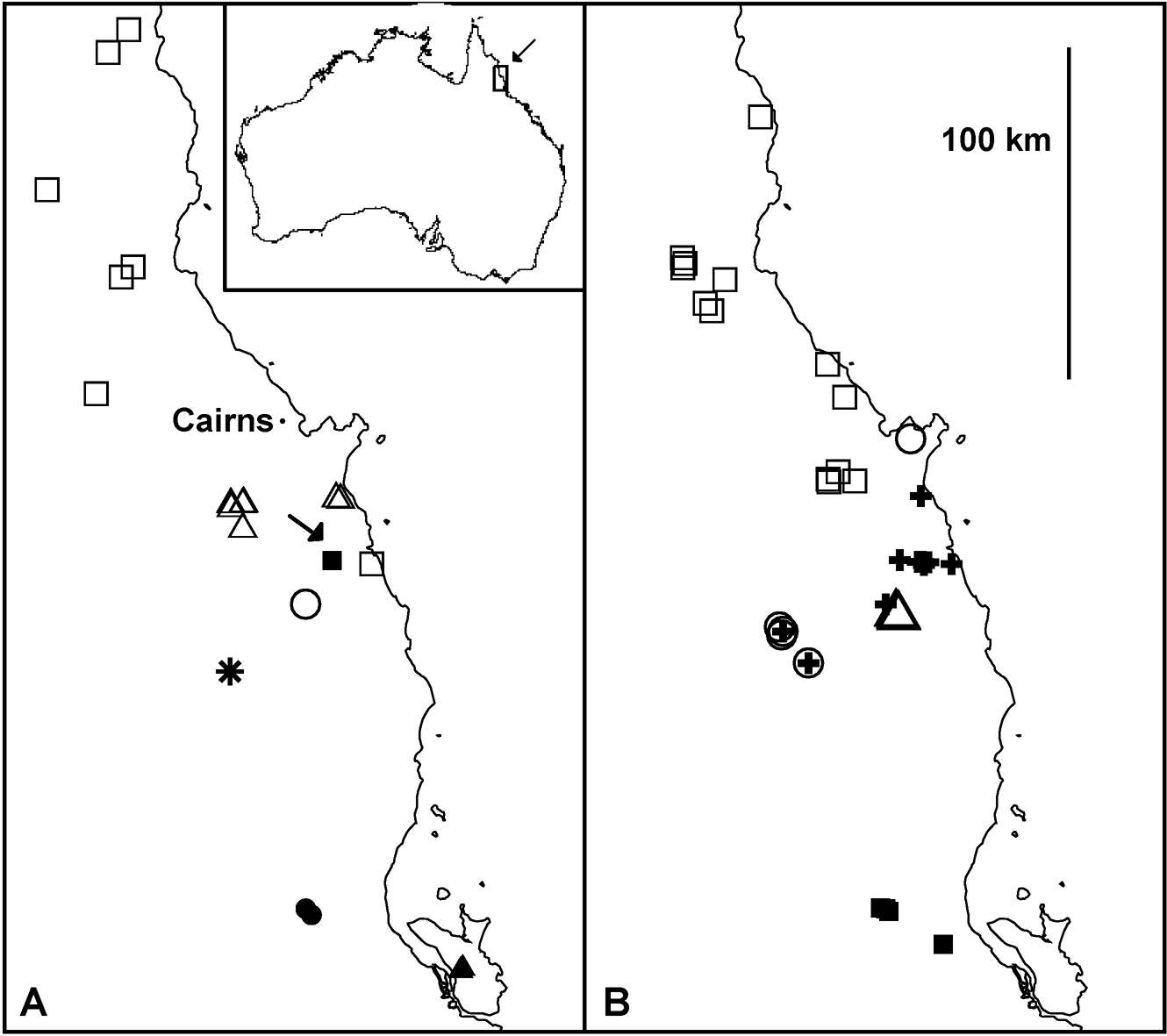
Figure 13.Localities in far north Queensland for A Agathodesmus adelphus sp. n. (open circle), Agathodesmus agnus sp. n. (open triangles), Agathodesmus gayundah sp. n. (filled triangle), Agathodesmus hahnensis sp. n. (open squares), Agathodesmus kerensis sp. n. (filled square), Agathodesmus kirrama sp. n. (filled circles), Agathodesmus millaa sp. n. (star) and B Agathodesmus parapholeus sp. n. (open circles), Agathodesmus quintanus sp. n. (crosses), Agathodesmus sagma sp. n. (open squares), Agathodesmus summus sp. n. (open triangles), and Agathodesmus yuccabinensis sp. n. (filled squares). Arrow in A indicates questionable, disjunct locality for Agathodesmus aenigmaticus sp. n. Mercator projections; inset shows location of main maps.
-
Sergei I. Golovatch, Jean-Jacques Geoffroy, Didier VandenSpiegel
Zookeys
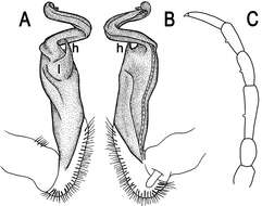
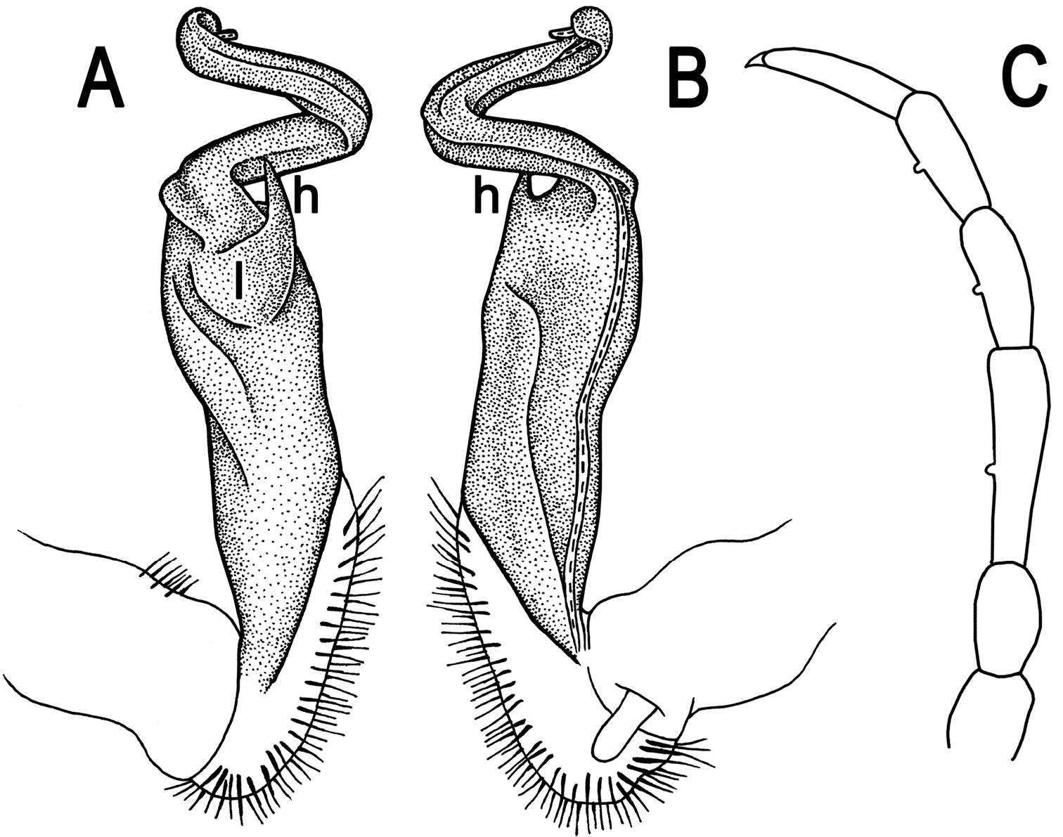
Figure 15.Helicodesmus anichkini sp. n., ♂ paratype; A, B right gonopod, lateral and mesal views, respectively. Scale bar: 0.1 mm. Designations of gonopod structures in text.
-
Natdanai Likhitrakarn, Sergei I. Golovatch, Somsak Panha
Zookeys
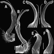
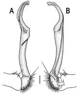
Figure 11.Tylopus hilaris (Attems, 1937), ♂ holotype; A, B right gonopod, lateral and mesal views, respectively C leg of segment 10, depicted not to scale.
-
Natdanai Likhitrakarn, Sergei I. Golovatch, Somsak Panha
Zookeys
Figure 13.Orthomorpha tuberculifera sp. n., ♂ holotype. A habitus, live coloration. B, C anterior part of body, dorsal and lateral views, respectively D, E segments 10 and 11, dorsal and lateral views, respectively F, G, H posterior part of body, dorsal, ventral and lateral views, respectively I, J sternal cones between coxae 4, subcaudal and sublateral views, respectively.
-
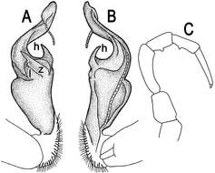
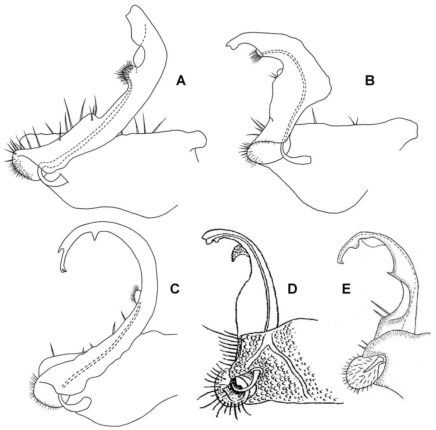
Figure 5.Medial views of right gonopod of Prosopodesmus species, not to same scale. A Prosopodesmus crater sp. n., paratype, ANIC 64–000214. B Prosopodesmus kirrama sp. n., paratype, QM S91626. C Prosopodesmus monteithi sp. n., QM 91635. D Prosopodesmus sinuatus (Miyosi, 1958), holotype, drawing scanned and modified from Fig. 1G in Miyosi (1958). E Prosopodesmus similis (Haga, 1968), holotype, drawing scanned and modified from Fig. 12B in Haga (1968).
-
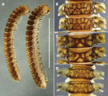
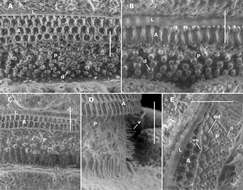
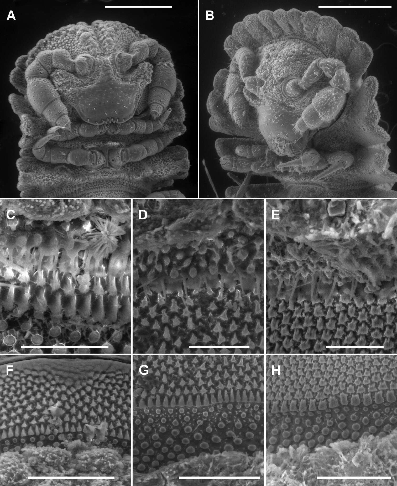
Figure 2.A, B Ventral views of head of Asticopyrgodesmus maiala sp. n., male paratype ex ANIC 64-000220 (A) and Notopyrgodesmus weiri sp. n., male paratype ANIC 64-000249 C, D, E Views of lobe-and-spike limbus on midbody rings (anterior at top) of Asticopyrgodesmus maiala sp. n., male paratype ex ANIC 64-000220 (C), Nephopyrgodesmus eungella sp. n., male paratype ex ANIC 64-000231 (D) and Notopyrgodesmus kulla sp. n., male paratype ex ANIC 64-000243 (E) F, G, H Views of prozonite sculpture on midbody rings (anterior at top) of Asticopyrgodesmus lamingtonensis sp. n., male paratype ex ANIC 64-000217 (F), Notopyrgodesmus eungella sp. n., male paratype ex ANIC 64-000231 (G) and Notopyrgodesmus kulla sp. n., male paratype ex ANIC 64-000243 (H). Scanning electron micrographs of uncoated specimens; scale bars: A = 0.5 mm, B = 0.2 mm, C, D, E, H = 0.05 mm, F, G = 0.1 mm.
-
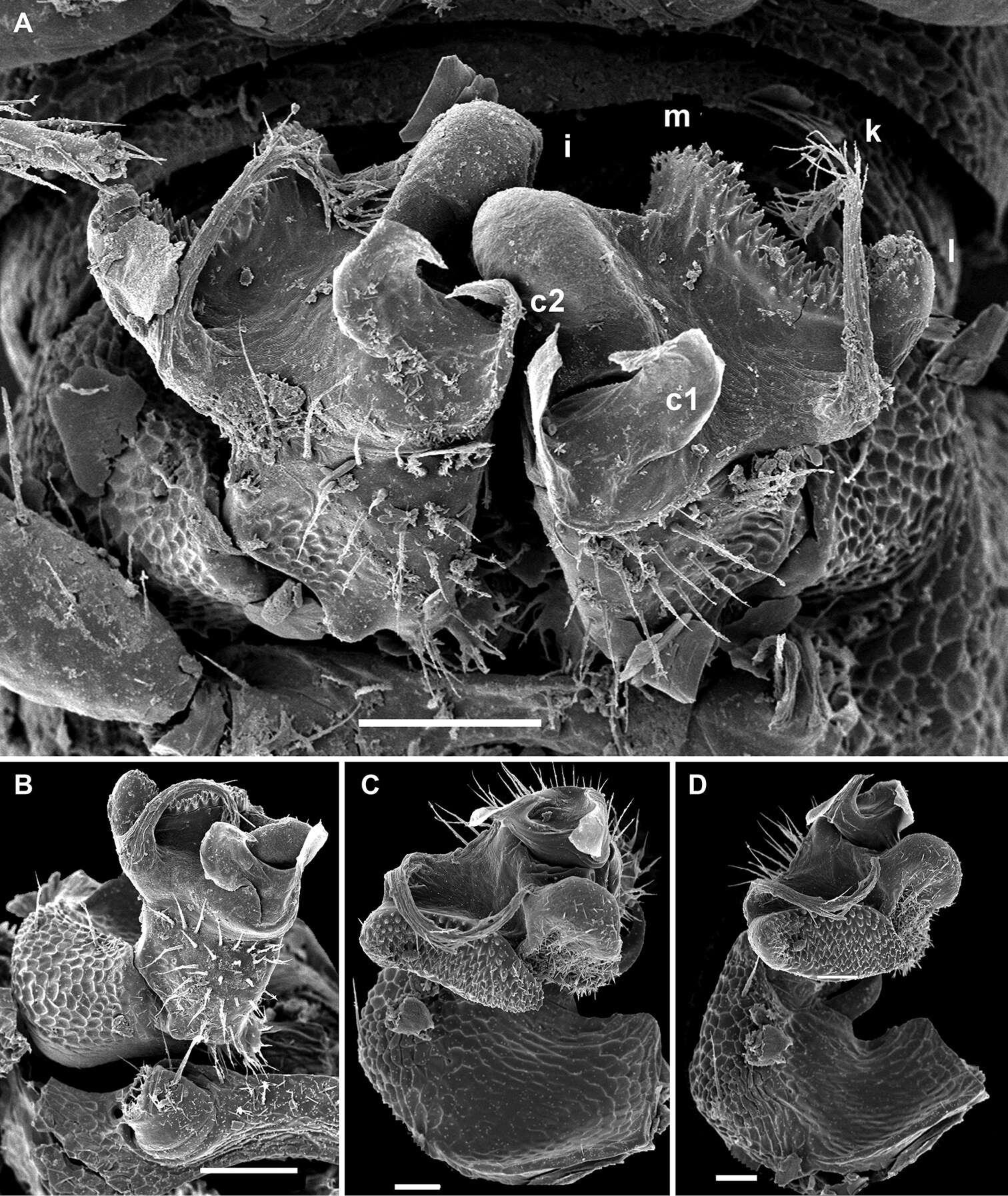
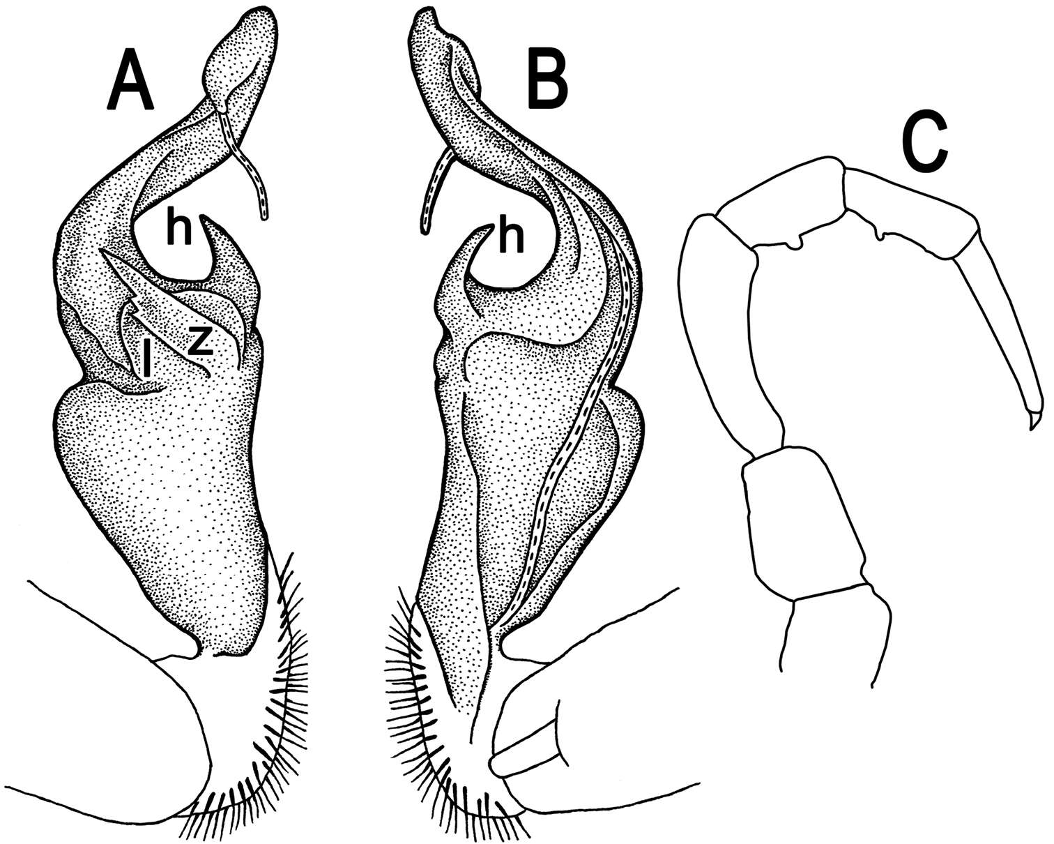
Figure 7.Posterior gonopod views. A Agathodesmus bonang sp. n., paratype ex series NMV K-11861-11869 B Agathodesmus carorum sp. n., NMV K-11884 C Agathodesmus chandleri sp. n., paratype ex series SAM OM-2004-2018 D Agathodesmus hahnensis sp. n., paratype ex QM S38962. B is uncoated specimen; scale bars: A, B, C = 0.1 mm, D = 0.2 mm.
-
Sergei I. Golovatch, Jean-Jacques Geoffroy, Didier VandenSpiegel
Zookeys
Figure 16.Monstrodesmus flagellifer sp. n., ♂ paratype; A, D anterior body part, lateral and dorsal views, respectively B, E, H midbody segments, lateral, dorsal and ventral views, respectively C, F, I posterior body part, lateral, dorsal and ventral views, respectively G, K head, frontal and ventral views, respectively J cross-section of a midbody segment, caudal view L antennomeres 4-8, sublateral view; tergal seta M tegument structure, limbus and stricture region N tergal seta, lateral view O ozopore region of a midbody segment, sublateral view. Scale bars: A–J 0.1 mm; K, L 0.05 mm; M–O 0.01 mm.
-
Natdanai Likhitrakarn, Sergei I. Golovatch, Somsak Panha
Zookeys
Figure 12.Tylopus sigma (Attems, 1953), ♂ paralectotype; A, B anterior part of body, dorsal and lateral views, respectively C segments 10 and 11, dorsal view D segments 9–11, lateral view E–G posterior part of body, lateral, dorsal and ventral views, respectively H, I sternal cones between coxae 4, caudal and lateral views, respectively.
-
Natdanai Likhitrakarn, Sergei I. Golovatch, Somsak Panha
Zookeys
Figure 14.Orthomorpha tuberculifera sp. n., ♂ paratype from Khao Rup Chang. A, B right gonopod, mesal and lateral views, respectively C-F distal part of right gonopod, mesal, lateral, suboral and subcaudal views, respectively. Scale bar: 0.2 mm.
-
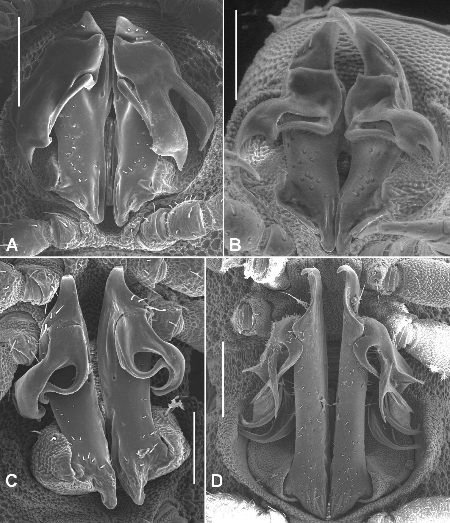
Figure 1.A Ventral view of head of Prosopodesmus crater sp. n., paratype, ANIC 64–000212, showing 12 lobes on anterior edge of collum, antennae retracted below edges of collum and ring 2 tergite, and textured frons with smooth clypeus. B Ventral view of telson of Prosopodesmus monteithi sp. n., QM S91632, showing 5+5 lobe pattern on edge of preanal ring and apical epiproct.
-
Figure 3.A–E Left lateral views of anterior end. A Asticopyrgodesmus maiala sp. n., holotype B Nephopyrgodesmus eungella sp. n., male paratype ex ANIC 64-000231 C Notopyrgodesmus kulla sp. n., male paratype ex ANIC 64-000242 D Nephopyrgodesmus lanosus sp. n., holotype E Nephopyrgodesmus weiri sp. n., holotype F Nephopyrgodesmus weiri sp. n., male paratype ANIC 64-000251, dorsal view of anterior end. Images not to same scale.
-
Figure 13.Localities in far north Queensland for A Agathodesmus adelphus sp. n. (open circle), Agathodesmus agnus sp. n. (open triangles), Agathodesmus gayundah sp. n. (filled triangle), Agathodesmus hahnensis sp. n. (open squares), Agathodesmus kerensis sp. n. (filled square), Agathodesmus kirrama sp. n. (filled circles), Agathodesmus millaa sp. n. (star) and B Agathodesmus parapholeus sp. n. (open circles), Agathodesmus quintanus sp. n. (crosses), Agathodesmus sagma sp. n. (open squares), Agathodesmus summus sp. n. (open triangles), and Agathodesmus yuccabinensis sp. n. (filled squares). Arrow in A indicates questionable, disjunct locality for Agathodesmus aenigmaticus sp. n. Mercator projections; inset shows location of main maps.
-
Sergei I. Golovatch, Jean-Jacques Geoffroy, Didier VandenSpiegel
Zookeys
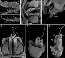
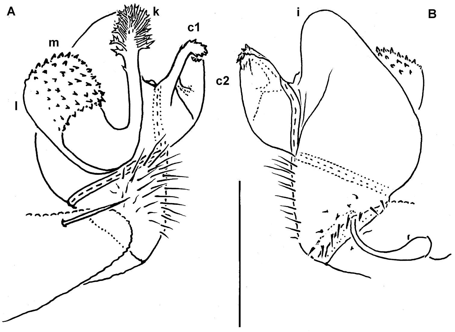
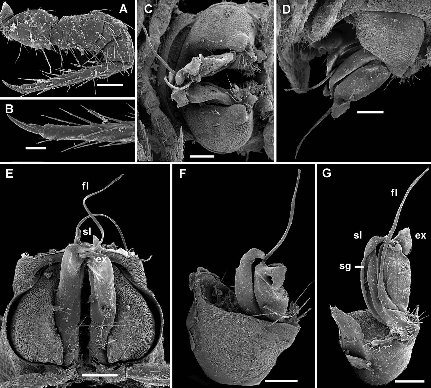
Figure 17.Monstrodesmus flagellifer sp. n., ♂ paratype; A midbody leg, lateral view B claw of a midbody leg C–E both gonopods in situ, ventral, lateral and ventrocaudal views, respectively F right gonopod, lateral view G left gonopod, mesal view. Scale bars: C–G 0.1 mm; A 0.05 mm; B 0.02 mm. Designations of gonopod structures in text.
-
Natdanai Likhitrakarn, Sergei I. Golovatch, Somsak Panha
Zookeys
Figure 13.Tylopus sigma (Attems, 1953), ♂ lectotype; A, B right gonopod, lateral and mesal views, respectively C leg of segment 10, depicted not to scale.
-
Natdanai Likhitrakarn, Sergei I. Golovatch, Somsak Panha
Zookeys
Figure 15.Orthomorpha tuberculifera sp. n., ♂ holotype. A, B right gonopod lateral and mesal views, respectively. Scale bar: 0.2 mm.
-
Figure 2.Midbody limbus and prozonite of Prosopodesmus species, dorsal views. A Prosopodesmus crater sp. n., paratype, QM S37593. B Prosopodesmus kirrama sp. n., paratype, QM S91627. C, D Prosopodesmus monteithi sp. n., QM S91632. E Prosopodesmus panporus Blower and Rundle, 1980, ANIC 64–000118. L limbus A anterior portion of prozonite P posterior portion of prozonite d disk mt microtubercles v villi ve microvillose extensions. Scale bars: A, B, E = 0.05 mm, C, D = 0.1 mm.