-
Saša Širca, Gregor Urek, Stela Lazarova, Milka Elshishka, Vlada Peneva
Zookeys
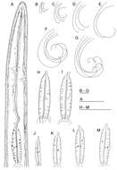
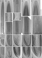
Figure 1.Longidorus carniolensis sp. n. Female: A Neck region F Habitus H Pharyngeal bulb Male: G Habitus I Pharyngeal bulb; Juveniles: B–E Habitus of first, second, third and forth juvenile stages J–M Pharyngeal bulb of first, second, third and forth juvenile stages. Scale bars: B–G 1 mm; A, H–M 100 μm.
-
Habibeh Jabbari, Gholamreza Niknam, Maria Teresa Vinciguerra, Shalaleh Moslehi, Joaquín Abolafia, Reyes Peña-Santiago
Zookeys
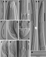
Figure 1.Crassolabium persicum sp. n. (all images are in lateral view) A Anterior region B Lip region and amphid fovea in surface C Pharyngeal expansion D Vagina E Spicules and lateral guiding piece F Female, posterior body region G Female, anterior genital branch H Male, posterior body region I Male, entire J Female, entire.
-
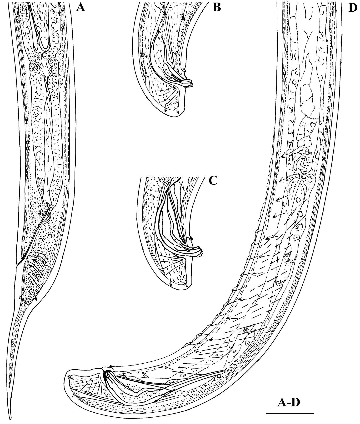
Vlada K. Peneva, Stela S. Lazarova, Francesca De Luca, Derek J. F. Brown
Zookeys
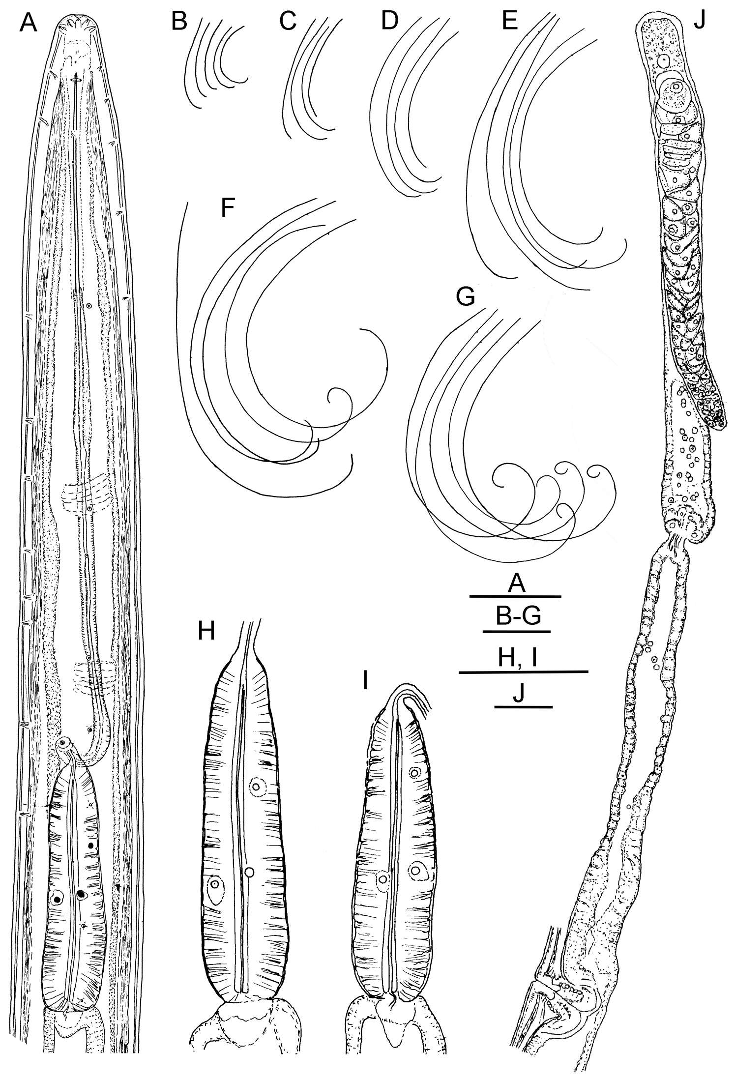
Figure 1.Longidorus cholevae sp. n. Female: A Anterior end F Habitus I Pharyngeal bulb J Anterior genital branch Male: G Habitus H Pharyngeal bulb Juveniles: B–E Habitus of first-, second-, third- and fourth-stage juveniles. Scale-bars: A, H, I, J 50 μm; B–G 1 mm.
-
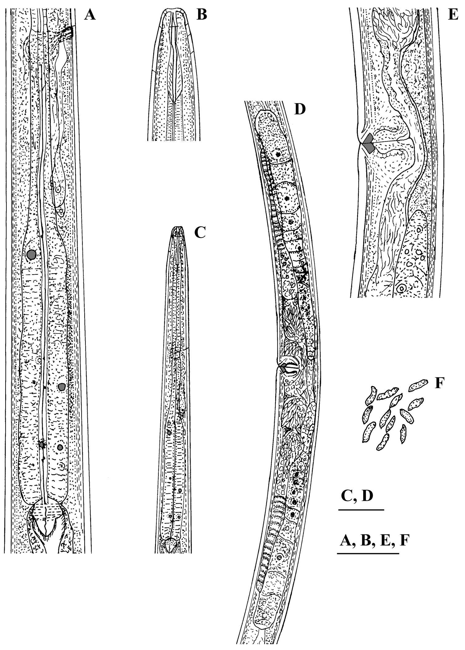
Sevdan Nedelchev, Milka Elshishka, Stela Lazarova, Georgi Radoslavov, Peter Hristov, Vlada Peneva
Zookeys
Figure 2.Calcaridorylaimus castaneae sp. n. Female: A Pharyngeal gland nuclei B Anterior region C Pharyngeal region D Genital system E Vulval region F Sperm cells in uterus. Scale bars: A, B, E, F – 30 μm; C, D – 50 μm.
-
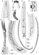
Figure 2.Longior longior Morffe & García sp. n. (female). A Esophageal region B Cephalic end C Tail, ventral view D Vulva E Egg F Genital tract G Entire nematode, lateral view.
-
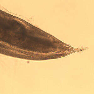
Enterobius vermicularis
-
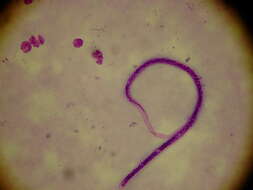
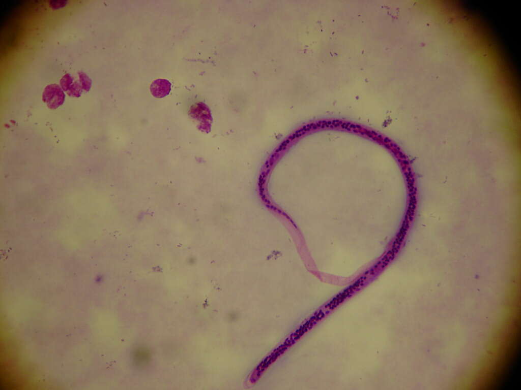
Wuchereria bancrofti
-
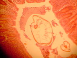
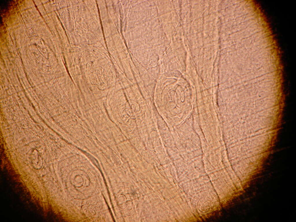
Trichinella spiralis
-
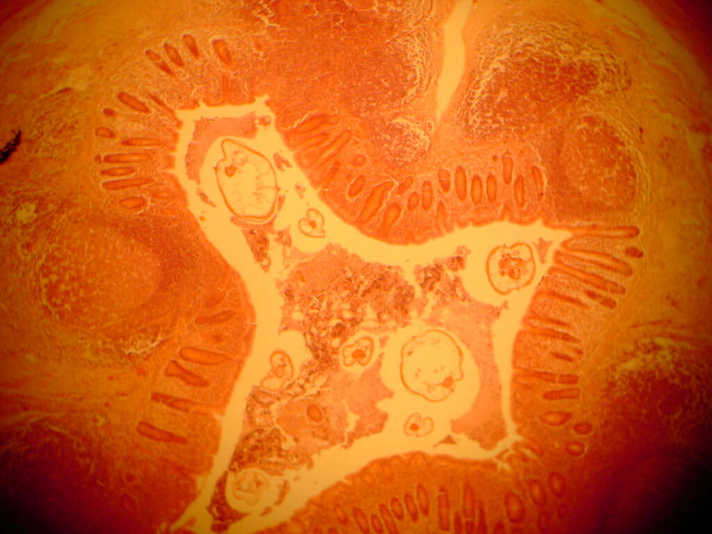
Öesophagostomum sp. adult femalePosterior end of a female Oesophagostomum sp., showing the pointed tail.From
CDC DPDx website
-
Saša Širca, Gregor Urek, Stela Lazarova, Milka Elshishka, Vlada Peneva
Zookeys
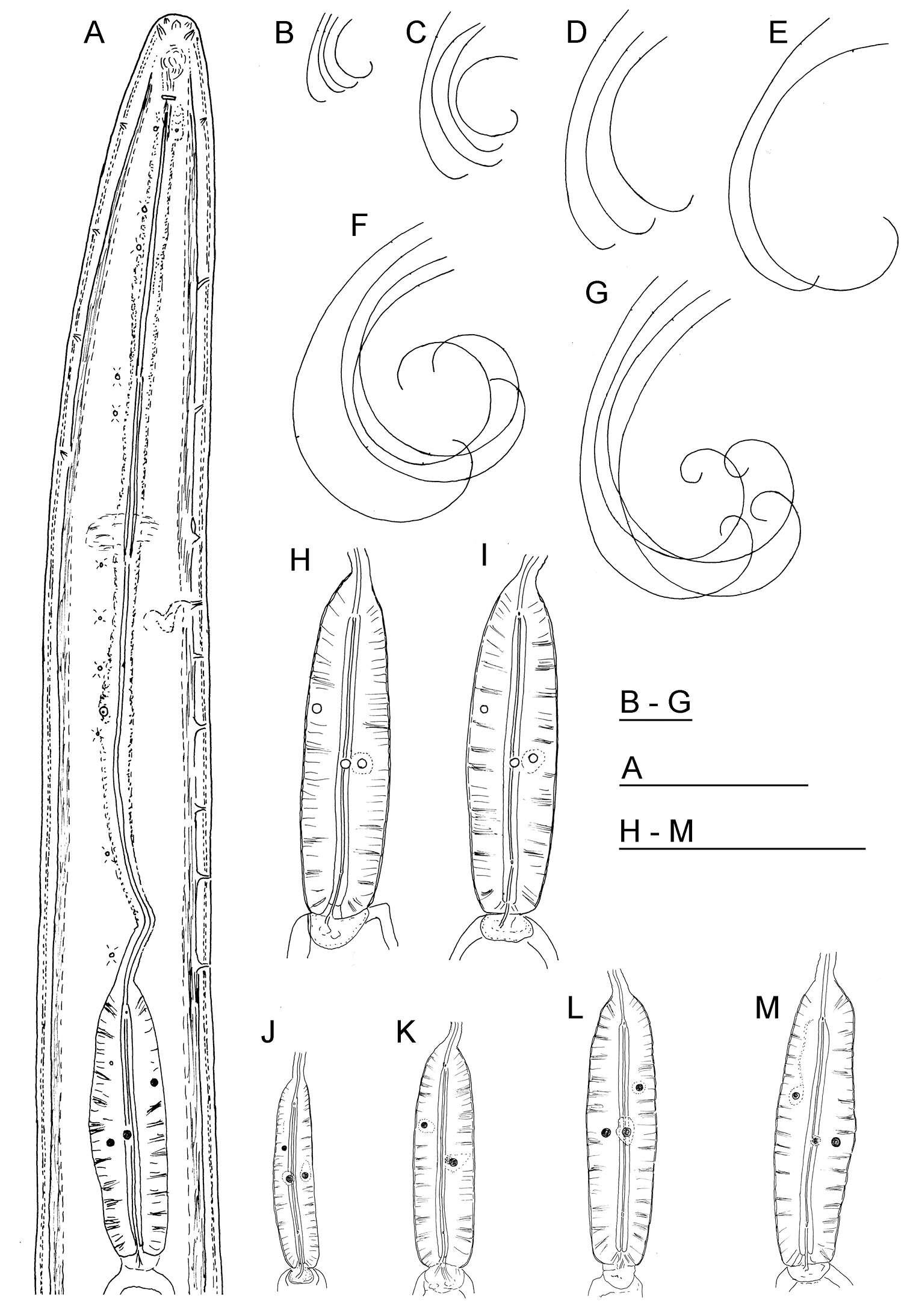
Figure 10.Longidorus carniolensis sp. n. Juvenile: A–D Anterior region of first, second, third and forth stages H–K Pharyngeal bulb of first, second, third and forth juvenile stages M, F, G, R genital primordium of first, second, third and forth stages N, S Tail shape of first stage O, T Tail shape of second stage P, U Tail shape of third stage Q, V Tail shape of forth stage Female: E Anterior region L Pharyngeal bulb W Tail shape. Scale bar: 50 μm.
-
Habibeh Jabbari, Gholamreza Niknam, Maria Teresa Vinciguerra, Shalaleh Moslehi, Joaquín Abolafia, Reyes Peña-Santiago
Zookeys
Figure 2.Crassolabium persicum sp. n. (light micrographs all in lateral view).A Female, entire B Anterior region C Pharyngo–intestinal junction D Female, genital system E Neck region F Male, entire G Male, posterior region H Vagina I Female, caudal region J Spicules K Lateral guiding piece L Oviduct–uterus junction M Lateral chord and pores. (Scale bars: A, F – 500 µm; B, H, K – 10 µm; C, G – 50 µm; D, E – 100 µm; I, J, L, M – 20 µm).
-
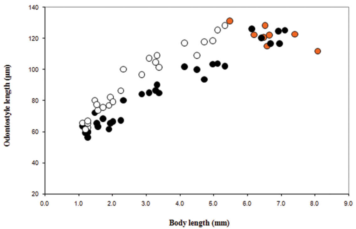
Vlada K. Peneva, Stela S. Lazarova, Francesca De Luca, Derek J. F. Brown
Zookeys
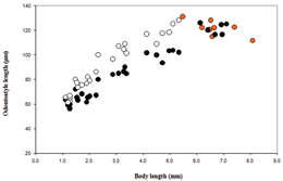
Figure 9.Longidorus cholevae sp. n. Scatter plot of the functional (˜, juveniles and adults, females in orange) and replacement (™, juveniles) odontostyle in relation to body length of the juvenile developmental stages and adults.
-
Sevdan Nedelchev, Milka Elshishka, Stela Lazarova, Georgi Radoslavov, Peter Hristov, Vlada Peneva
Zookeys
Figure 3.Calcaridorylaimus castaneae sp. n. Female: A Posterior region. Male: B, C Extruded spicules with supplements D Posterior region. Scale bar: A–D – 30 μm.
-
Figure 3.Longior longior Morffe & Garcíasp. n. (male). A Entire nematode, lateral view B Esophageal region, lateral view C Cephalic end D Tail, lateral view.
-
Enterobius vermicularis
-
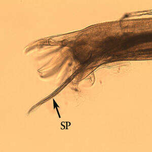
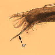
Male Oesophagostomum sp. Posterior end of male Oesophagostomum sp. showing the bursa. Note the spicule (SP).From
CDC DPDx Website
-
Saša Širca, Gregor Urek, Stela Lazarova, Milka Elshishka, Vlada Peneva
Zookeys
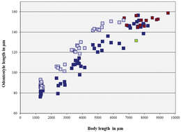
Figure 11.Longidorus carniolensis sp. n. Scatter plot of the functional (, dark blue) and replacement odontostyle (, light blue) in relation to the body length of the juvenile stages and adults: females (, dark blue) and males (, red), female with very short odontostyle (, green).
-
Vlada K. Peneva, Stela S. Lazarova, Francesca De Luca, Derek J. F. Brown
Zookeys
Figure 10.Phylogenetic relationships of Longidorus cholevae sp. n. and its closest species for the D2-D3 rDNA. Bayesian Inference strict consensus tree acquired under GTR+G model. Numbers at the nodes indicating posterior probabilities higher that 0.8 and bootstrap values more that 70% for ML and NJ are presented.
-
Sevdan Nedelchev, Milka Elshishka, Stela Lazarova, Georgi Radoslavov, Peter Hristov, Vlada Peneva
Zookeys
Figure 4.Calcaridorylaimus castaneae sp. n. Female: A Anterior end C Amphid F–I Tail shapes Male B Anterior end D Posterior end with extruded spicules, arrow indicating the spur E Posterior end. Scale bars: A, B, D–I – 20 μm; C – 6 μm.
-
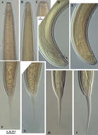
Figure 5. Longior longior Morffe & García sp. n. (female). A Cephalic end B Egg. Longior longior Morffe & García sp. n. (male) C Cephalic end D Tail, lateral view E Pre-cloacal median mammiform papilla, lateral view F Post-cloacal dorso-lateral papilla. Longior similis Morffe, García & Ventosa, 2009 (male) G Cephalic end H Tail, lateral view. Scale bars: A, B, C, D, G, H. 0.05 mm. E, F. 0.020 mm.
-
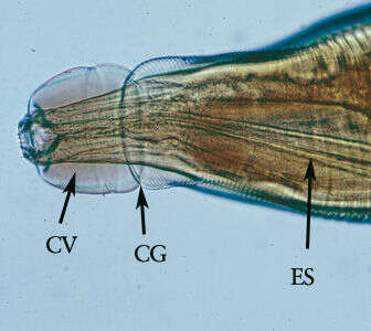
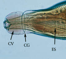
Öesophagostomum adultMagnification of the anterior end. Note the presence of the cephalic vesicle (CV), cephalic groove (CG) and esophagus (ES).From
CDC DPDx website
-
Saša Širca, Gregor Urek, Stela Lazarova, Milka Elshishka, Vlada Peneva
Zookeys
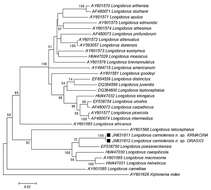
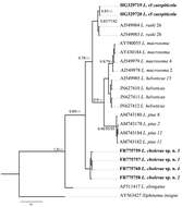
Figure 12.Phylogenetic tree of rDNA D2/D3 expansion region sequences of Longidorus carniolensis sp. n. from Slovenia (square mark) and sequences of closely related Longidorus species (NCBI GenBank). Sequences were analysed using Neighbour Joining Method. Bootstrap support values higher than 50% are presented.
-
Vlada K. Peneva, Stela S. Lazarova, Francesca De Luca, Derek J. F. Brown
Zookeys
Figure 11.Phylogenetic relationships of Longidorus cholevae sp. n. and its closest species for the partial 18S-ITS1 rDNA regions. Bayesian Inference strict consensus tree acquired under K2+G model. Numbers at the nodes indicating posterior probabilities higher that 0.8 and bootstrap values more that 70% for ML and NJ are presented.
-
Sevdan Nedelchev, Milka Elshishka, Stela Lazarova, Georgi Radoslavov, Peter Hristov, Vlada Peneva
Zookeys
Figure 5.Calcaridorylaimus castaneae sp. n. Female: A Entire body B Lip region D Prerectum, arrow pointing tongue-like valve E Pharyngeal bulb F Vulval region with posterior uterus G Vulval region with egg in posterior uterus H, I Vulval region J Cardia L Lateral field. Male: A Entire body C Lip region K Cardia M Supplements. Scale bars: A – 200 μm; B, C, M – 6 μm; E–L – 20 μm.