-

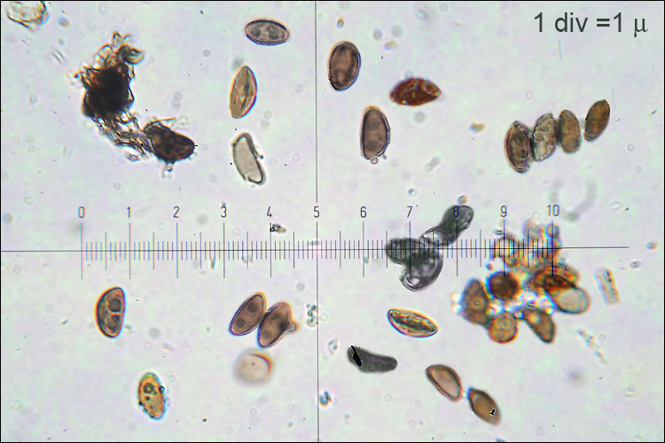
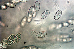
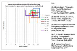
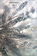
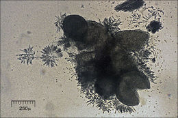
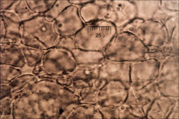
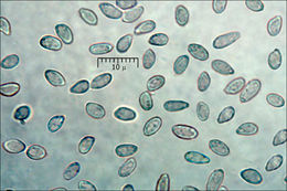
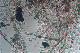
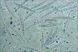
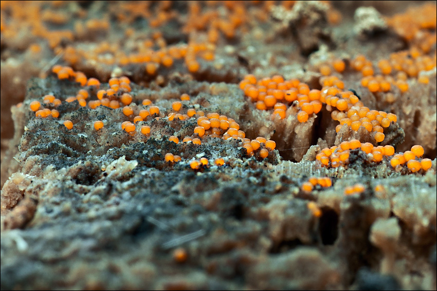
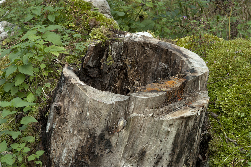
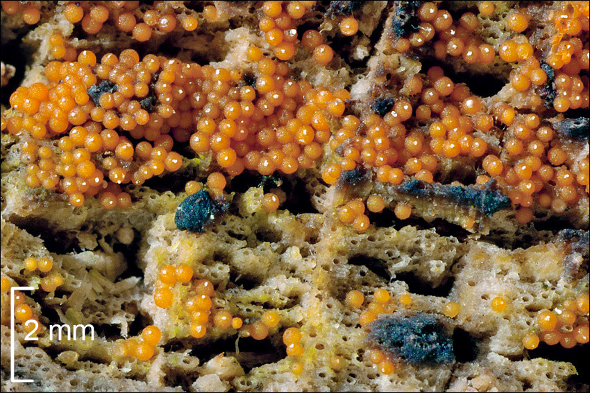
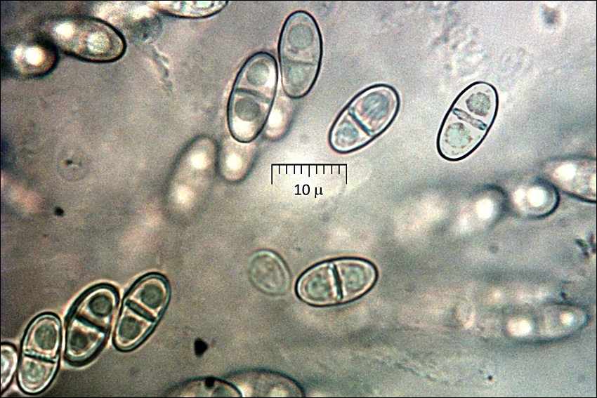
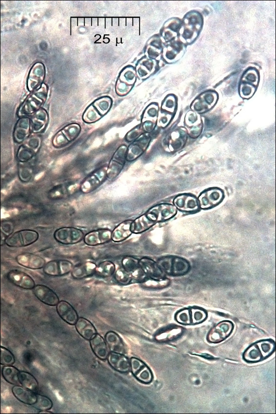
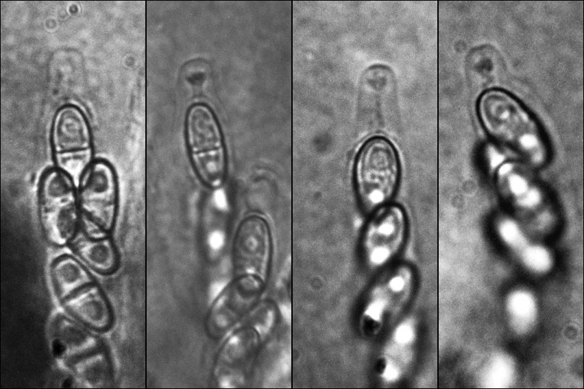
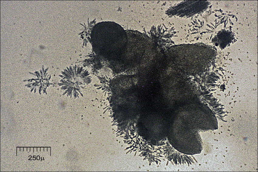
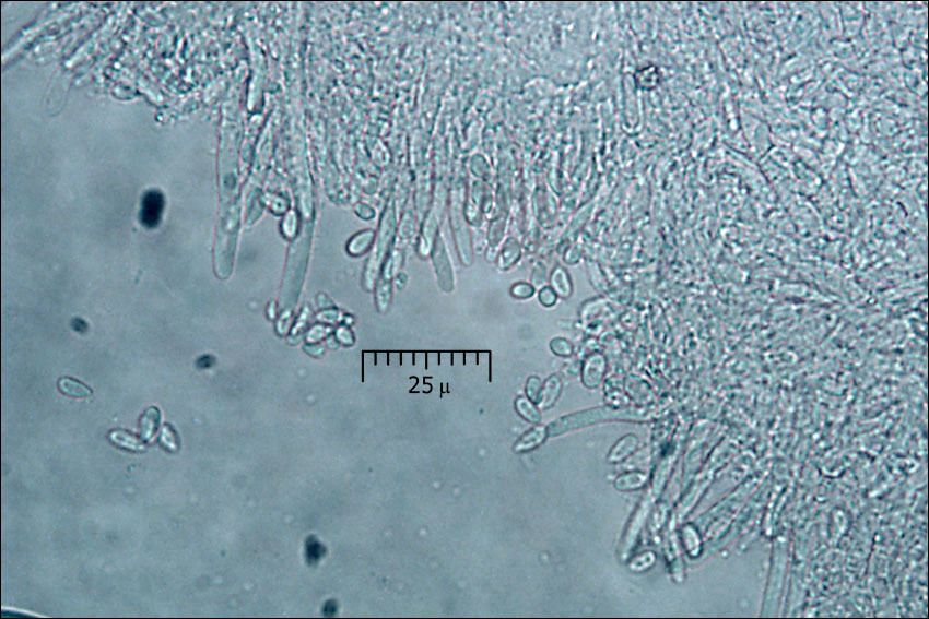
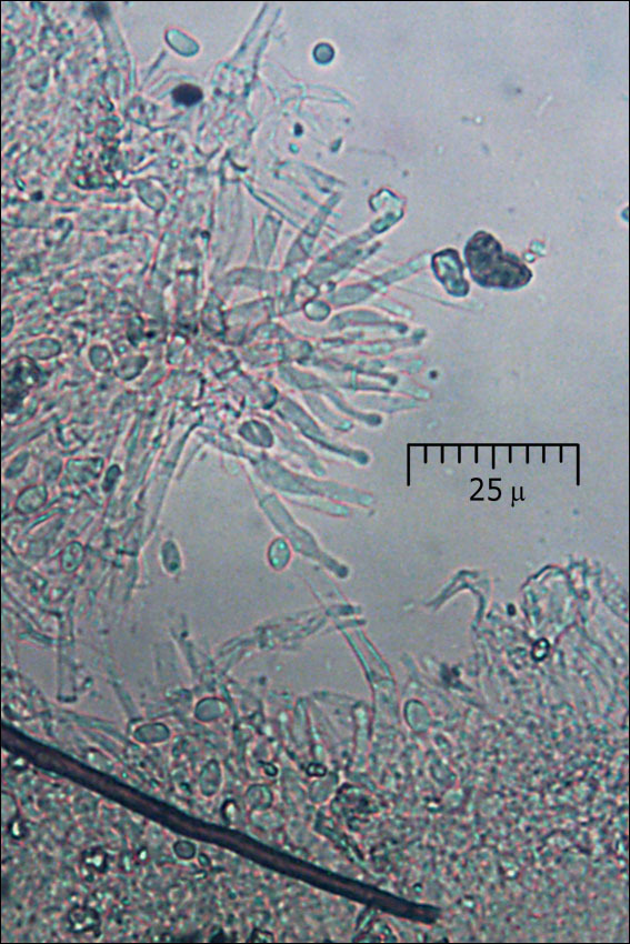
Habitat: Path side surrounded by pastures and mixed wood, near a farm house, locally flat terrain on southeast oriented mountain slope, open place, half sunny, mixed overgrown old scree and alluvial deposits, calcareous ground, exposed to direct rain, average precipitations ~ 3.000 mm/year, average temperature 7-9 deg C, elevation 560 m (1.850 feet), alpine phytogeographical region. - Substratum: old, debarked, hollow stump of a cut down Juglans regia in its final stage of disintegration. - Comments: First I thought this is a myxomicete, however, microscope immediately revealed that it belongs to ascomycetes. Two species (within the scope of the literature available to me) Nectria peziza and Nectria coccinea were the closest candidates. Based on fruit body size, ostioles and oil drops N. peziza seems a better fit. - Fruit body diameter: 0.39 (SD = 0.04) mm, n = 15 (in Ref.: (2) two observations cited: 0.34 mm and 0.3 to 0.5 mm). Ostioles clearly visible. Spores are septated and not warty. When observed slightly out of focus, longitudinal groves are barely visible on some (with my equipment), very slightly constricted, always with two large oil drops. Dimensions of free spores: 11.9(SD = 0.3) x 6.0 (SD = 0.14) μ, Q= 2.00 (SD = 0.14), n= 15. Dimensions of spores measured within asci: 11.7 (SD = 0.9) x 6.2 (SD = 0.5) μ, Q = 1.91 (SD = 0.24), n = 20, (data from Ref.: (2): 11.9 (SD = 1.1) x 5.9 (SD = 0.5) μ, n = 7). Asci dimensions: 88.1 (SD = 10.3) x 9.6 (SD = 1.0) μ, n = 9, (data from Ref.: (2): 70 - 90/7-9 μ; in Ref.:(1) = 60 - 80/7-10 μ). Olympus CH20 NEA 100x/1.25, magnification 1.000 x, oil (pictures of spores, apical ring), NEA 40x/0.65, magnification 400x (asci, trama), Bausch & Lomb 4x/0.10, magnification 40x (perithecia), in water. AmScope MA500 digital camera. - Herbarium: Mycotheca and lichen herbarium (LJU-Li) of Slovenian Forestry Institute, Večna pot 2, Ljubljana, Index Herbariorum LJF - Ref.: (1) J. Breitenbach, F. Kraenzlin, Eds., Fungi of Switzerland, Vol.1. Ascomycetes, Verlag Mykologia (1984), p 260. (2) H.O. Baral & O. Baral, G. Marson, In vivo veritas, Hypocreales, 2xCD, 2nd edition (2003) (3) http://www.rogersmushrooms.com/gallery/DisplayBlock~bid~6524~gid~~source~gallerydefault.asp (4) F. J. Seaver, Notes on North American Hypocreales-II. Nectria Peziza, Bulletin of the Torrey Botanical Club, p203. (available at http://www.jstor.org/stable/2479115?seq=3 ) 5) T. Matsushima, Icones Microfungorum a Matsushima lectorum(1975), p179 (available at http://www.mycobank.org )
-
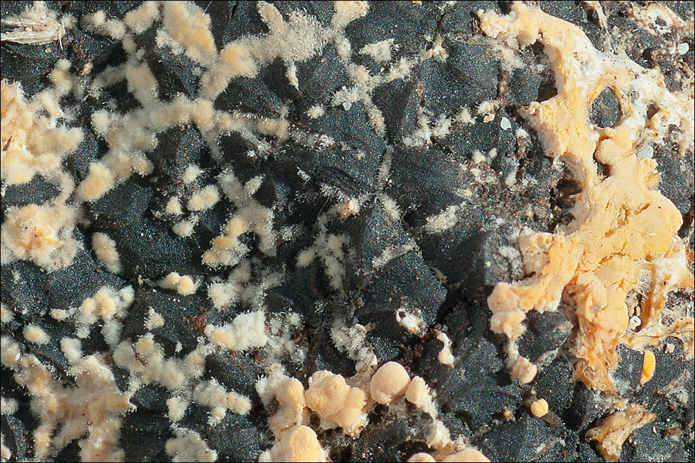
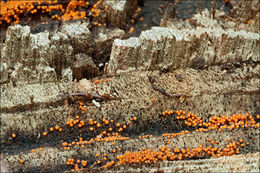
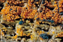
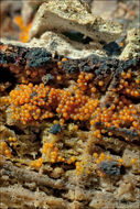
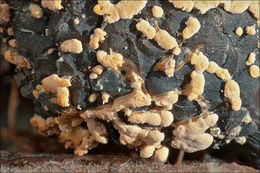
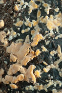
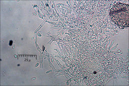
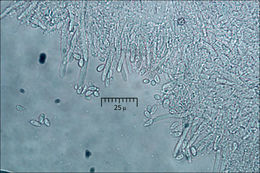
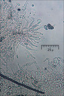
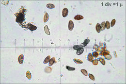
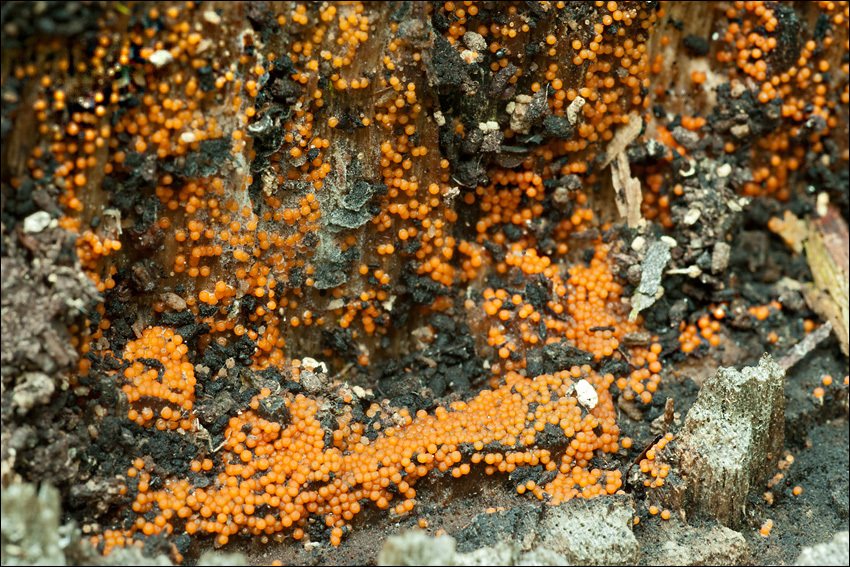
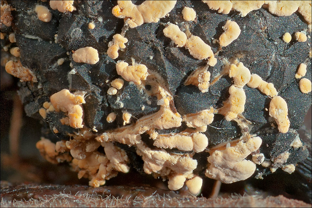
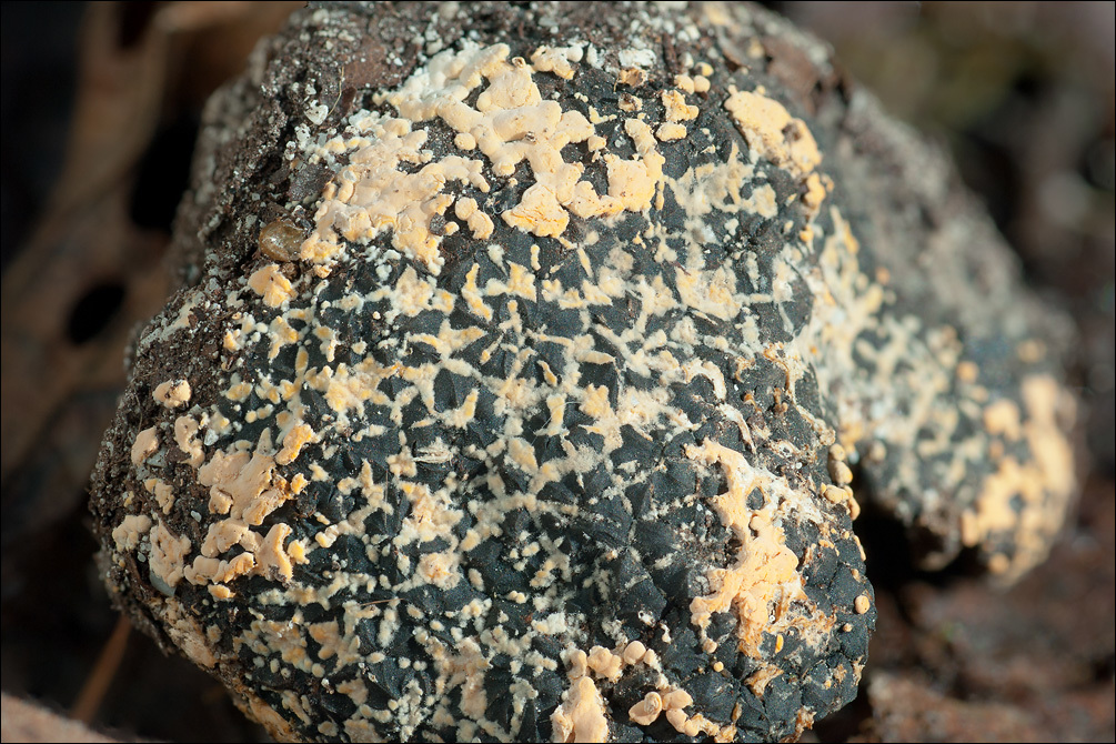
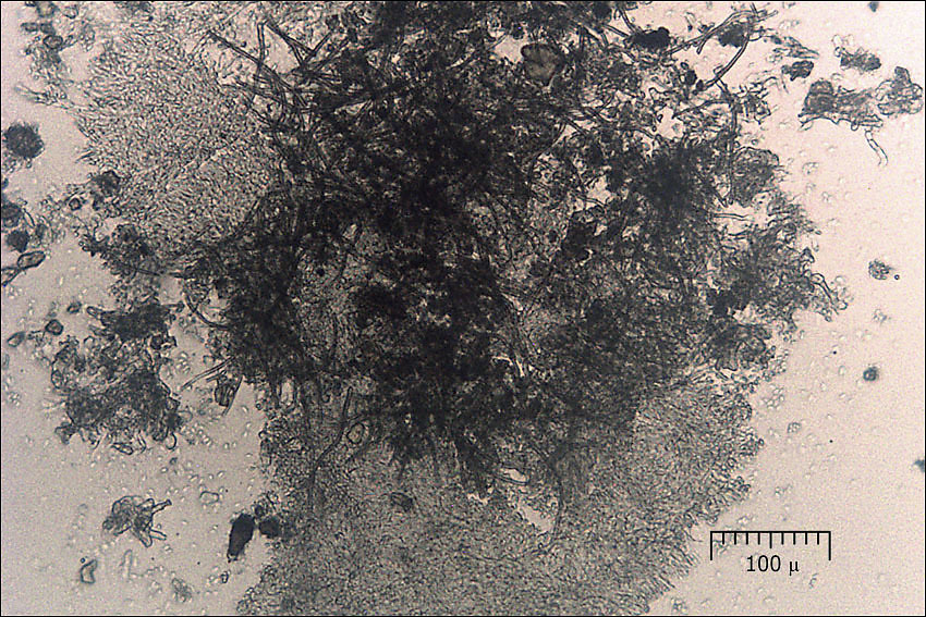
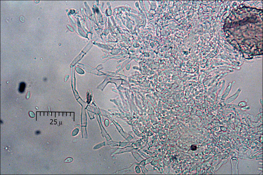
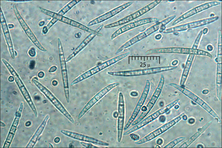
Syn.: Penicillium roseum Link, Gliocladium roseum Bainier - Habitat: mixed wood, Fagus sylvatica, Picea abies dominant trees; moderately inclined mountain slope, southeast aspect; skeletal, colluvial, calcareous ground; in shade; relatively warm and dry place; partly protected from direct rain by tree canopies; average precipitations ~ 3.000 mm/year, average temperature 7-9 deg C, elevation 560 m (1.840 feet), alpine phytogeographical region.Substratum: on somewhat over-mature Rhizopogon aestivus fruitbody.Comments: Orange blobs on the surface of almost black sporocarp of Rhizopogon aestivus are anamorph form of a parasitic fungus Clonostachys rosea (teleomorph formerly known as Bionectria). This was determined by growing fungus in culture from herbarium sample (Ref.:1). Clonostachys rosea is a common species found on several substrates including other fungi (fungicolous species). It colonizes living plants as an endophyte, can live in ground living as a saprophyte, or lives as a parasite on other fungi or nematodes. It produces several kinds of mycotoxins. It is used in biological pest control on vegetable, mostly for treatment of grey mold (Botrytis cinerea) of tomato and strawberries.The long fusiform and septated conidia also observed belong most probably to another fungus belonging to genus Fusarium. It has not been determined to species level. Small conidia of Clonostachys rosea smooth; dimensions: 4.7 [5.5 ; 5.9] 6.8 x 2.9 [3.3 ; 3.5] 3.8 microns; Q = 1.4 [1.6 ; 1.7] 2; N = 40; C = 95%; Me = 5.7 x 3.4 microns; Qe = 1.7. Fusiform, long, narrowly spindle-shaped, slightly bend conidia dimensions; 35.9 [46.9 ; 51.8] 62.8 x 4.5 [5.2 ; 5.5] 6.2 microns; Q = 6.2 [8.8 ; 9.9] 12.4; N = 31; C = 95%; Me = 49.3 x 5.3 microns; Qe = 9.3; number of septa: AVG = 4.9 (SD = 1.4), N=34.Olympus CH20, NEA 100x/1.25, magnification 1.000 x, oil (small conidia), NEA 40x/0.65, magnification 400x (large conidia, hypha, conidiophores), NEA 10x/0.25, magnification 100x (squash); fresh material; in water. AmScope MA500 digital camera.Herbarium: Mycotheca and lichen herbarium (LJU-Li) of Slovenian Forestry Institute, Vena pot 2, Ljubljana, Index Herbariorum LJFRef.:(1) Personal communication with Dr.Walter Gams, http://www.ascofrance.com , who has grown a culture and determined the species.(2) https://en.wikipedia.org/wiki/Clonostachys_rosea_f._roseaNikon D700/Nikkor Micro 105mm/f2.8
-
Syn.: Penicillium roseum Link, Gliocladium roseum Bainier - Habitat: mixed wood, Fagus sylvatica, Picea abies dominant trees; moderately inclined mountain slope, southeast aspect; skeletal, colluvial, calcareous ground; in shade; relatively warm and dry place; partly protected from direct rain by tree canopies; average precipitations ~ 3.000 mm/year, average temperature 7-9 deg C, elevation 560 m (1.840 feet), alpine phytogeographical region. Substratum: on somewhat over-mature Rhizopogon aestivus fruitbody. Comments: Orange blobs on the surface of almost black sporocarp of Rhizopogon aestivus are anamorph form of a parasitic fungus Clonostachys rosea (teleomorph formerly known as Bionectria). This was determined by growing fungus in culture from herbarium sample (Ref.:1). Clonostachys rosea is a common species found on several substrates including other fungi (fungicolous species). It colonizes living plants as an endophyte, can live in ground living as a saprophyte, or lives as a parasite on other fungi or nematodes. It produces several kinds of mycotoxins. It is used in biological pest control on vegetable, mostly for treatment of grey mold (Botrytis cinerea) of tomato and strawberries. The long fusiform and septated conidia also observed belong most probably to another fungus belonging to genus Fusarium. It has not been determined to species level. Small conidia of Clonostachys rosea smooth; dimensions: 4.7 [5.5 ; 5.9] 6.8 x 2.9 [3.3 ; 3.5] 3.8 microns; Q = 1.4 [1.6 ; 1.7] 2; N = 40; C = 95%; Me = 5.7 x 3.4 microns; Qe = 1.7. Fusiform, long, narrowly spindle-shaped, slightly bend conidia dimensions; 35.9 [46.9 ; 51.8] 62.8 x 4.5 [5.2 ; 5.5] 6.2 microns; Q = 6.2 [8.8 ; 9.9] 12.4; N = 31; C = 95%; Me = 49.3 x 5.3 microns; Qe = 9.3; number of septa: AVG = 4.9 (SD = 1.4), N=34. Olympus CH20, NEA 100x/1.25, magnification 1.000 x, oil (small conidia), NEA 40x/0.65, magnification 400x (large conidia, hypha, conidiophores), NEA 10x/0.25, magnification 100x (squash); fresh material; in water. AmScope MA500 digital camera. Herbarium: Mycotheca and lichen herbarium (LJU-Li) of Slovenian Forestry Institute, Vena pot 2, Ljubljana, Index Herbariorum LJF Ref.: (1) Personal communication with Dr.Walter Gams, http://www.ascofrance.com , who has grown a culture and determined the species. (2) https://en.wikipedia.org/wiki/Clonostachys_rosea_f._rosea Nikon D700/Nikkor Micro 105mm/f2.8
-
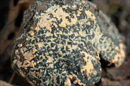
Slo.: jesenov (?) skorjeder - Habitat: Harwood forest, almost flat calcareous terrain, northeast oriented humid place, in shade, partly protected from direct rain, average precipitations ~ 3.000 mm/year, average temperature 8-10 deg C, elevation 480 m (1.600 feet), alpine phytogeographical region. - Substratum: fallen, partly rotten branch of a hardwood tree, still in bark, probably Fraxinus sp. (F. excelsior or F. ornus)- Comments: Spores smooth, with longitudinal slit (some ?), ellipsoid in front view, almond shaped in side view, somewhat flattened, some slightly falcate; dimensions: 10.4 (SD = 0.7) x 5.7 (SD = 0.7) micr., Q = 1.86 (SD = 0.18), n = 30. Motic B2-211A, magnification 1.000 x, oil, in water. Stromata show in 5% KOH deep orange-brown pigments. - Ref.: (1) http://pyrenomycetes.free.fr/hypoxylon/html/Hypoxylon_petriniae.htm . (2) http://www.asturnatura.com/fotografia/setas-hongos/hypoxylon-petriniae-m-stadler-j-fourn-2004-1/6811.html .