-
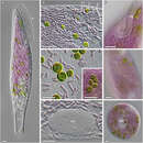
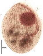
Blepharisma, pink heterotrich ciliate. From a freshwater pond near Boise, Idaho.
-
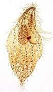
Infraciliature (dorsal side) of the medium-size pigmented heterotrich ciliate, Blepharisma lateritium (Ehrenberg,1831; Stein, 1859). The cell is teardrop-shaped and pale pink in color. The peristome extends about 2/3 the cell length along the left side. The peristome is bordered on the left by serial polykinetids forming an adoral zone of membranelles and on the right by a narrow undulating membrane which is less conspicuous. The longitudinal somatic kineties bend to the left margin on the dorsal surface (seen here). The single ellipsoid to spherical macronucleus is located left of midline in the cell center. There are multiple very small micronuclei which are difficult to discern in vivo. These become swollen and densely stained in silver impregnated specimens (seen here at edge of macronucleus). Stained by silver carbonate techniique (see Foissner, W.Europ. J. Protistol.27,313-330;1991). Collected from an artificial freshwater dredge pond near Boise, Idaho October 2004. Brightfield optics.
-
Infraciliature (ventral side) of the medium-size pigmented heterotrich ciliate, Blepharisma lateritium (Ehrenberg,1831; Stein, 1859). The cell is teardrop-shaped and pale pink in color. The peristome extends about 2/3 the cell length along the left side. The peristome is bordered on the left by serial polykinetids forming an adoral zone of membranelles and on the right by a narrow undulating membrane which is less conspicuous. The longitudinal somatic kineties bend to the left margin on the dorsal surface. The single ellipsoid to spherical macronucleus is located left of midline in the cell center. There are multiple very small micronuclei which are difficult to discern in vivo. These become swollen and densely stained in silver impregnated specimens (seen here). Stained by silver carbonate techniique (see Foissner, W.Europ. J. Protistol.27,313-330;1991). Collected from an artificial freshwater dredge pond near Boise, Idaho October 2004. Brightfield optics.
-
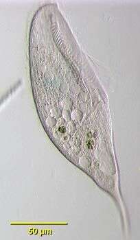
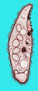
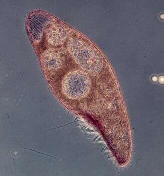
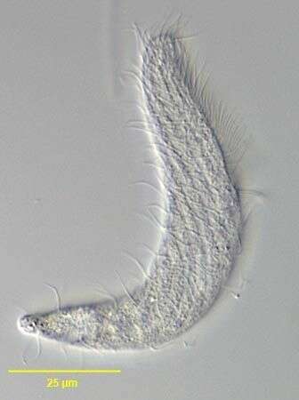
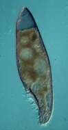
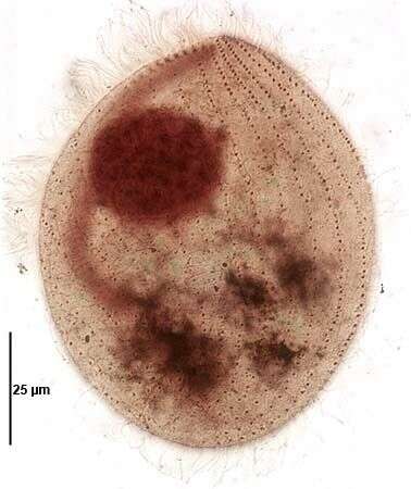
Portrait of the medium-size pigmented heterotrich ciliate, Blepharisma lateritium (Ehrenberg,1831; Stein, 1859). The cell is teardrop-shaped and pale pink in color. The peristome extends about 2/3 the cell length along the left side. The peristome is bordered on the left by serial polykinetids forming an adoral zone of membranelles and on the right by a narrow undulating membrane which is less conspicuous. The longitudinal somatic kineties bend to the left margin on the dorsal surface. The single ellipsoid to spherical macronucleus is located left of midline in the cell center. There are multiple very small micronuclei which are difficult to discern in vivo. These become swollen and densely stained in silver impregnated specimens. The contractile vacuole is at the posterior end. There are rows of pink cortical pigment granules between the somatic kineties. The pigment, blepharismin, is structurally similar to hypericin. When exposed to light, blepharismin causes a change in the cell's membrane potential and thus direction of ciliary beat causing light avoidance or photodispersal. Exposure to bright light for even short periods causes cell lysis. This is often observed in illuminated Blepharisma under the microscope. Blepharismin probably has a defensive function against predators such as the haptorid ciliate, Dileptus. Collected from an artificial freshwater dredge pond near Boise, Idaho October 2004. DIC optics.
-
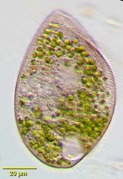
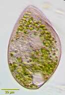
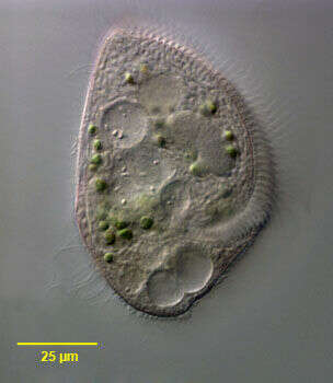
Infraciliature (ventral side) of the medium-size pigmented heterotrich ciliate, Blepharisma lateritium (Ehrenberg,1831; Stein, 1859). The cell is teardrop-shaped and pale pink in color.Green endosymbiotic zoochlorellae are visible in the cytoplasm.A zoochlorellae-bearing Blepharisma species otherwise indistinguishable from B. lateritium was reported from freshwater in Iowa (Johnson,L.P.A symbiotic Blepharisma.Proc. Iowa Acad. Sci. 55:391-393,1948). The peristome extends about 2/3 the cell length along the left side. The peristome is bordered on the left by serial polykinetids forming an adoral zone of membranelles and on the right by a narrow undulating membrane. The longitudinal somatic kineties bend to the left margin on the dorsal surface. The single ellipsoid to spherical macronucleus is located left of midline in the cell center. There are multiple very small micronuclei which are difficult to discern in vivo. These become swollen and densely stained in silver impregnated specimens (seen here). Stained by silver carbonate techniique (see Foissner, W.Europ. J. Protistol.27,313-330;1991). Collected from a freshwater pond near Boise, Idaho October 2004. Brightfield optics.
-
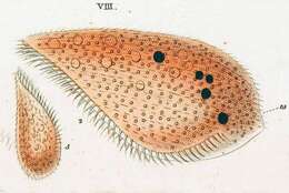
Originally described by Ehrenberg under the name Bursaria lateritia.
-
Originally described by Ehrenberg under the name Bursaria lateritia.
-
Originally described by Ehrenberg under the name Bursaria lateritia.
-
-
Phase contrast portrait of Blepharisma containing symbiotic green algae (zoochlorellae). An isolated report describes a species of Blepharisma resembling B. lateritium that had symbiotic algae (Johnson,L.P. A symbiotic Blepharisma. Proc. Iowa Acad. Sci. 55:391-93,1948).Specimen from freshwater pond near Boise, Idaho
-
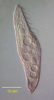
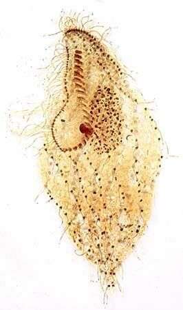
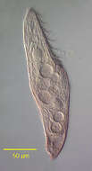
Portrait of Blepharisma americanum (Suzuki, 1954).The pink coloration is due to the pigment blepharismin.The metachronously beating adoral membranelles are seen along the large peristome to the viewer's right anteriorly. Gigantism is common in this species. The three macronuclear nodules are visible here (the smaller middle nodule is adjacent to the larger anterior nodule. The contractile vacuole is at the posterior end.DIC.
-
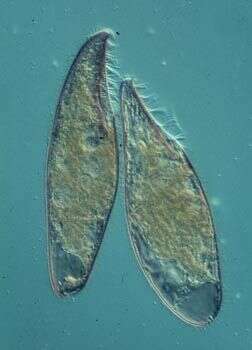
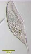
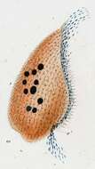
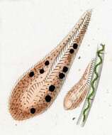
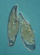
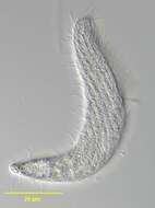
Two individuals of this 'pink' ciliate. This is the normal form the the cell. The pointed end is anterior, and an adoral zone of membranelles extends from the front to the cytostome near the middle of the cell. The large vacuole at the back of the cell is the contractile vacuole.
-
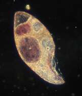
Dark ground image of Blepharisma americanum. The anterior is pointed towards the bottom of the image. The silk like structure leading from the front of the cell is the adoral zone of memb ranelles and the undulating membrane. This species becomes cannibalistic under some circumstances, and the large red inclusions are food vacuoles containing the remnants of other cells of the same species.
-
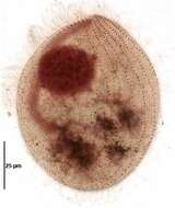
Phase contrast image of a cannibalistic (large) form of this species. The anterior is to bottom right. Under some circumstances, this species changes its feeding preferences and starts to eat other members of the same species. The large food vacuoles contain other blepharismas that are being digested.
-
This image shows that the different emmbranelles in the adoral zone of membranelles beat slightly out of phase with one another. This creates a wave-like appearance. Anterior is to the lower wide of the image.
-
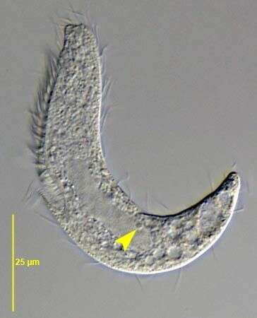
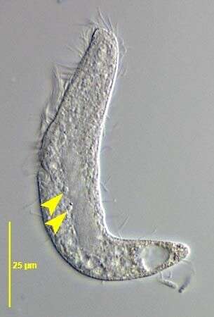
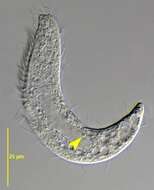
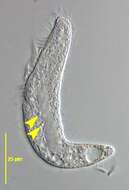
Blepharisma hyalinum (PERTY,1852) has 2-3 micronuclei one of which is seen here (yellow arrowhead). The oblong macronucleus is also visible in the cell center.DIC.
-
-
Blepharisma hyalinum (PERTY,1852) has 2-3 micronuclei two of which are seen here (yellow arrowhead). The other micronucleus is just out of the focal plane at the posterior end of the oblong macronucleus in the cell center.DIC.
-
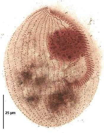
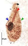
infraciliature of Blepharisma hyalinum (PERTY,1852).The blue arrowhead indicates the paraoral membrane at the right side of the peristome. The red arrowhead indicates the adoral membranelles (~25) each composed of one short and two longer kineties.One of the approximately 12 longitudinal somatic kineties is indicated by the green arrowhead. Stained by the silver carbonate technique (see Foissner, W.Europ. J. Protistol.27:313-330;1991).Brightfield.
-
-
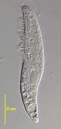
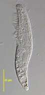
Righ dorsolateral infraciliature of Blepharisma hyalinum (PERTY,1852).Stained by the silver carbonate technique (Foissner, W.Europ. J. Protistol.27:313-330;1991).Brightfield.
-
Ventral infraciliature of Blepharisma hyalinum (PERTY,1852).Stained by the silver carbonate technique (Foissner, W.Europ. J. Protistol.27:313-330;1991).Brightfield.