-
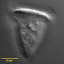
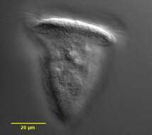
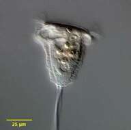
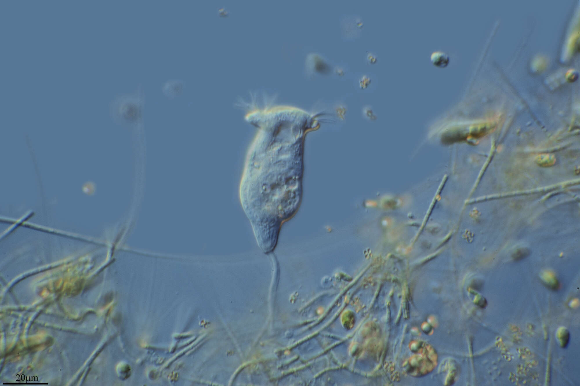
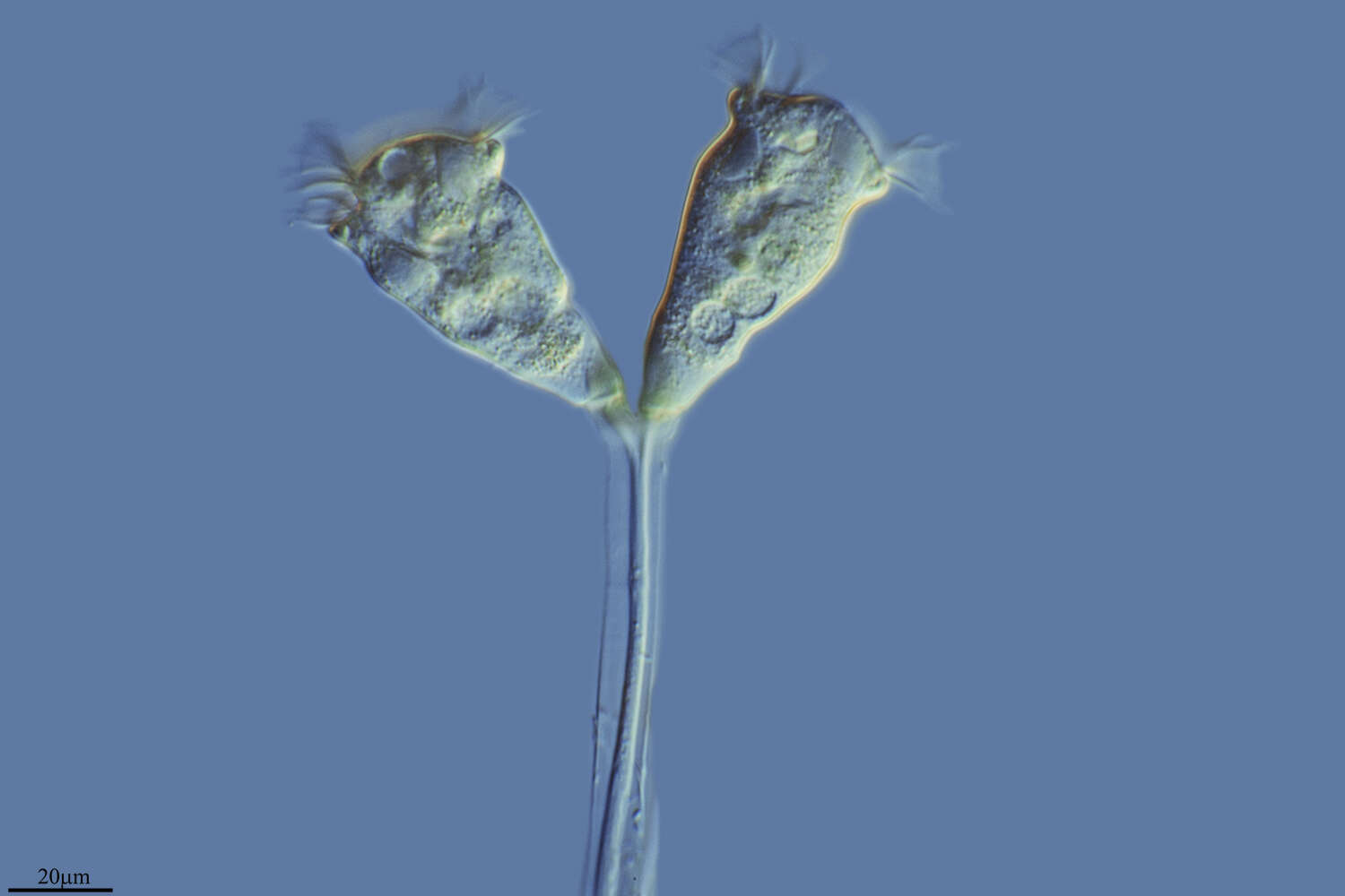
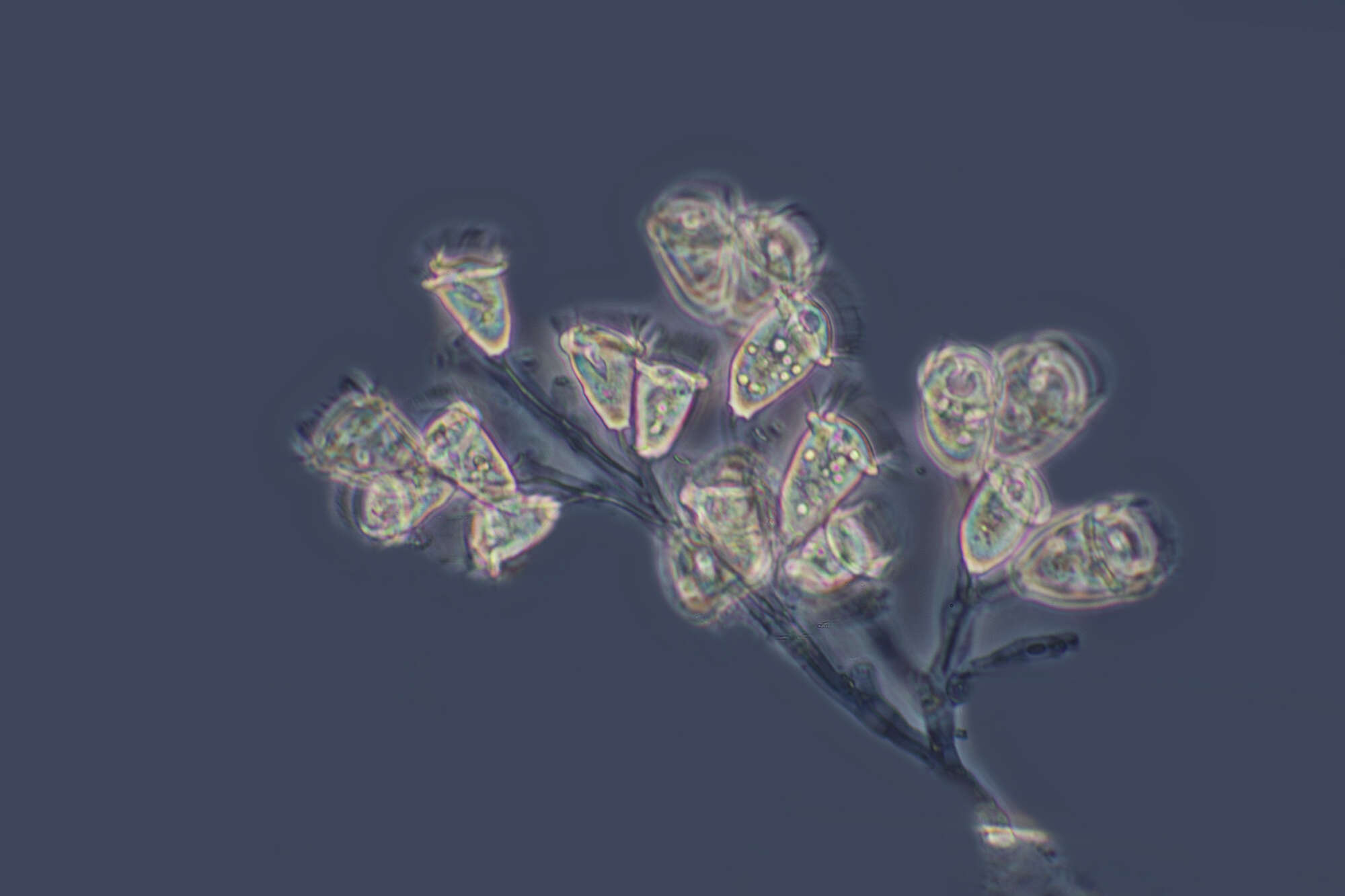
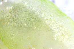
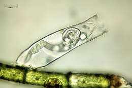
Surface detail of the peritrich ciliate, Pseudovorticella chlamydophora (Penard, 1922) Jankowski, 1976. Pseudovorticella is distinguished from Vorticella by silver staining which reveals a lattice-like silver line system in the former and circumferential lines without vertical connections in the latter. Pseudovorticella also has two contractile vacuoles. P. chlamydophora is distinguished by a distinct hyaline layer consisting of large cuboid pellicular blebs. The lattice-like pattern of these blebs is visible here. Feeds on bacteria. From freshwater pond near Boise, Idaho. DIC.
-
-
-
-
Almind Sø, Jylland, Danmark
-
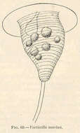
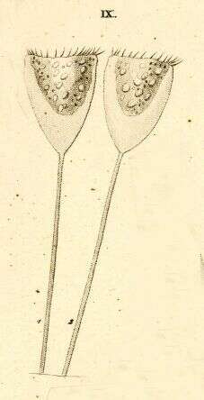
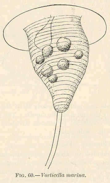
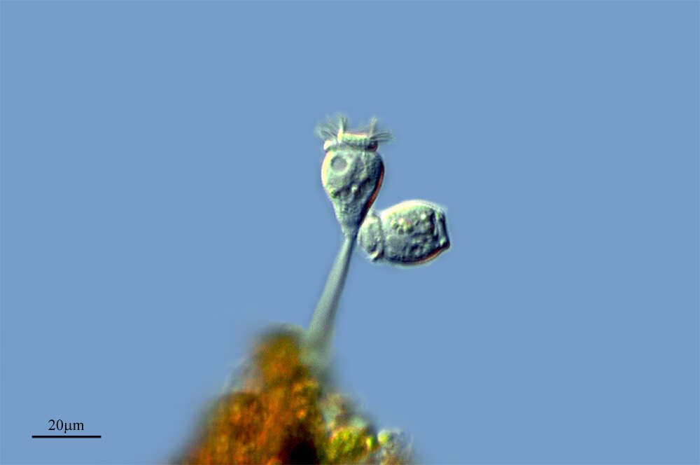
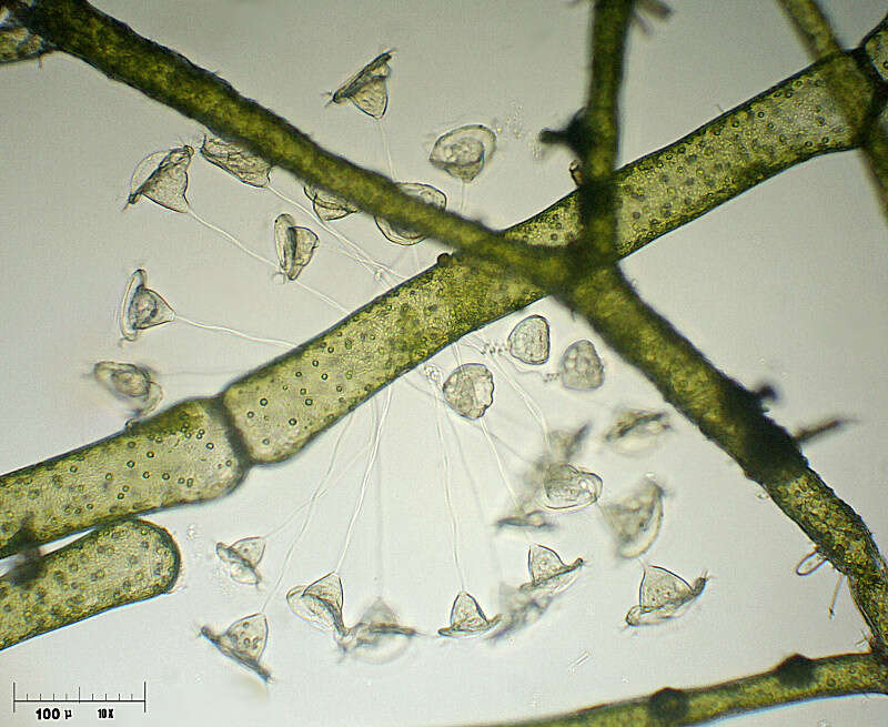
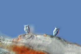
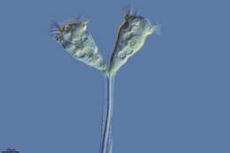
Vorticella marina.
-
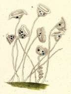
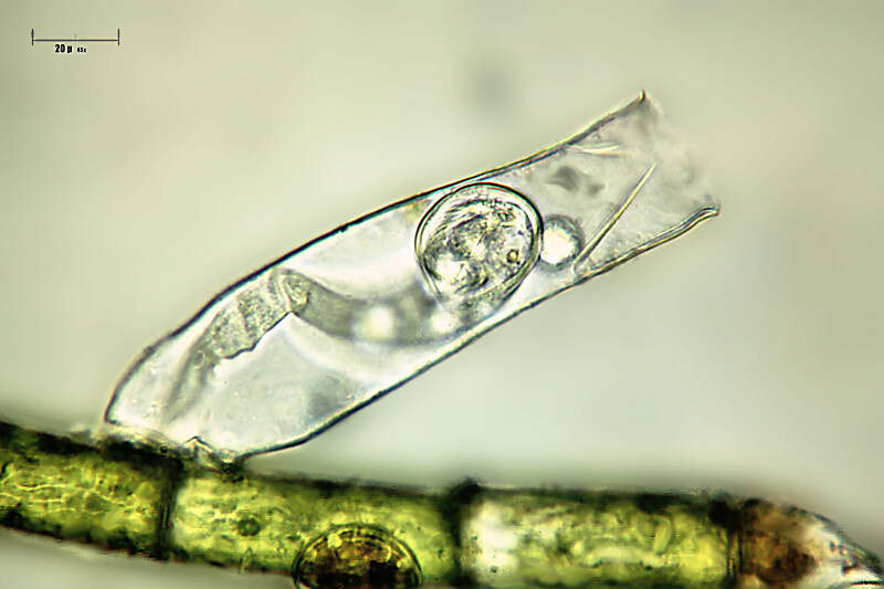
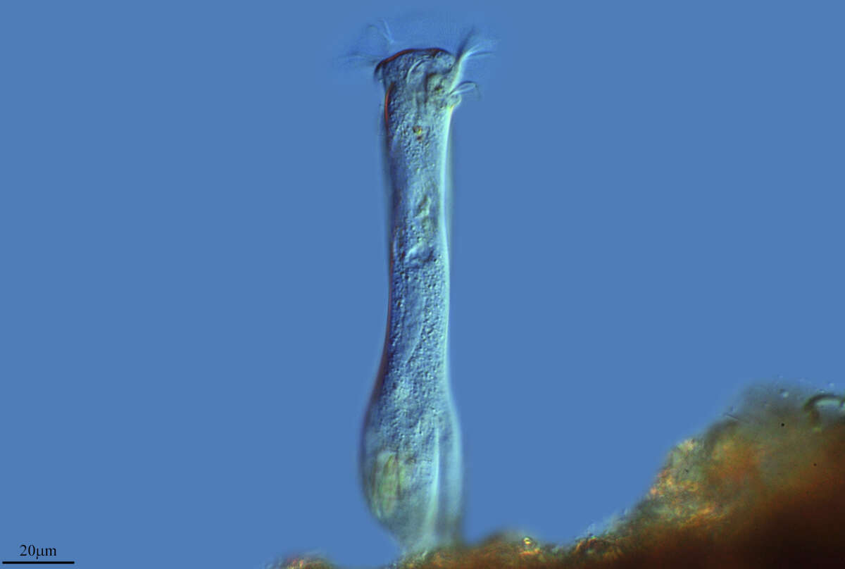
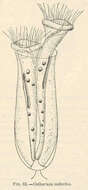
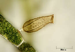
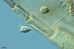
Cothurnia imberbis.
-
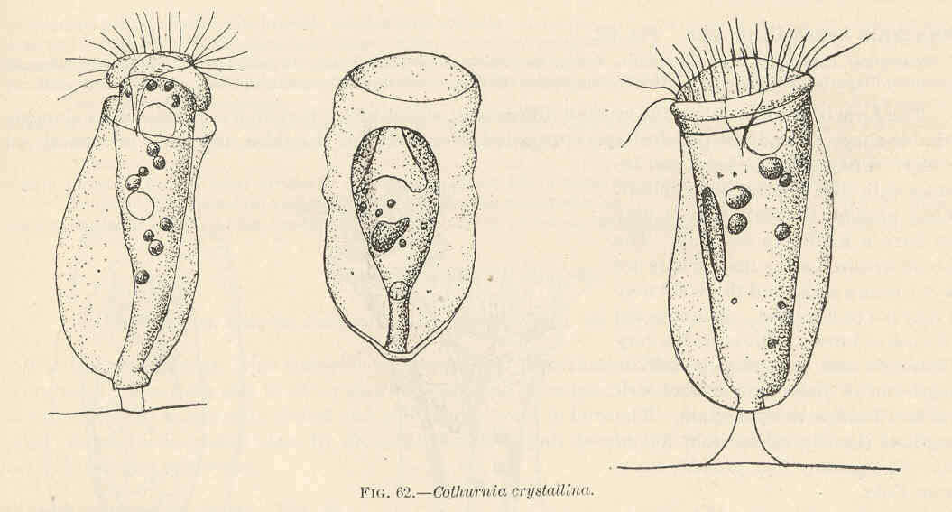
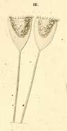
Cothurnia crystallini.
-
-
Miranda do Douro Municipality, Braganca, Portugal
-
Canada Del Hoyo, Castille la Mancha, Spain
-
Casa Blanca, Castille la Mancha, Spain
-
San Andres Y Sauces, Canary Islands, Spain
-
Los Lmites, La Rioja, Espaa
-
-
Urbanitzacio El Lledoner, Catalonia, Spain
-
Ribadelago de Franco, Castilla y Len, Espaa
-
Ontgola, Castile-La Mancha, Spain
-
Arboli, Catalonia, Spain
-
Logrono, La Rioja, Spain
-
Mahide, Castille and Leon, Spain
-
Grove, O, Galicia, Spain
-
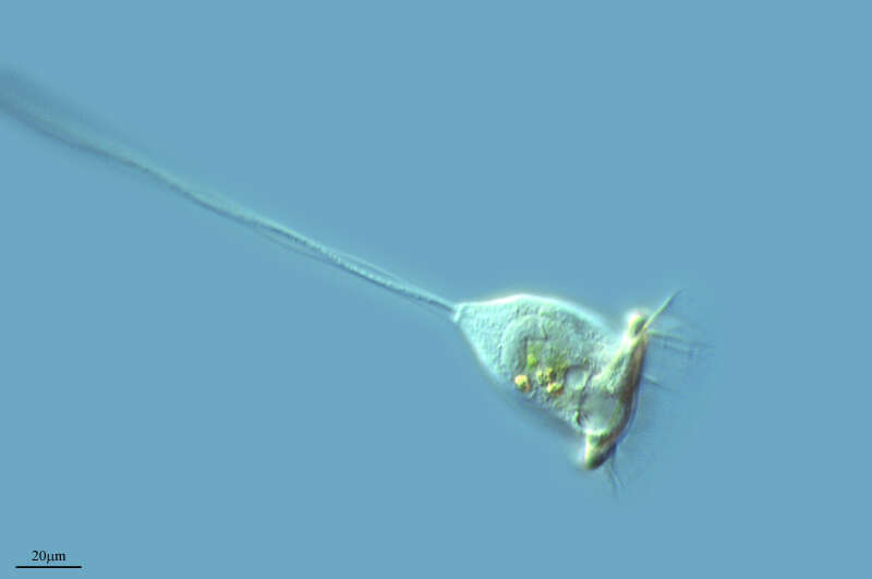
-
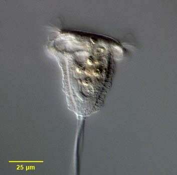
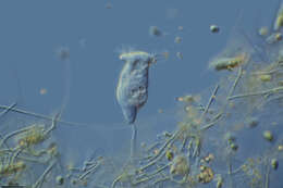
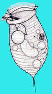
Portrait of the peritrich ciliate, Pseudovorticella chlamydophora (Penard,1922) Jankowski, 1976. This genus is distinguished from the genus Vorticella by its grid-like silver line system. The transverse components of the silverline system of Vorticella species have no vertical connections. P. Chlamydophora has a thick clear pellicular layer composed of cuboid units, which give the cell surface a distinctive quilted appearance. The extended cell is an inverted bell shape connected at the aboral scopula to a contractile stalk. The cell is spherical when contracted. The stalk contracts as a coil rather than a zigzag (e.g. Haplocaulus). The peristomal disc is almost flush. The ciliature is reduced to two rows of peristomal cilia, which beat counterclockwise toward the funnel-shaped buccal cavity (seen here to the viewers left anteriorly). The roughly C-shaped macronucleus is oriented in the long axis (to the viewers left of midline here). A single contractile vacuole is seen adjacent to the buccal cavity. The otherwise identical P. vestita has two contractile vacuoles. Multiple yellowish food vacuoles are seen here. P. chlamydophora may be gregarious but does not form true colonies. Collected from a freshwater pond near Boise, Idaho May 2004. DIC optics.