These parasites are usually not preyed on directly, but are ingested from host to host.
Toxocaara cati, as well as most nematodes, do not have appendages, eyes, segments, or other features that would cause these small creatures to stand out. The hydroskeleton is a single chamber, similar to a balloon filled with water. Movement depends on the pressure exerted in the hydroskeleton over the parasite's body. By contracting its longitudinal muscles, which are separated into four quadrants, the worm squeezes its "balloon" and moves through the hosts body in "contractile waves" (Schierenberg, 1997). Adult T. cati also have a complete digestive system with a mouth at one end, made up of three fleshy lips, and an anus near the posterior end (Roberts and Janovy, 2000).
Toxocara cati are dioecious, and like most nematodes, the females in this species, at about 10 cm, are larger than the males which can be up to 6 cm (Uga et al., 2000). The posterior end of the males is curved with paired spicules, small pointy structures that help him to "feel" a female with which he can mate (Roberts and Janovy, 2000).
The rough, pitted eggs produced, on average, measure 75 X 65 µm and may survive for years in the environment (Uga et al., 2000). These appear very dark under the microscope because the one-celled zygote fills the entire interior of the egg, but due to their ovoid shape, the albuminous shell of the egg becomes visible as well.
Four larval instars precede the well-known "arrowhead" adult nematode. Both males and females have clear thickened cuticles, alae, along the sides of the anterior end of the worm, which end abruptly and give them the appearance of an arrow. These alae may provide stability and help in its movement through the host's body.
Range length: 6 to 10 cm.
Other Physical Features: ectothermic ; heterothermic ; bilateral symmetry
Sexual Dimorphism: female larger; sexes shaped differently
Toxocara cati hosts are predominantly within the genus Felis, the cats. Most research, however, has been focused on the house cat, Felis domesticus because of its close contact with humans. Studies excluding strays cited 8% of the cats with Toxocara cati while another study including strays found 42% of cats had the parasite (O'Lorcain, 1994).
There are also many other paratenic hosts. These include earthworms, cockroaches, birds, rodents, dogs and humans (Schierenberg, 1997). Within the paratenic hosts, T. cati remain juveniles and, depending on where they migrate, cause immune reactions throughout their host's body. Therefore during the other stages of its life, T. cati is found in limited areas of the cat. As J1s, they remain in the egg, in the environment outside of potential hosts. If ingested, as J2s they may be found in the stomach wall, or if they undergo further migration - in the liver, lungs, bronchi, trachea, pharynx, stomach, and small intestine. Finally, as J3s, J4s, and adults, T. cati is found in the lumen of the intestine of cats only. Eggs are produced within this area, and excreted with the cat's feces (Prociv, 1989).
Toxocara cati are mainly found in kittens because of the transmammary route of transmission. In female kittens, they are most prevalent from 24-48 weeks, while in male kittens they are mainly found in those aging 12-24 weeks. The reason for this sexual variation is possibly due to the different timing of their immune system development (O'Lorcain, 1994).
Habitat Regions: temperate ; tropical ; terrestrial
Terrestrial Biomes: desert or dune ; savanna or grassland ; chaparral ; forest ; rainforest ; scrub forest ; mountains
Other Habitat Features: urban ; suburban ; agricultural
Toxocara cati are found in domestic as well as wild cats around the world. Because cats have become more domesticated, this species is also more concentrated in those areas where humans reside. However, the spread of T. cati is dependent on the ingestion of its eggs, which are excreted with the hosts feces. Climate is a major factor in not only maintaining the eggs, but the feces that surrounds it. Therefore it has been observed that areas with mild temperate climates are the most favorable environment for T. cati.
Biogeographic Regions: nearctic ; palearctic ; oriental ; ethiopian ; neotropical ; australian
Other Geographic Terms: cosmopolitan
Toxocara cati reside in the lumen of the small intestines. However, because they do not attach to the wall of its host's gut, it becomes apparent that these worms do not suck blood. Instead they feed primarily off of the liquid contents in the intestine (Roberts and Janovy, 2000), often resulting in malnutrition for the host.
Pharyngeal glands and intestinal epithelium produce digestive enzymes to feed on the hosts’ body fluids. Extracellular digestion begins within the lumen and is finished intracellularly (Barnes, 1987).
Animal Foods: body fluids
Primary Diet: carnivore (Eats body fluids)
Toxocara cati are found in domestic as well as wild cats around the world.
Ecosystem Impact: parasite
Species Used as Host:
Anywhere from 8-42% of cats may be infected with T. cati (O'Lorcain, 1994). Routes of transmission are either transmammary (directly to the newborn kittens), or by accidental ingestion of eggs found within paratenic hosts, or within the environment.
Cats are not the only source of infection. Every year 3,000-4,000 cases of human infection are also reported to the Centers for Disease Control and Prevention and state public health departments (CDC, 2000). Children are most often the recipient of these worms by accidently swallowing the eggs when they lick their fingers, or orally clean out the dirt underneath their fingernails. As with other paratenic hosts, the J2 larvae hatch and migrate throughout the human's body. The tissues they migrate to determine the symptoms of the child's infection.
Infection with the worm, T. cati, is called Toxocariasis. The two forms of this disease are ocular larva migrans (OLM) and visceral larva migrans (VLM). OLM is an eye disease caused when the worm enters the eye causing inflammation and scaring of the retina. This may lead to blindness. VLM, on the other hand, is due to repeated infections of T. cati, and effects of its continual movement throughout the body. The organs and tissues swell from the immune response to this foreign invader, and the central immune system may be affected. Symptoms of VLM include fever, coughing, ashma, or pneumonia. VLM is easily treated with antiparasitic drugs and anti-inflammatory medications. OLM, on the other hand is more difficult to treat, and the focus is usually on prevention versus cure (CDC, 2000).
Negative Impacts: injures humans (causes disease in humans ); causes or carries domestic animal disease
Adult T. cati migrate to the intestine, and begin producing eggs after just a few weeks. The eggs are excreted with the host's feces, and require a variable amount of time in the environment to mature. Depending on temperature and weather conditions, maturation takes on average about 2-3 weeks during the summer (Prociv, 1989). During this time, the juvenile passes through its first stage (J1), and into its second infective stage (J2) (Roberts and Janovy, 2000). After this time, any organism (from worms and rodents to humans) that ingests these eggs is at risk of infection. Stimulated by the stomach acid, the eggs hatch and the J2 larvae begin their migration through the host's body. Many move from the stomach into the stomach wall, to the liver and lungs, and then back into the stomach wall again. After having passed through four juvenile stages, the mature T. cati migrate to the small intestine where they reproduce and begin shedding eggs (Prociv, 1989).
Nematodes within the Secernentea have phasmids, which are unicellular glands. Phasmids likely function as chemoreceptors. Females may produce pheromones to attract males.
Nematodes in general have papillae, setae and amphids as the main sense organs. Setae detect motion (mechanoreceptors), while amphids detect chemicals (chemoreceptors).
Communication Channels: tactile ; chemical
Other Communication Modes: pheromones
Perception Channels: tactile ; chemical
Adult T. cati migrate to the intestine, and begin producing eggs after just a few weeks. The eggs are excreted with the host's feces, and require a variable amount of time in the environment to mature. Depending on temperature and weather conditions, maturation takes on average about 2-3 weeks during the summer (Prociv, 1989). During this time, the juvenile passes through its first stage (J1), and into its second infective stage (J2) (Roberts and Janovy, 2000). After this time, any organism (from worms and rodents to humans) that ingests these eggs is at risk of infection. Stimulated by the stomach acid, the eggs hatch and the J2 larvae begin their migration through the host's body. Many move from the stomach into the stomach wall, to the liver and lungs, and then back into the stomach wall again. After having passed through four juvenile stages, the mature T. cati migrate to the small intestine where they reproduce and begin shedding eggs (Prociv, 1989).
Paratenic hosts (those hosts that carry the worm, but the worm will not further develop) are especially important in T. cati infection of cats because of their predatory nature. If a mouse, for example, ingests a T. cati egg - the egg hatches, but the J2 larvae does not undergo further development. When this mouse is eaten by the cat, the J2 enters the wall of the stomach, and molts (by shedding its external cuticle and replacing it with the new one formed underneath) into J3 (Schrierenberg, 1997). When the juvenile procedes to migrate into the intestine lumen, it undergoes its last molt, into J4. These juveniles then molt into immature adults, and finally mature into adults capable of reproducing (Prociv, 1989).
Another important method of transmission is through the mammary glands of a pregnant female cat. Because T. cati do not migrate through the bloodstream and into the placenta, kittens are born free of worms, even if their mother was infected. However, after about 3-4 weeks, the newly hatched J2s migrate to the mother's mammary glands, where they are ingested by the feeding kittens (O'Lorcain, 1994). These enter the stomach, and then intestine of the kitten where they undergo the rest of their development into mature adults (Prociv, 1989).
Key Reproductive Features: sexual ; fertilization (Internal ); oviparous
Parental Investment: pre-fertilization (Provisioning)
Toxocara cati is a common nematode (roundworm) parasite of domestic cats. Although Toxocara canis is the most common cause of the human parasitic disease known as toxocariasis (including the clinical syndromes of ocular larva migrans [OLM] and visceral larva migrans [OLM]), the importance of T. cati in human disease may have been underestimated to date (Fisher 2003). Although T. canis infections in dogs are often acquired by puppies transplacentally, this is not so for T. cati in kittens. Both dogs and cats can acquire their respective nematode parasites at any age by ingesting eggs or paratenic hosts. The most widely recognized source of infection by Toxocara in humans is ingestion of contaminated soil, often by toddlers, but infection is also possible via the consumption of partial or whole paratenic hosts ("transport hosts"), such as earthworms or raw livers of domestic animals (chickens, ducks, cows, and pigs). Uncooked vegetables have also been reported as a possible source of infection, especially those from farms that utilize animal or human excrement as fertilizer. One additional possible source of infection reported is contact with embryonated eggs on a dog’s hair coat. (Lee et al. 2010 and references therein)
Škrkavka kočičí (Toxocara cati) je kosmopolitně rozšířený parazit koček a kočkovitých šelem. Jedná se o nejběžnější hlístici koček v evropských podmínkách. U dospělých koček nepůsobí výrazné klinické příznaky, avšak u koťat může způsobit vážné zdravotní problémy a vést až k úhynu. Na rozdíl od příbuzné škrkavky psí se nepřenáší transplacentárně (tedy z matky na plod), nýbrž jen mlékem či pozřením paratenického hostitele.
Škrkavka kočičí (Toxocara cati) je kosmopolitně rozšířený parazit koček a kočkovitých šelem. Jedná se o nejběžnější hlístici koček v evropských podmínkách. U dospělých koček nepůsobí výrazné klinické příznaky, avšak u koťat může způsobit vážné zdravotní problémy a vést až k úhynu. Na rozdíl od příbuzné škrkavky psí se nepřenáší transplacentárně (tedy z matky na plod), nýbrž jen mlékem či pozřením paratenického hostitele.
Toxocara cati, also known as the feline roundworm, is a parasite of cats and other felids. It is one of the most common nematodes of cats, infecting both wild and domestic felids worldwide. Adult worms are localised in the gut of the host. In adult cats, the infection – which is called toxocariasis – is usually asymptomatic. However, massive infection in juvenile cats can be fatal.
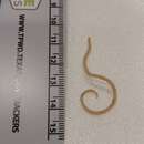
Feline roundworms are brownish-yellow to cream-colored to pink and may be up to 10 cm in length. Adults have short, wide cervical alae giving their anterior ends the distinct appearance of an arrow (hence their name, toxo, meaning arrow, and cara, meaning head). Eggs are pitted ovals with a width of 65 μm and a length of about 75 μm making them invisible to the human eye. The larvae are so small that they are easily transmitted from an adult female to her nursing kittens through her milk.[1][2][3][4]
Wild felids can become infected from a variety of sources; the primary source is infected fecal matter. The eggs of the roundworm become infective in three to four weeks after being passed out in fecal matter.[5] Contact with the soil, licking fur near feet, and eating a host animal (such as rodents) can also lead to infection of the felines.[5] The consumption of infected carrion also leads to contraction of the parasites, which is some of the food that members of Felidae consume.[6] The eggs hatch in the intestines and the larvae are then released into the cat's digestive tract.[5] The larvae are capable of migrating through the tissues and are found in the liver, lungs, tracheal washings and muscles as well as in the digestive tract.[7][5] From there, they move up to the trachea where they are swallowed, causing hacking and other problems.[5] The larvae can also move throughout the body and cause more damage to the infected individuals. The worms can even go into the mother's milk and infect the young.[5]
There are numerous clinical signs when dealing with feline roundworm. Some clinical signs that can be detected easily are vomiting, decreased appetite, and poor growth.[8] Like many diseases, changes in behavior can also be attributed to toxocariasis. Decreased appetite will result in a scrawny, mangy, and sickly appearance. Toxocariasis is exceptionally detrimental to kittens, as appetite loss and poor growth can ultimately lead to mortality. Additional clinical signs that can be identified include a pot bellied appearance, abdominal discomfort, and diarrhea.[8] Those with a small worm burden, however, may not show the clinical signs of being infected with worms,[8] and not receive treatment.
Treatment for Toxocara cati infections in cats is rather simple. There are a number of anthelmintics that will kill the adult worms, including Ivermectin, emodepside, fenbendazole, milbemycin, and moxidectin. However, most drugs are ineffective against the immature parasites. Consequently, infected cats will usually need multiple doses administered in two or three week intervals in order to fully eradicate the worms.[8][9]
It is possible for Toxocara cati to be transmitted to humans, usually as a consequence of humans consuming the larval stage of the parasite, resulting in a condition known as toxocariasis.[2] Typically, this happens when an individual pets an infected cat, picks up the parasite off of the fur and touches their face before washing their hands. The larvae migrate through the viscera in humans. Depending on the location and number of the larva in the human host, the disease can either be asymptomatic or cause conditions such as fever, cough, pneumonia, and vision loss.[3][4]
The two more severe forms of the disease are visceral toxocariasis and ocular toxocariasis. Visceral toxocariasis typically occurs in children, but can infect persons of any age. Signs and symptoms can include fever, wheezing, hepatomegaly, abdominal pain, anorexia, or skin reaction. Rarely, the migrating larvae can cause eosinophilic meningitis or encephalitis, myelitis, optic neuritis, radiculitis, cranial nerve palsy, or myocarditis. In lab findings, there is almost always a marked peripheral eosinophilia and often, anemia and a hypergammaglobulinemia.[10]
Ocular toxocariasis typically occurs in 5 to 10-year-olds resulting in significant damage to the eye.[11] Usually only one eye is affected, and manifestations can include strabismus, decreased vision, and leukocoria. Eye exam may show a subretinal granulomatous mass or posterior pole granuloma.[10] Even in relatively healthy people, the roundworm larvae infect organs such as the liver, lungs, eyes or brain and cause severe symptoms, such as:
Toxocara cati, also known as the feline roundworm, is a parasite of cats and other felids. It is one of the most common nematodes of cats, infecting both wild and domestic felids worldwide. Adult worms are localised in the gut of the host. In adult cats, the infection – which is called toxocariasis – is usually asymptomatic. However, massive infection in juvenile cats can be fatal.
Feline roundworms are brownish-yellow to cream-colored to pink and may be up to 10 cm in length. Adults have short, wide cervical alae giving their anterior ends the distinct appearance of an arrow (hence their name, toxo, meaning arrow, and cara, meaning head). Eggs are pitted ovals with a width of 65 μm and a length of about 75 μm making them invisible to the human eye. The larvae are so small that they are easily transmitted from an adult female to her nursing kittens through her milk.
Toxocara cati es un parásito de gatos y otros felidos de distribución mundial. Es uno de los parásitos nemátodos más comunes de los gatos.
Los gusanos adultos se localizan en el tracto digestivo del hospedador. En gatos adultos, la infección presenta síntomas como decaimiento, falta de apetito y vómitos acompañados del propio parásito, mientras que en gatos recién nacidos, la infección masiva puede ser mortal.
Toxocara cati Schrank (1788), es el ascarídeo de los gatos. Su morfología es muy similar a la de T. canis, los machos adultos miden de 3 a 6 cm de longitud y las hembras de 4 a 7 cm de longitud. Los huevos miden aproximadamente 75 x 70 μm, siendo de menor tamaño que los de T. canis, pero mayores que los de A. lumbricoides.
La toxocariasis es el término clínico aplicado a la infección en seres humanos producida por Toxocara canis (T. canis) y en menor grado por Toxocara cati (T. cati) (codificadas en la Clasificación Internacional de Enfermedades como CIE-9 128.0; CIE-10 B83.0(Despommier, 2003; Heymann & American Public Health Association, 2004; Overgaauw, 1997a). El hábitat definitivo de T. catis es en el intestino delgado del gato.[1]
Toxocara cati es un parásito de gatos y otros felidos de distribución mundial. Es uno de los parásitos nemátodos más comunes de los gatos.
Los gusanos adultos se localizan en el tracto digestivo del hospedador. En gatos adultos, la infección presenta síntomas como decaimiento, falta de apetito y vómitos acompañados del propio parásito, mientras que en gatos recién nacidos, la infección masiva puede ser mortal.
Toxocara cati Schrank (1788), es el ascarídeo de los gatos. Su morfología es muy similar a la de T. canis, los machos adultos miden de 3 a 6 cm de longitud y las hembras de 4 a 7 cm de longitud. Los huevos miden aproximadamente 75 x 70 μm, siendo de menor tamaño que los de T. canis, pero mayores que los de A. lumbricoides.
La toxocariasis es el término clínico aplicado a la infección en seres humanos producida por Toxocara canis (T. canis) y en menor grado por Toxocara cati (T. cati) (codificadas en la Clasificación Internacional de Enfermedades como CIE-9 128.0; CIE-10 B83.0(Despommier, 2003; Heymann & American Public Health Association, 2004; Overgaauw, 1997a). El hábitat definitivo de T. catis es en el intestino delgado del gato.
Toxocara cati est une espèce de nématodes parasites de l'intestin des félins, et notamment du chat domestique. Il s'agit d'un gros ver rond de couleur rosée mesurant de 4 à 10 cm de long[1]. Ce parasite est très courant chez le chat domestique, notamment chez le chat ne vivant pas confiné dans une maison[2]. Il se transmet par ingestion des œufs présents dans l'environnement ou par l'ingestion d'hôtes intermédiaires comme les oiseaux ou les rongeurs, proies du chat ou par le lait maternel[3],[2].
L'humain peut être infecté par Toxocara cati : il s'agit de la toxocarose, généralement bénigne. L'ingestion du parasite se produit généralement par l’ingestion des œufs présents sur les légumes[3]. L'influence de Toxocara cati dans la toxocarose est encore débattue, car le ver impliqué dans cette maladie est beaucoup plus généralement Toxocara canis[2].
Ce ver infecte d'autres félins que le chat domestique, comme le Guigna par exemple[4].
Toxocara cati est une espèce de nématodes parasites de l'intestin des félins, et notamment du chat domestique. Il s'agit d'un gros ver rond de couleur rosée mesurant de 4 à 10 cm de long. Ce parasite est très courant chez le chat domestique, notamment chez le chat ne vivant pas confiné dans une maison. Il se transmet par ingestion des œufs présents dans l'environnement ou par l'ingestion d'hôtes intermédiaires comme les oiseaux ou les rongeurs, proies du chat ou par le lait maternel,.
L'humain peut être infecté par Toxocara cati : il s'agit de la toxocarose, généralement bénigne. L'ingestion du parasite se produit généralement par l’ingestion des œufs présents sur les légumes. L'influence de Toxocara cati dans la toxocarose est encore débattue, car le ver impliqué dans cette maladie est beaucoup plus généralement Toxocara canis.
Ce ver infecte d'autres félins que le chat domestique, comme le Guigna par exemple.
De kattenspoelworm (Toxocara cati) is een vrij lange parasitaire rondworm (Nematode) die voorkomt in de darmen van een aantal soorten katachtigen. Het is een van de meest voorkomende spoelwormen bij katten. De levenscyclus lijkt sterk op die van de hondenspoelworm. De besmettelijke eitjes van deze spoelworm kunnen ook ziektes veroorzaken bij de mens (toxocariasis).
Bronnen, noten en/of referentiesGlista kocia (Toxocara cati) – nicień bytujący w jelicie cienkim kota. U człowieka występuje rzadko, do zakażenia może dojść w wieku dziecięcym lub w przypadku rażącego zaniedbania higieny.
Samce miewają od 3 do 7 cm, średnica 1 mm, samice 4–10 cm, średnica 2 mm.
Jaja prawie kuliste, otoczone gruba warstwa oskórka, kot zaraża się przez połknięcie jaj, okres osiągnięcia dojrzałości 6 tygodni, jaja mogą zarażać ptaki, dżdżownice, karaluchy, jagnięta, gryzonie.[potrzebny przypis]
Glista kocia (Toxocara cati) – nicień bytujący w jelicie cienkim kota. U człowieka występuje rzadko, do zakażenia może dojść w wieku dziecięcym lub w przypadku rażącego zaniedbania higieny.
Toxocara cati é uma espécie de nematódeo do gênero Toxocara comumente encontrado parasitando gatos e outros felinos.
Toxocara cati, còn được gọi là giun tròn mèo, là ký sinh trùng của mèo và các loài thuộc họ mèo khác. Nó là một trong những giun tròn phổ biến nhất ở mèo, lây nhiễm cho cả loài hoang dã và các giống thuộc họ Mèo nhà, trên toàn thế giới. Giun trưởng thành ký sinh trong ruột của vật chủ. Ở mèo trưởng thành, nhiễm trùng - được gọi là bệnh toxocara - thường không có triệu chứng. Tuy nhiên, nhiễm trùng nặng ở mèo vị thành niên có thể dẫn đến tử vong.
Giun tròn mèo có màu vàng nâu, màu kem đến màu hồng và có thể dài tới 10 cm. Giun trưởng thành có cổ alae ngắn - rộng và phân đuôi của giun có hình dáng bề ngoài như một mũi tên. Trứng giun có hình dạng bầu dục có chiều rộng 65 μm và chiều dài khoảng 75 μm khiến chúng trở nên vô hình đối với mắt người. Ấu trùng quá nhỏ đến nỗi chúng dễ lây truyền từ một con cái trưởng thành sang mèo con của nó qua sữa.[1][2][3][4]
Điều trị nhiễm trùng Toxocara cati ở mèo khá đơn giản. Có một số loại thuốc diệt giun sẽ giết chết giun trưởng thành; tuy nhiên, hầu hết các loại thuốc đều không hiệu quả đối với các ký sinh trùng chưa trưởng thành. Do đó, mèo bị nhiễm bệnh thường sẽ cần nhiều liều dùng trong hai hoặc ba tuần để loại bỏ hoàn toàn giun.[5]
Toxocara cati, còn được gọi là giun tròn mèo, là ký sinh trùng của mèo và các loài thuộc họ mèo khác. Nó là một trong những giun tròn phổ biến nhất ở mèo, lây nhiễm cho cả loài hoang dã và các giống thuộc họ Mèo nhà, trên toàn thế giới. Giun trưởng thành ký sinh trong ruột của vật chủ. Ở mèo trưởng thành, nhiễm trùng - được gọi là bệnh toxocara - thường không có triệu chứng. Tuy nhiên, nhiễm trùng nặng ở mèo vị thành niên có thể dẫn đến tử vong.
Giun tròn mèo có màu vàng nâu, màu kem đến màu hồng và có thể dài tới 10 cm. Giun trưởng thành có cổ alae ngắn - rộng và phân đuôi của giun có hình dáng bề ngoài như một mũi tên. Trứng giun có hình dạng bầu dục có chiều rộng 65 μm và chiều dài khoảng 75 μm khiến chúng trở nên vô hình đối với mắt người. Ấu trùng quá nhỏ đến nỗi chúng dễ lây truyền từ một con cái trưởng thành sang mèo con của nó qua sữa.

猫回虫 (ねこかいちゅう、Toxocara cati) とは、世界中に分布するネコおよびネコ科動物を宿主とするもっとも一般的な回虫。成虫は宿主の消化管に寄生する。成猫では通常は無症状であるが、幼猫への大量感染では致死的となり得る。


