-
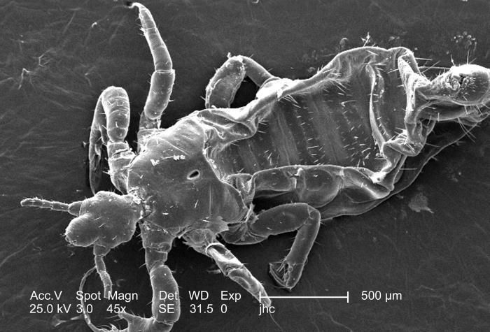
Under a relatively low magnification of 45x, this 2006 scanning electron micrograph (SEM) depicted a dorsal, or superior view, of a male louse, Pediculus humanus var. corporis. The head, or cephalic region is at the left, from which its two antennae were extended. The head was attached to the thoracic region, which gave rise to its three pairs of jointed legs. While the abdominal region, towards the far right, is the region in which was housed the stomach, and intestines. The jointed nature of its extremities, designates this organism as a member of the phylum Arthropoda, and the fact that there are three pairs of legs, the louse is, thereby, placed into the class, Insecta. Note the small, hair-like structures adorning the exoskeletal surface of this insect. These are known as setae, and are not hairs at all, but extensions of the chitinous exoskeletal surface, which provide the organism with sensorial data about its surroundings.Created: 2006
-
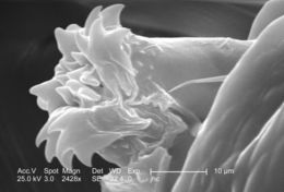
Under a high magnification of 2428x, this 2006 scanning electron micrograph (SEM) depicted an enlarged inferior view of the mouthparts found on the cephalic, or head region of a male body louse, Pediculus humanus var. corporis. The floral-like structure in this field of view represents the labial funnel, or haustellum, which is a tube-like structure, formed from what is believed to be a modification of the labium, but is armed with teeth, which act to grab, and hold on to the skin surface of its host while obtaining its blood meal. The insect pierces the hosts skin with its sharply-pointed set of three stylets, and together, form what is termed the fascicle, which is hidden from view in this image. Note PHIL# 9214, 9216, in which case a louse is in its feeding mode on the skin surface of its host.Created: 2006
-
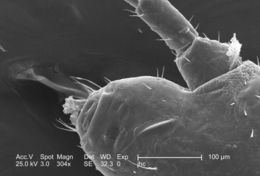
Under a moderately low magnification of 304x, this 2006 scanning electron micrograph (SEM) depicted an enlarged inferior view of the mouthparts found on the cephalic, or head region of a male body louse, Pediculus humanus var. corporis. The floral-like structure in this field of view represents the labial funnel, or haustellum, which is a tube-like structure, formed from what is believed to be a modification of the labium, but is armed with teeth, which act to grab, and hold on to the skin surface of its host while obtaining its blood meal. The insect pierces the hosts skin with its sharply-pointed set of three stylets, and together, form what is termed the fascicle, which is hidden from view in this image. Note PHIL# 9214, 9216, in which case a louse is in its feeding mode on the skin surface of its host. Also note the scape and pedicle, or the respective proximal segments of the insect's left antenna in this view as well.Created: 2006
-
Under a high magnification of 2189x, this 2006 scanning electron micrograph (SEM) depicted an enlarged inferior-oblique view of the mouthparts found on the cephalic, or head region of a male body louse, Pediculus humanus var. corporis. The floral-like structure in this field of view represents the labial funnel, or haustellum, which is a tube-like structure, formed from what is believed to be a modification of the labium, but is armed with teeth, which act to grab, and hold on to the skin surface of its host while obtaining its blood meal. The insect pierces the hosts skin with its sharply-pointed set of three stylets, and together, form what is termed the fascicle, which is hidden from view in this image. Note PHIL# 9214, 9216, in which case a louse is in its feeding mode on the skin surface of its host.Created: 2006
-
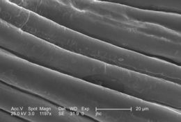
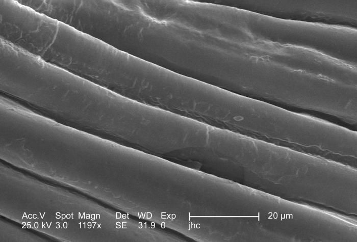
Under a moderately high magnification of 1197x, this 2006 scanning electron micrograph (SEM) depicted an enlarged view of the chitinous, exoskeletal surface of a male louse, Pediculus humanus var. corporis. In this particular view, there are no visible exoskeletal adnexae, which are quite often complex in shape, and variable in size, with forms following functions, which are just as varied. The exoskeleton is composed of chitin, a molecule made up of bound units of acetylglucosamine, which is joined in such a way as to allow for increased points at which hydrogen bonding can occur. In this way chitin provides increased strength, and durability as an exoskeletal foundation.Created: 2006
-
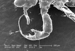
Under a relatively low magnification of 97x, this 2006 scanning electron micrograph (SEM) depicted a dorsal view of a male body louse, Pediculus humanus var. corporis. Occupying the field of view, was the insects left leg of its first pair of legs. There was a piece of debris attached to the distal claw tip of this appendage. The jointed nature of its extremities, designates this organism as a member of the phylum Arthropoda, and the fact that there are three pairs of legs, the louse is, thereby, placed into the class, Insecta. Note the small, hair-like structures adorning the exoskeletal surface of this insect. These are known as setae, and are not hairs at all, but extensions of the chitinous exoskeletal surface, which provide the organism with sensorial data about its surroundings.Created: 2006
-
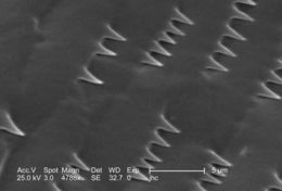
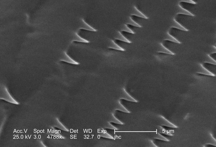
Magnified 4788x, this 2006 scanning electron micrograph (SEM) depicted an enlarged view of the chitinous, exoskeletal surface of a male louse, Pediculus humanus var. corporis. In this particular view, the exoskeletal adornments are few, consisting of small, spike-like structures. Quite often the shape and size of these skeletal adnexae are quite complex, with forms following functions, which are just as varied. The exoskeleton is composed of chitin, a molecule made up of bound units of acetylglucosamine, which is joined in such a way as to allow for increased points at which hydrogen bonding can occur. In this way chitin provides increased strength, and durability as an exoskeletal foundation.Created: 2006
-
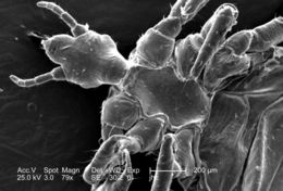
Under a relatively low magnification of 79x, this 2006 scanning electron micrograph (SEM) depicted a ventral, or inferior view, of a male louse, Pediculus humanus var. corporis. The head, or cephalic region is at the left, from which its two antennae were extended. The head attached to the thoracic region, which gave rise to its three pairs of jointed legs. While the abdominal region, towards the far right, is the region in which was housed the stomach, and intestines. The jointed nature of its extremities, designates this organism as a member of the phylum Arthropoda, and the fact that there are three pairs of legs, the louse is, thereby, placed into the class, Insecta. Note the small, hair-like structures adorning the exoskeletal surface of this insect. These are known as setae, and are not hairs at all, but extensions of the chitinous exoskeletal surface, which provide the organism with sensorial data about its surroundings.Created: 2006
-
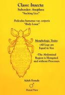
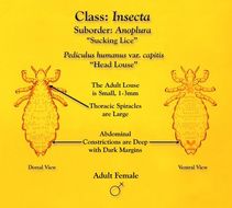
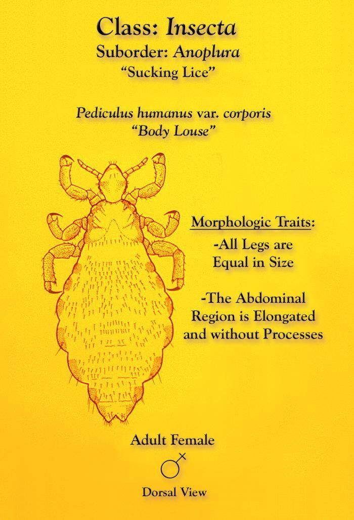
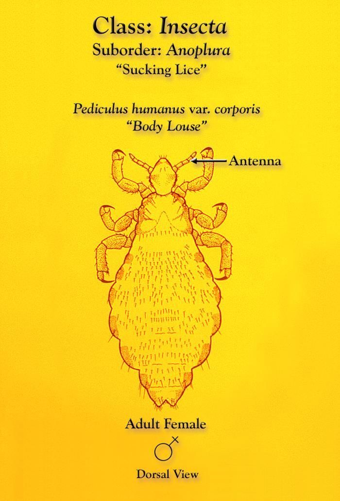
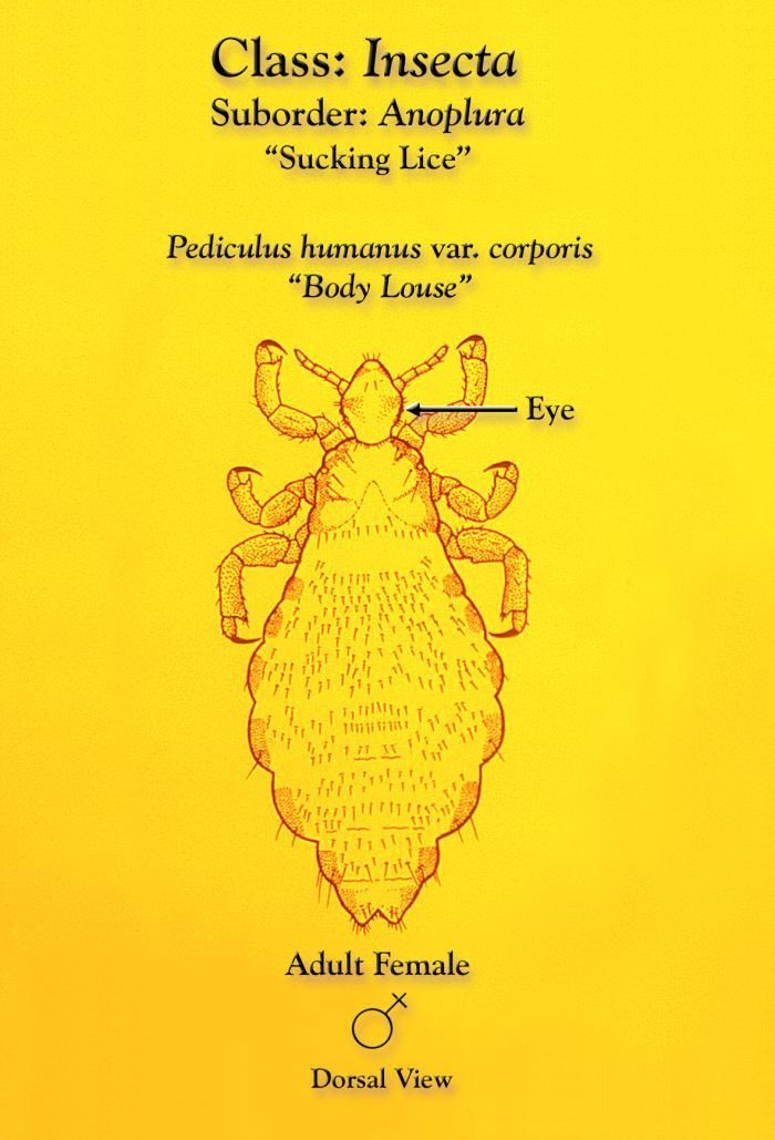
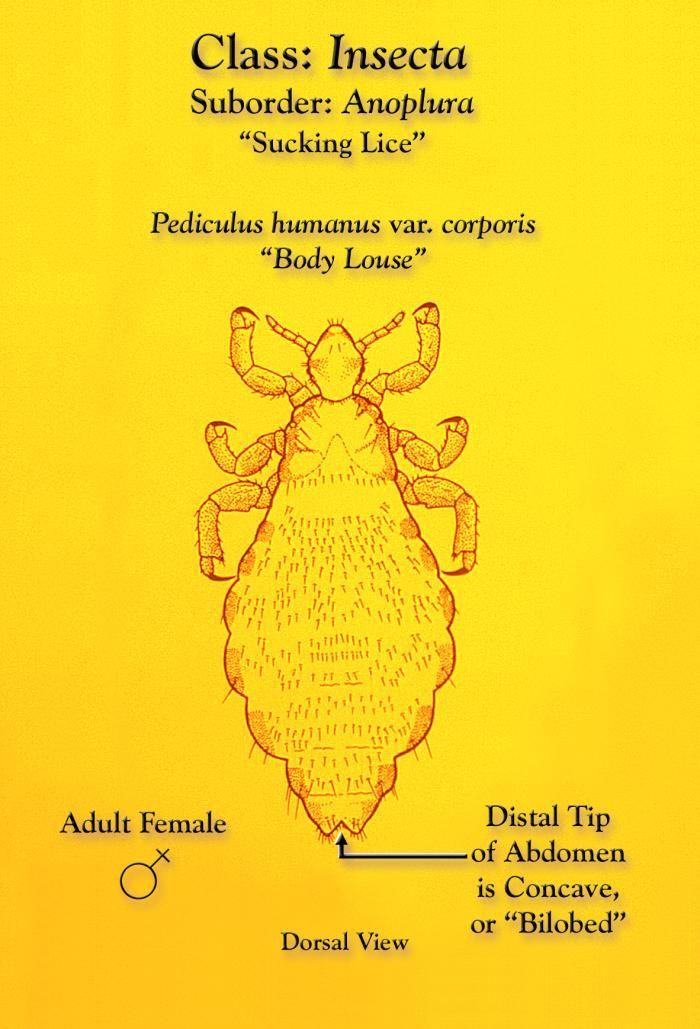
This illustration depicts a dorsal view of a female body louse, Pediculus humanus var. corporis.Created: 1975
-
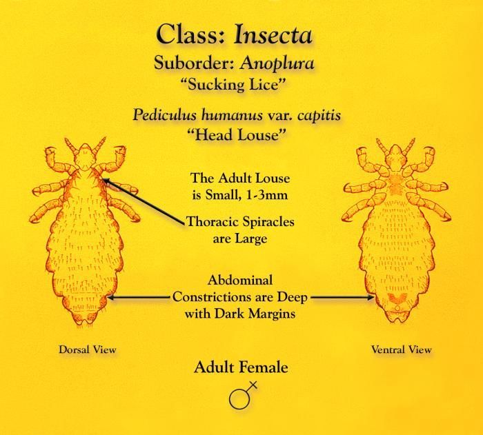
This illustration depicts some of the morphologic characteristics of a female head louse, Pediculus humanus var. capitis from dorsal (Lt) and ventral (Rt) views.Created: 1975
-
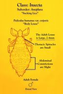
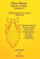
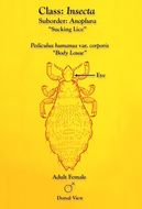
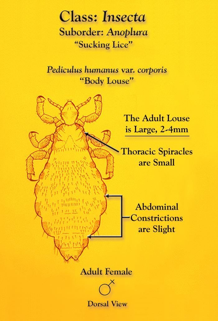
This illustration depicts some of the morphologic characteristics of a female body louse, Pediculus humanus var. corporis from a dorsal view.Created: 1975
-
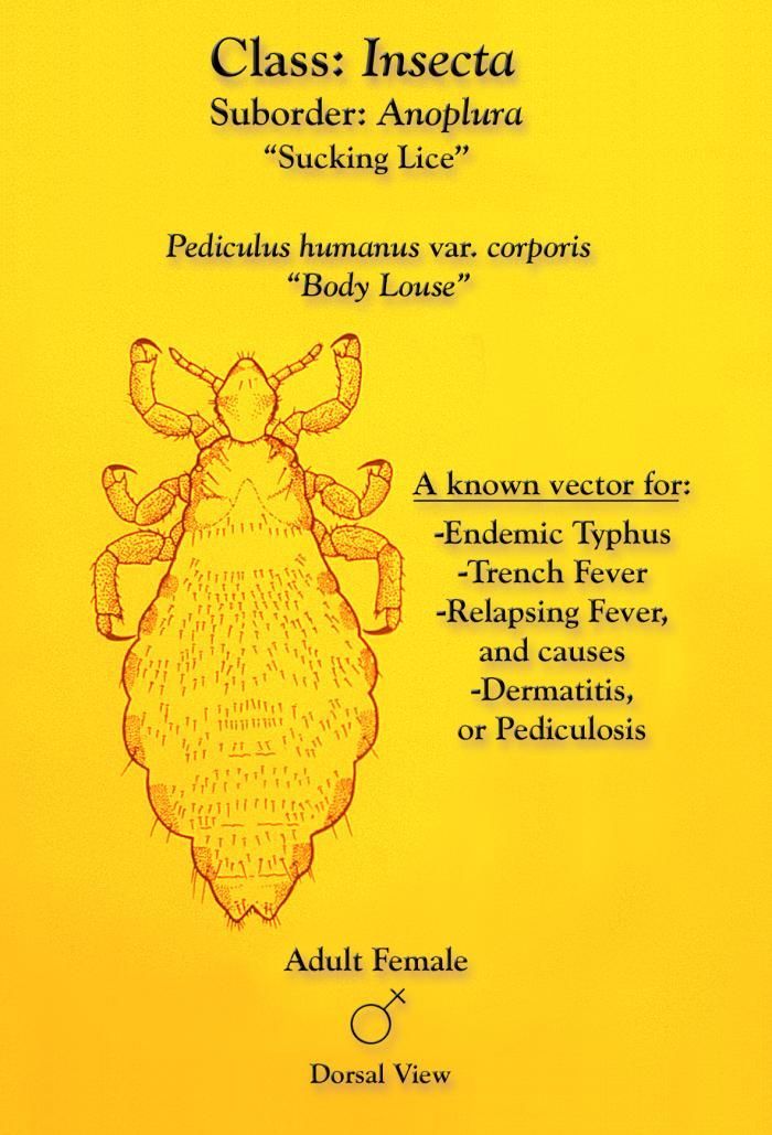
This illustration depicts a dorsal view of a female body louse, Pediculus humanus var. corporis.Created: 1975
-
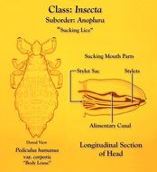
This illustration depicts a dorsal view (Lt), and an enlarged longitudinal mid-sagittal view (Rt) of the head region of a Pediculus humanus var. corporis louse, which is a member of the Order Anoplura, or sucking lice.Created: 1975
-
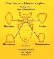
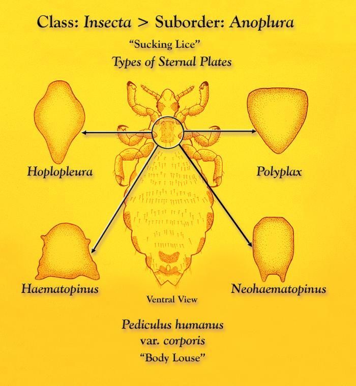
This illustration depicts a ventral view of a female body louse, Pediculus humanus var. corporis, which reveals the size, location, and shape of the thoracic sternal plate of this genus, as well as four other genera of Anoplural sucking lice, Hoplopleura, Polyplax, Haematopinus, and Neohaematopinus.Created: 1975
-
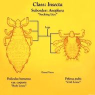
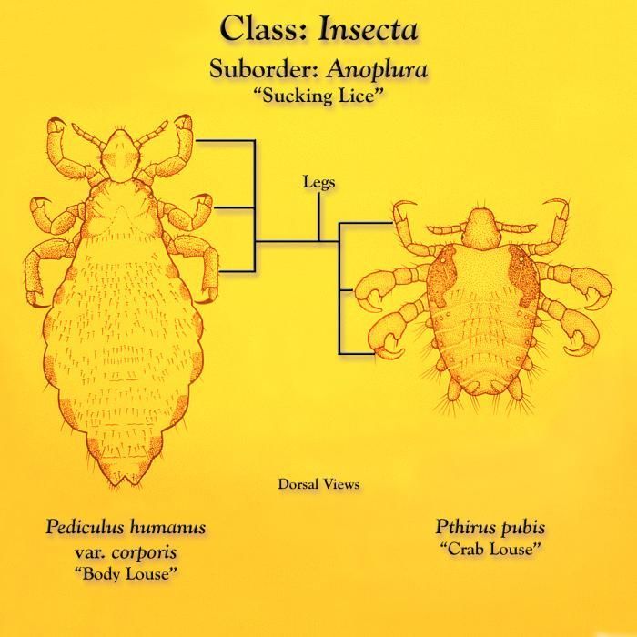
This illustration depicts a dorsal view of a body louse, Pediculus humanus var. corporis (Lt), and a crab louse, Pthirus pubis (Rt).Created: 1975
-
This illustration depicts a dorsal view of a female body louse, Pediculus humanus var. corporis.Created: 1975
-
This illustration depicts a dorsal view of a female body louse, Pediculus humanus var. corporis.Created: 1975
-
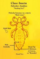
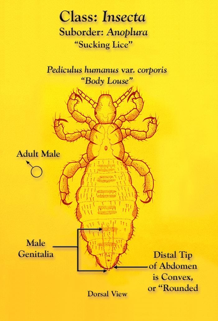
This illustration depicts a dorsal view of a male body louse, Pediculus humanus var. corporis.Created: 1975
-
This illustration depicts a dorsal view of a female body louse, Pediculus humanus var. corporis.Created: 1975
-
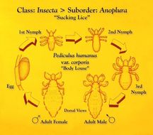
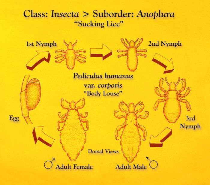
This illustration depicts the life cycle of a sucking louse, Pediculus humanus var. corporis, and the morphologic changes that take place at each successive developmental plateau reached.Created: 1975
-
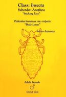
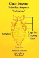
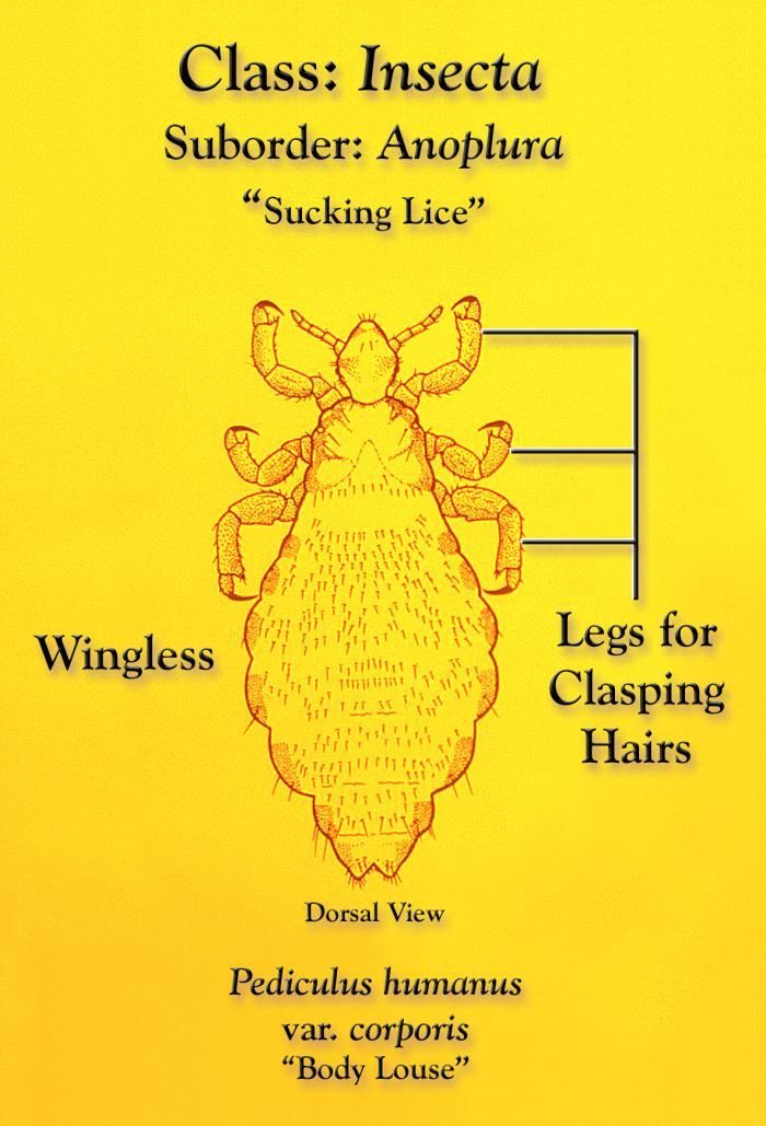
This illustration depicts some of the morphologic characteristics found in a sucking louse, Pediculus humanus var. corporis, of the Order Annoplura.Created: 1975
-
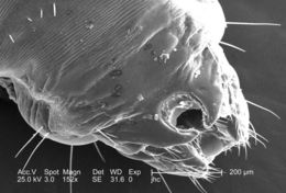
At double the magnification of PHIL #9239, this 2006 scanning electron micrograph (SEM), magnified 152x, revealed the distal tip of the abdominal region of a female body louse, Pediculus humanus var. corporis from a dorsal perspective. Some of the morphologic characteristics seen in this image include the two gonopodia, which are located dorsal to the larger two setae-bearing claspers. It is into this notch that the male would insert the aedeagus, or penis during the process of copulation. This notch, identifying the louse as a female is observable to the naked eye, whereas, in the male louse, the distal abdomen is rounded, and not concave.Created: 2006
-
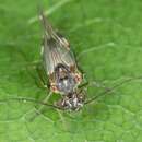
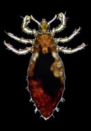
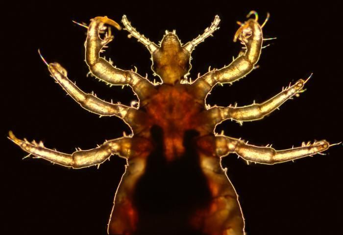
This 2006 photograph depicted a magnified ventral view of a male body louse, Pediculus humanus var. corporis, focusing on the insects cephalic and thoracic regions. Its counterpart, the dorsal view may be seen in PHIL# 9217. Some of the external morphologic features displayed by members of the genus Pediculus include an elongated abdominal region without any processes, and three pairs of legs, all equal in length and width. The distal tip of the males abdomen is rounded, whereas, the females (PHIL# 9202) is concave.Body lice are parasitic insects that live on the body, and in the clothing or bedding of infested humans. Infestation is common, found worldwide, and affects people of all races. The dark mass inside the abdomen is a previously ingested blood meal, obtained by the louse when the photographer offered his arm to the insect on which it fed.Created: 2006
-
This 2006 photograph depicted a ventral view of a male body louse, Pediculus humanus var. corporis. Its counterpart, the dorsal view may be seen in PHIL# 9217. Some of the external morphologic features displayed by members of the genus Pediculus include an elongated abdominal region without any processes, and three pairs of legs, all equal in length and width. The distal tip of the males abdomen is rounded, whereas, the females (PHIL# 9202) is concave.Body lice are parasitic insects that live on the body, and in the clothing or bedding of infested humans. Infestation is common, found worldwide, and affects people of all races. The dark mass inside the abdomen is a previously ingested blood meal, obtained by the louse when the photographer offered his arm to the insect on which it fed.Created: 2006