dcsimg

Diagnostic Description

provided by EOL authors
Description Morphology

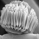
Colonies on corn-meal agar light yellow, growing sparsely; vegetative hyphae colorless, sep- tate, thin walled; rhizoids present; sporangiophores scattred, yellowish, erect, irregularly branched, mostly 10-17.5 μm in diam, up to 1 cm or more high, smooth, thick-walled, with conspicuous septa, branching several times, tapering above and terminating in a single sporo- cladium, usually over severn sporocladia present on one sporophore; sporocladia usually arranged in a zigzag pattern on the sporangiophore axes, 13-39 × 18-47 μm, sessile, non-septate, dome-shaped, bearing numerous pseudophialides crowded over the upper surfaces; pseudophialides ellipsoidal, 6-8× 2-3 μm, with small apical projections bearing the sporangiola; sporangiola nearly hyaline, elongate-clavate, 19-25 (aver. 22) × 3.8-4.2 μm, enveloped in liquid at maturity ; sporangial wall projecting about 2-3 μm beyond the apex of the spore as a hyaline enlargement of the tip; sporangiospores lanceolate, 16-22 (aver. 19) × 3.8-4.2 μm near the base, uniformly tapered to the acute apices, round below. Zygospores not observed.
Colonies on potato-dextrose agar maize yellow, vegatative hyphae branching irregularly, growing luxiously, sporangiophores rare, bearing relatively less sporocladia than on CMA, usually less than three sporocladia forming on one sporangiophore.

Notes This species is distinguished from the relative species L. .macrospora by it’s smaller (16-22 × 3.8-4.2 μm) sporangiospores, which is larger (26-36 x 3-5 μm) in the later one (Chang, 1967).


Ecology and Distribution Specimens examined

YMS0602,isolated from soil, Yangmingshun National Park, Taipei. Sep. 2006.

(BiotaTaiwanica myco.biota,biodiversity 2015)

References

  • H.M. Ho.
  • (Accessed site 13 July 2015)

license
cc-by-nc
copyright
Biota Taiwanica Mycobiota & Mycobiologica
bibliographic citation
H.M. HO
author
C Robertson
original
visit source
partner site
EOL authors

Brief Summary

provided by EOL authors

Both species ofLinderinahave been isolated from soil. The colonies of both species are yellow and they grow well on relatively nutrient-rich culture media.Linderina pennisporafills the Petri dish with aerial hyphae and sporocladia are produced from the upper portion of the sporangiophore.Linderina macrosporaproduces fewer sporocladia. Both species have been isolated at least one other time from soil in addition to the original isolation. Cole and Samson (1979) and O’Donnel (1979) both have publishedSEMphotographs ofLinderinaspp. (Zygomycetes 2015)

References

  • Updated Feb 26, 2007 (Accessed site 13 July 2015)
  • Baijal, U. 1963. Linderina pennispora Raper & Fennell from India. Mycolpath. Mycol. Appl. 21:109-111.
  • Benjamin, R.K. 1966. The merosporangium. Mycologia 58:1-42.
  • Benny, G.L, and H.C. Aldrich. 1975. Ultrastructural observations on septal and merosporangial ontogeny in Linderina pennispora (Kickxellales; Zygomycetes). Can. J. Bot. 53:2325-2335.
  • Chang, Y. 1967. Linderina macrospora sp. nov. from Hong Kong. Trans. Brit. Mycol. Soc. 50:311-314.
  • Chien, C.-Y. 1971. Linderina macrospora from forest soil of the southeastern Unitied States. Mycologia 63:410-412.
  • Cole, G.T., and R.A. Samson. 1979. Patterns of Development in Conidial Fungi. Pitman Publishing Limited, London, United Kingdom.
  • Raper, K.B, and D.I. Fennell. 1952. Two noteworthy fungi from Liberian soil. Amer. J. Bot. 39:79-86.
  • Young, T.W.K. 1970. Ultrastructure of the spore wall of Linderina. Trans. Brit. Mycol. Soc. 54:15-25.
  • Young, T.W.K. 1971. Ultrastructure of the wall of the germinating sporangiospore of Linderina pennispora (Mucorales). Ann. Bot. 35:183-191.

license
cc-by-3.0
copyright
Gerald L. Benny
bibliographic citation
Baijal, U. 1963. Linderina pennispora Raper & Fennell from India. Mycolpath. Mycol. Appl. 21:109-111. Benjamin, R.K. 1959. The merosporangiferous Mucorales. Aliso 4:321-433. Benjamin, R.K. 1966. The merosporangium. Mycologia 58:1-42. Benny, G.L, and H.C. Aldrich. 1975. Ultrastructural observations on septal and merosporangial ontogeny in Linderina pennispora (Kickxellales; Zygomycetes). Can. J. Bot. 53:2325-2335. Chang, Y. 1967. Linderina macrospora sp. nov. from Hong Kong. Trans. Brit. Mycol. Soc. 50:311-314. Chien, C.-Y. 1971. Linderina macrospora from forest soil of the southeastern Unitied States. Mycologia 63:410-412. Cole, G.T., and R.A. Samson. 1979. Patterns of Development in Conidial Fungi. Pitman Publishing Limited, London, United Kingdom. O’Donnell, K.L. 1979. Zygomycetes in Culture. Palfrey Contributions in Botany No. 2. Department of Botany, University of Georgia, Athens, Georgia. Raper, K.B, and D.I. Fennell. 1952. Two noteworthy fungi from Liberian soil. Amer. J. Bot. 39:79-86. Young, T.W.K. 1968. Electron microscopic study of the asexual structures in Kickxellaceae. New Phytologist 67:823-836. Young, T.W.K. 1970. Ultrastructure of the spore wall of Linderina. Trans. Brit. Mycol. Soc. 54:15-25. Young, T.W.K. 1971. Ultrastructure of the wall of the germinating sporangiospore of Linderina pennispora (Mucorales). Ann. Bot. 35:183-191.
author
C Robertson
original
visit source
partner site
EOL authors