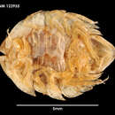
“Serolis (Serolis) exigua n. sp.
Pl. I Fig. 2; Text Figs. 4 a--c, 6 e, 17 a—i, i8 a—h.
Diagnosis. Head with anterio-lateral angles slightly prolonged in a lateral direction; greatest width of the head at the front margin. Coxal plates marked off by dorsal sutures on the second to fourth pereion segments. The posterio-lateral angles of the coxal plates of the pereion segments and pleural plates of the second and third abdominal segments all reaching beyond those of their preceding segments. Anterior masticatory process on the left mandible not expanded and without cutting edge. Inner lobe of first maxilla expanded distally. Outer lappets of outer lobe of the second maxilla each provided with two apical setae. Maxilliped with the distal epipodite fused with the basipodite in its entire length. Pa1p of maxilliped consisting of three joints, of which the second is approximately cordiform. Basipodites of the first three pairs of pleopods with their inner proximal angles projecting and furnished with setae. Endopodite of fourth pleopod entire (not bifid). Exopodite of the fifth pleopod with the lateral margin furnished with two long plumose setae.
Description.
Type. Female with young, about 7 mm. in length.
Shape of body and sculpturing (P1. I Fig. 2, Text fig. 17 a). The head, except a submarginal part at its front margin, the tergites of the pereion segments and the free abdominal segments, except the greater part of their pleurae, are elevated as compared with the lateral parts of the body. A dorsal longitudinal carina along the mid-line passes from the centre of the head (= from a spot between the front margins of the eyes) along the body to the distal end of the pleotelson. Posterior margin of the tergites of the pereion segments and the free segments of abdomen all with a small backward-directed triangular tip in the middle line, (very indistinct on the first pereion segment).
Head (Pl. I Fig. z, Text fig. 17 a). Slightly broader than it is long, posteriorly in the middle continuous with the first pereion segment without trace of any suture. The longitudinal keel in the middle line is faint, yet distinct in the posterior part of the head, as well as on the first pereion segment. The lateral parts of the posterior margin of the head are divided from the first pereion segment only by a slight groove, which develops anteriorly into a distinct suture. Front margin, in the middle, with a very small rostrum; laterally from the rostrum on either side it is somewhat concave; between the first and second peduncular joints of the antennulae the front margin forms a small triangular point, which extends somewhat further in front than the rostrum. The anteriolateral angles of the head are triangularly prolonged and slightly bent downwards. The eyes are small, reniform; the distance between the eye and the anterior margin of the head is about the length of one eye. Between the eyes and the longitudinal keel in the middle line there is, on either side, a short indistinct longitudinal ridge.
Pereion (Pl. I Fig. 2, Text fig. 17 a). The first pereion segment, ventrally, with a distinct longitudinal suture passing through the sockets for the first pereiopods. The ventral part of the segment is covered by the marsupium.
Second, third, and fourth segments, in the middle, of about equal length, the fifth and sixth in the middle each about half as long as the fourth. Posterior margin of the tergites of all the segments with a small backward-directed triangular tip in the mid-line (indistinct on the first segment).
Coxal plates demarcated by dorsal sutures on the second to fourth segment; the sutures are curved and not quite in a line with each other; on the second, third, and fourth segments there is a convexity of the posterior margins medially from the coxal plates; a similar convexity is also to be observed on the first segment. Lateral margins of the epimera of the first to fourth segments continuous with each other; only the extreme tips of the coxal plates of the fourth, fifth, and sixth segments protrude freely.
The ventral surface of the pereion segments is almost entirely covered by the marsupium.
Abdomen. (Pl. I Fig. 2, Text fig. 17 a). On the first three segments the longitudinal keel along the mid-line is very distinct, whilst the triangular points in the middle of the posterior margins are small and indistinct.
From the median keel the dorsal surface slopes slightly; on the first segment to its lateral margins, on the second and third segments to their pleural plates. The lateral parts of the pleural plates of second and third segments have their posterior margins somewhat elevated compared with their anterior margins. The posterio-lateral angles of the pleural plates of the third abdominal segment extend further back than those of the second abdominal segment, reaching to about two-thirds the length of pleotelson.
The sternites of the first three abdominal segments are posteriorly protracted into one long point in the middle, and two short lateral points, one on each side; the sternites are sculptured, with a longitudinal ridge along the middle line.
Pleotelson, broadly cordiform. The longitudinal carina along the middle line is distinct. Anterior parts of the lateral margins each with a marginal ridge extending somewhat further back than the pleural plates of the second abdominal segment. Between these ridges and the longitudinal carina in the middle there is on either side yet another, longitudinal and somewhat curved, ridge, slightly concave on its inner side. These ridges and the marginal ridges are connected by a short ridge. The part of the pleotelson which is marked off distally by the latter ridge, medially by the ridge situated laterally from and nearest to the middle keel, and laterally and proximally delimited by the marginal ridge, is subtriangular and somewhat elevated.
Antennulae. The peduncle is longer than the flagellum and consists of four joints. Second peduncular joint slightly longer than the first. Third joint narrower and about as long as the second plus half the first. Fourth joint short, about two-fifths as long as the third. The flagellum consists of 16 joints, each joint carrying a sensory filament.
Antennae. (Fig. 17 b). First joint of the peduncle short and visible only from below. Second and third joints of about equal length, forming an angle with each other. Second peduncular joint proximally with a faint incomplete suture on the ventral side, but without suture dorsally. Fourth peduncular joint half again to twice as long as the third; fifth joint slightly longer and narrower than the fourth. Ventral surface of the third, fourth, and fifth joints, near the rostral margin, exhibits groups of setae forming transverse rows. On the distal part of the third joint there are two such rows, each consisting of two groups, on the fourth three rows, each, as a rule, with three groups of setae, on the fifth joint there are five rows.
The flagellum is slightly longer than the last peduncular joint and consists of ten joints. There is a row of prolonged triangular scale-processes along the rostral margin of the ventral surface on the central joints of the flagellum.
Mandibles (Figs. 17 c and d). Left mandible (fig. 17 c) with the rostral masticatory process stronger than the weak, caudal one, which is prolonged distally into bristle-like processes. Right mandible with two weak masticatory »setae».
Setae on the second and third joints of the mandibular palp with oblong-ovate end-knobs.
First pair of maxillae (Fig. 17 e). Distal margin of outer lobe with eleven setae, situated in two rows. Distal end of inner lobe elliptically expanded.
Second pair of maxillae (Fig. 17 f). Inner lobe much broader and longer than both lappets of outer lobe; its distal margin with about fifteen setae, situated in two rows. Lappets of outer lobe each with two apical setae.
Maxillipeds (Fig. 17 g). Distal epipodite fused with the basipodite in its entire length. Composite setae on the distal margin of the basipodite approximately as in S. paradoxa.lvl Upper lip (Fig. 17 h). Normal.
First pair of pereiopods (Fig. 17 i). Basipodite longer than ischium, merus and carpus together. Ischium markedly widening towards its distal end, about as long as the meral and carpal joints together. The merus is short, almost trapezoidal and somewhat broader than the carpus.
For the two composite setae at the upper distal angle of the carpus see Fig. 6 e. Close to the upper distal angle of the carpus there is a group of simple setae on the caudal surface, and on the distal margin there are hair-like points devoid of a setal canal (see Fig. 6 e).
The propodal joint is somewhat shorter than the ischium, merus and carpus together. On the lower margin of propodus there is only one row of composite setae, each seta consisting of a triangular scale which, proximally, is fused with a simple seta (see Figs. 4 a, b, c). The usual caudal row of leaf-like setae is replaced by a row of projecting, anteriorly rounded scales (Fig. 4 c). Close to the lower margin of the propodus on the caudal side there is a submarginal row of simple setae. The caudal and rostral surfaces of the propodus exhibits scattered shorter setae of the same non-composite kind.
The other pereiopods (Figs. 18 a, b). Carpal and propodal joints increasing in length from the second to the seventh pereiopod. Together they are shorter on the third pereiopod than the basipodite, but on the seventh longer than that joint. Upper margin of the basipodite on the second to fourth pereiopods has delicate »hairs» lacking a setal canal; on the fifth to seventh pereiopods such »hairs» occur also on the upper margin of the ischium. The »hairiness» of the limbs increases from the second to seventh, where the hairs cover almost the whole upper margin of the basipodite and the ischium.
First three pairs of pleopods (Figs. 18 c, d). Inner angle of the basipodite of the first pair exhibits three (two in one specimen) setae; on the second and third pairs two setae. Endopodite on the first and second pairs oval in outline, on the third almost circular.
Fourth pair of pleopods (Fig. 18 e). Exopodite and endopodite triangular, the endopodite somewhat smaller than the exopodite; both are divided by an almost transverse suture, somewhat distally from the middle. Lateral margin of the exopodite provided with plumose setae, its proximal margin and distal part of its inner margin furnished with hair-like setae. Margins of endopodite smooth.
Fifth pair of pleopods (Fig. 18 f). Exopodite divided by a transverse suture somewhat distally from the middle; its lateral margin close to the distal end furnished with two long plumose setae. Endopodite likewise divided by a transverse suture; about one-third of the joint is situated posteriorly from the suture.
Uropods (Figs. 18 g and h). Exopodite and endopodite subequal in length, but the exopodite does not extend quite as far back as the endopodite. Both the rami are broadly oval in outline and have their distal margins broadly rounded. Outer and inner margins serrate, distal margins (Fig. 18 h) serrate and in addition provided with some large incisions.
Remarks. In its general shape the new species most resembles S. glacialis, septemearinata and polita, but is easily distinguishable, especially by the different sculpturing of the head and the pleotelson.
The single row of composite setae on the lower margin of the propodus of the first pereiopods and the two long plumose setae on the lateral margin of the exopodite of the fifth pleopod are features peculiar to the species, which have not been found in any other member of the genus.
Localities and Material.
St. 39. Falkland Islands, Port William, lat. 51° 4o' S., long. 57° 41' W. 4o m. Sand and small stones with algae. 417 19o2. a females possessing marsupia. Length of the specimens 6.8 and 6.5 mm.
St. 49. Falkland Islands, Berkeley Sound, lat. 51° 35' S., long. 57° 56' W. 25-30 m. Shells and stones. 1°4 1902. Mature female with its marsupium filled with young. Length about 7 mm (type specimen).,
Distribution:: Falkland Islands (Sw. Ant. Exped.).
(Nordenstam, 1933)