-
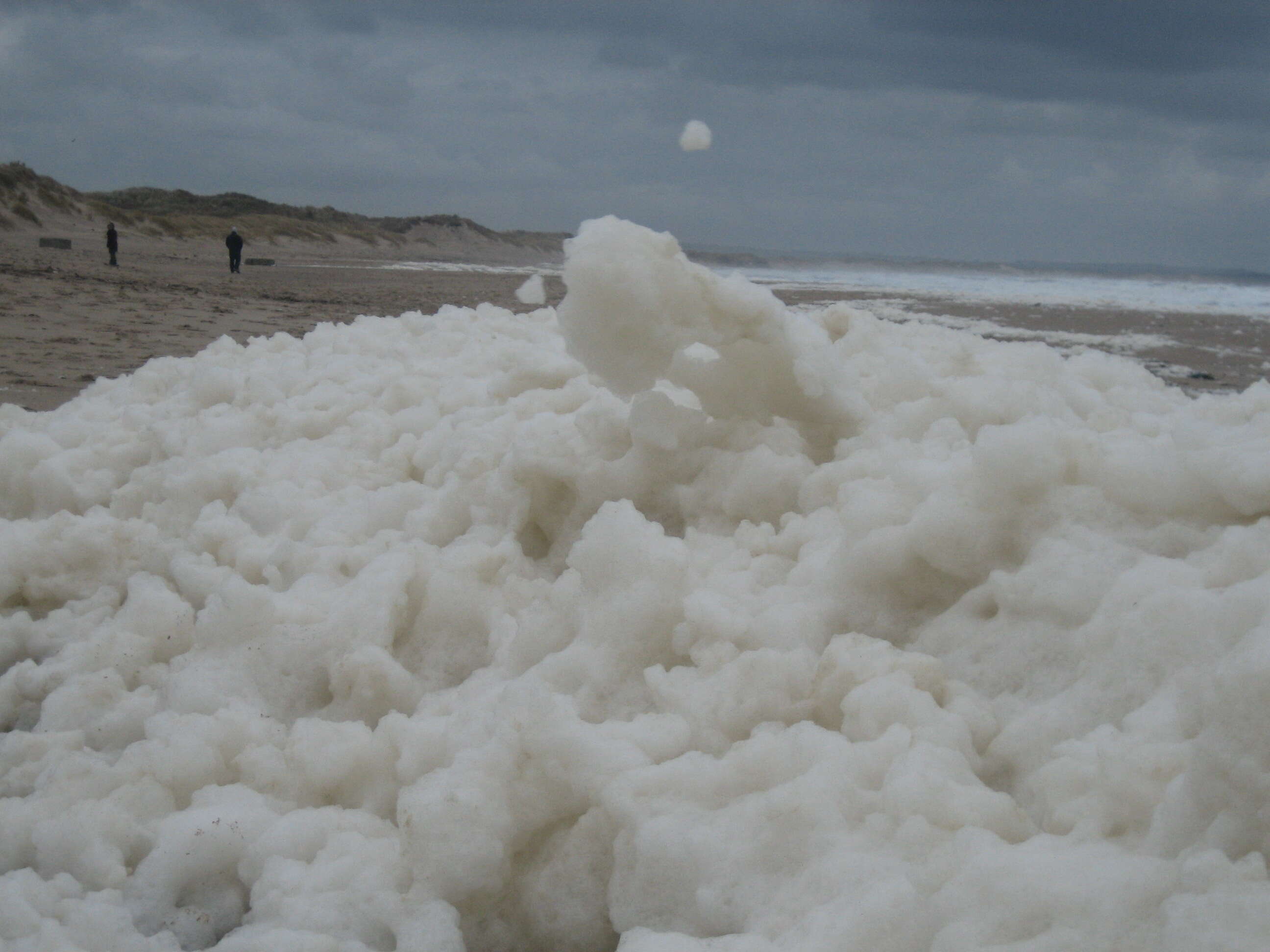
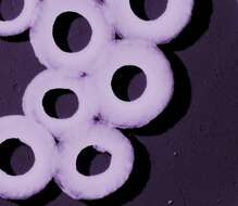
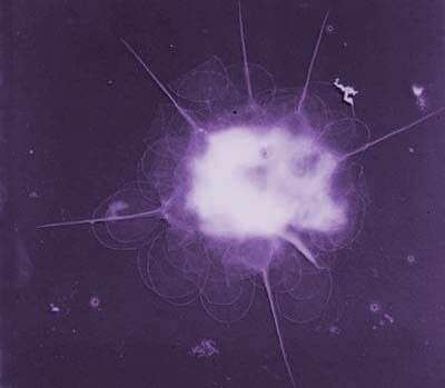
Summary.mw-parser-output table.commons-file-information-table,.mw-parser-output.fileinfotpl-type-information{border:1px solid #a2a9b1;background-color:#f8f9fa;padding:5px;font-size:95%;border-spacing:2px;box-sizing:border-box;margin:0;width:100%}.mw-parser-output table.commons-file-information-table>tbody>tr,.mw-parser-output.fileinfotpl-type-information>tbody>tr{vertical-align:top}.mw-parser-output table.commons-file-information-table>tbody>tr>td,.mw-parser-output table.commons-file-information-table>tbody>tr>th,.mw-parser-output.fileinfotpl-type-information>tbody>tr>td,.mw-parser-output.fileinfotpl-type-information>tbody>tr>th{padding:4px}.mw-parser-output.fileinfo-paramfield{background:#ccf;text-align:right;padding-right:0.4em;width:15%;font-weight:bold}.mw-parser-output.commons-file-information-table+table.commons-file-information-table,.mw-parser-output.commons-file-information-table+div.commons-file-information-table>table{border-top:0;padding-top:0;margin-top:-8px}@media only screen and (max-width:719px){.mw-parser-output table.commons-file-information-table,.mw-parser-output.commons-file-information-table.fileinfotpl-type-information{border-spacing:0;padding:0;word-break:break-word;width:100%!important}.mw-parser-output.commons-file-information-table>tbody,.mw-parser-output.fileinfotpl-type-information>tbody{display:block}.mw-parser-output.commons-file-information-table>tbody>tr>td,.mw-parser-output.commons-file-information-table>tbody>tr>th,.mw-parser-output.fileinfotpl-type-information>tbody>tr>td,.mw-parser-output.fileinfotpl-type-information>tbody>tr>th{padding:0.2em 0.4em;text-align:left;text-align:start}.mw-parser-output.commons-file-information-table>tbody>tr,.mw-parser-output.fileinfotpl-type-information>tbody>tr{display:flex;flex-direction:column}.mw-parser-output.commons-file-information-table+table.commons-file-information-table,.mw-parser-output.commons-file-information-table+div.commons-file-information-table>table{margin-top:-1px}.mw-parser-output.fileinfo-paramfield{box-sizing:border-box;flex:1 0 100%;width:100%}} Description: Sea foam blown onto Druridge Bay beach, Northumberland, by a strong onshore wind. I've never seen anything like this amount of the stuff before. While it's a novel thing to see, and kind of fun to watch dogs play in, it's disturbing to find that it may be a harbinger of changes afoot in the North Sea and in the global climate. The foam is formed (according to the site below) by a micro-organism called phaeocystis. With excessive nutrients, it blooms out of control and forms large gelatinous blobs. When whipped up by wind and waves, they become sea foam. The blooms also produce a volatile sulphur compound that contributes to climate change.
europa.eu.int/comm/research/rtdinfsup/en/world4.htm. Date: Taken on 26 February 2006, 14:14. Source:
Sea Foam - Harbinger of Change. Author:
Akuppa John Wigham from Newcastle upon Tyne, England. Flickr tagsInfoFieldphaeocystis, north sea, climate change, northumberland, northumbria, england, druridge bay, global warming, cresswell, sea, sea foam, foam.
-
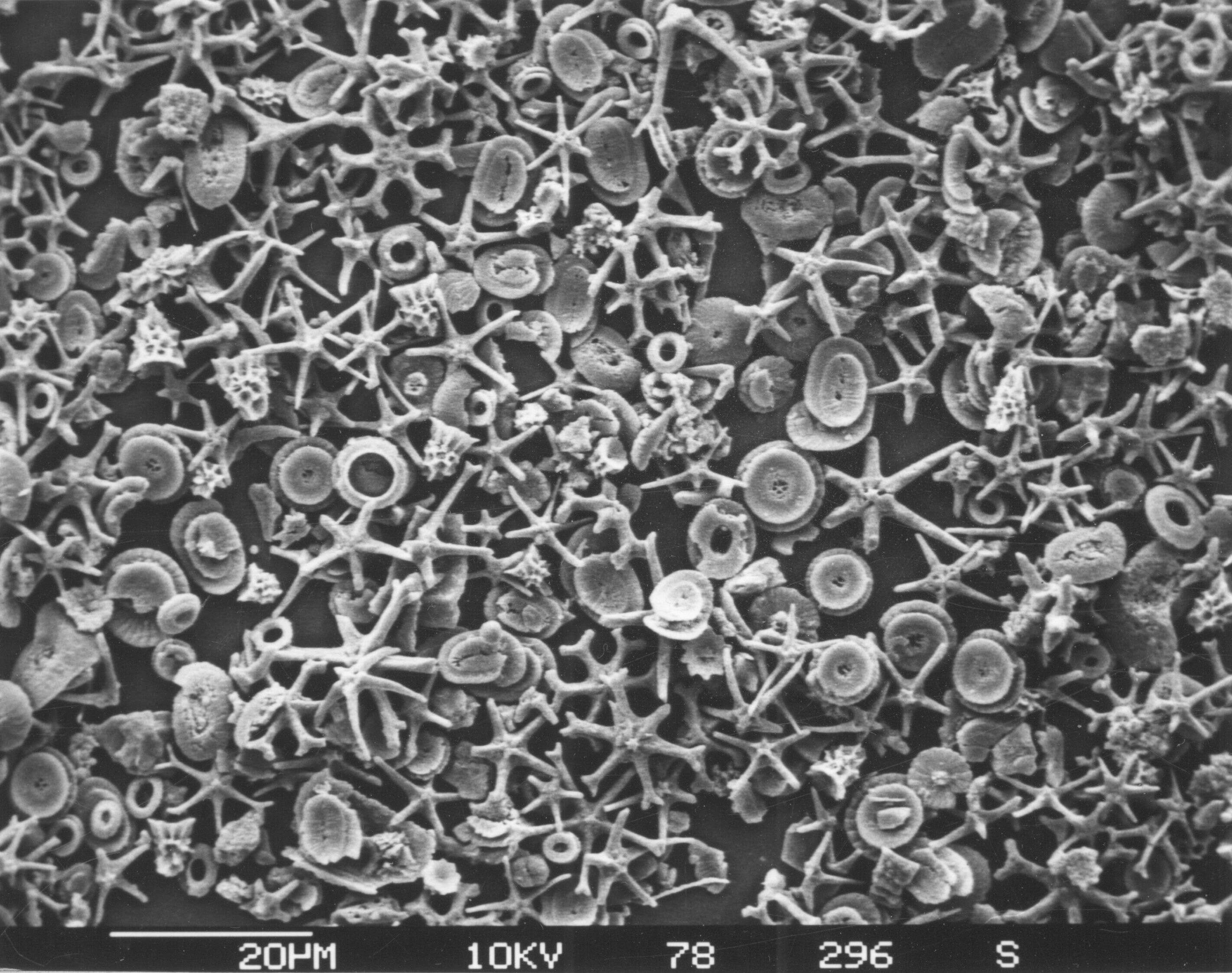
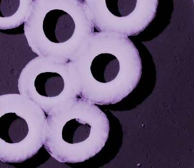
Description: Nannoplankton microfossils from marine sediment consisting of coccoliths and discoasteridae. Date: 8 October 2013, 11:20:34. Source: Own work. Author: Hannes Grobe.
-
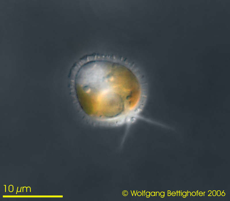
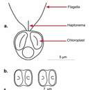
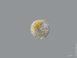
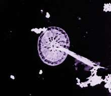
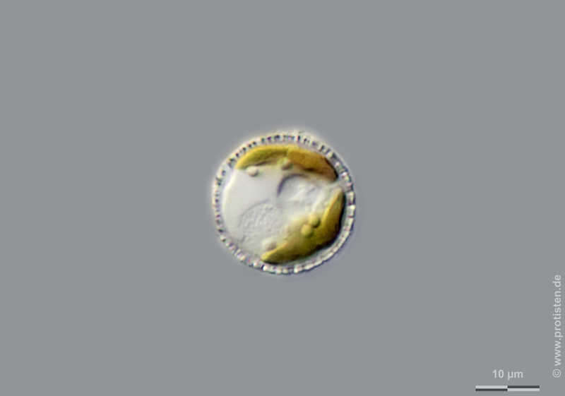
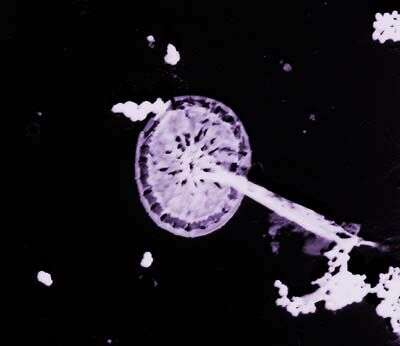
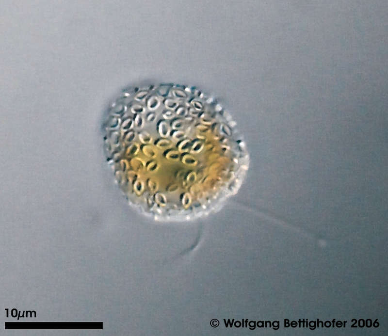
Hymenomonas roseola This tiny prymnesiomonad belongs to group of Coccolithophoraceae. In a mucous cover they wear lots of calcified scales (coccoliths). The depth of focus picture shows coccoliths covering the monad cell. Picture generated from 7 shots using CombineZ. Collected from littoral region (reed bed by Phragmites) of a rain storage reservoir in Kiel (Schleswig-Holstein, Germany). Images were taken using Zeiss Universal with Olympus C7070 CCD camera.Image under Creative Commons License V 3.0 (CC BY-NC-SA). Place name: Pond Demühlen, rain storage reservoir in Kiel-Russee (Schleswig-Holstein, Germany) Latitude: 54.304095 Longitude: 10.086073 Diese winzige Prymnesiomonade gehört zur Gruppe der Coccolithophoraceae. In der unhüllenden Gallertschicht sie viele verkalkte Schuppen (Coccolithen) eingelagert. Tiefenschärfe durch Multiebenenabbildung aus 7 Bildebenen, automatisch gestapelt mit CombineZ. Aus der von Schilf bewachsenen Uferzone eines Regenrückhaltebeckens in Kiel. Mikrotechnik: Zeiss Universal, Kamera: Olympus C7070. Creative Commons License V 3.0 (CC BY-NC-SA). For permission to use of (high-resolution) images please contact postmaster@protisten.de.
-
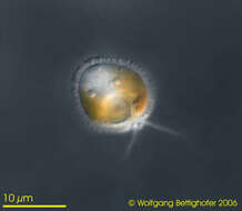
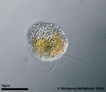
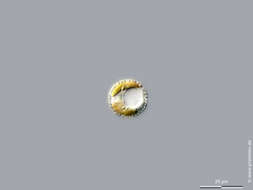
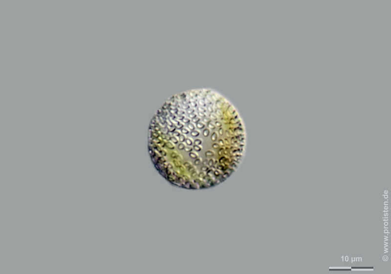
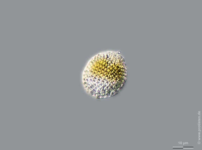
Hymenomonas roseola Scale bar indicates 10 µm. The specimen was gathered in a pond in the forest of Altenholz-Stift near Kiel (Schleswig-Holstein, Germany). Sampling date 5/2017. The image was built up using several photomicrographic frames with manual stacking technique. Images were taken using Zeiss Axioskop with Olympus OM-D M5 MKII. Image under Creative Commons License V 3.0 (CC BY-NC-SA). Place name: Pond in the forest of Altenholz-Stift (Schleswig-Holstein, Germany) Latitude: 54.384913 Longitude: 10.125691 Der Messbalken markiert eine Länge von 10 µm. Die Probe wurde in einem Waldteich bei Altenholz-Stift (nahe Kiel) im Mai 2017 gesammelt. Mikrotechnik: Zeiss Axioplan, Kamera: Olympus OM-D M5 MKII. Creative Commons License V 3.0 (CC BY-NC-SA). For permission to use of (high-resolution) images please contact postmaster@protisten.de.
-
Hymenomonas roseola Scale bar indicates 10 µm. The specimen was gathered in a pond in the forest of Altenholz-Stift near Kiel. Sampling date 5/2017. The image was built up using several photomicrographic frames with manual stacking technique. Images were taken using Zeiss Axioskop with Olympus OM-D M5 MKII. Image under Creative Commons License V 3.0 (CC BY-NC-SA). Place name: Pond in the forest of Altenholz-Stift (Schleswig-Holstein, Germany) Latitude: 54.384913 Longitude: 10.125691 Der Messbalken markiert eine Länge von 10 µm. Die Probe wurde in einem Waldteich bei Altenholz-Stift (nahe Kiel) im Mai 2017 gesammelt. Mikrotechnik: Zeiss Axioplan, Kamera: Olympus OM-D M5 MKII. Creative Commons License V 3.0 (CC BY-NC-SA). For permission to use of (high-resolution) images please contact postmaster@protisten.de.
-
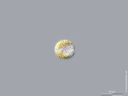
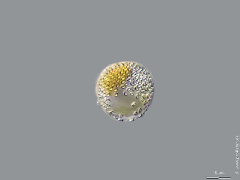
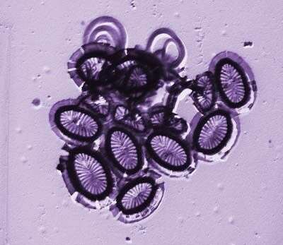
Hymenomonas roseola Scale bar indicates 25 µm. The specimen was gathered in a pond in the forest of Altenholz-Stift near Kiel. Sampling date 5/2017. The image was built up using several photomicrographic frames with manual stacking technique. Images were taken using Zeiss Axioskop with Olympus OM-D M5 MKII. Image under Creative Commons License V 3.0 (CC BY-NC-SA). Place name: Pond in the forest of Altenholz-Stift (Schleswig-Holstein, Germany) Latitude: 54.384913 Longitude: 10.125691 Der Messbalken markiert eine Länge von 25 µm. Die Probe wurde in einem Waldteich bei Altenholz-Stift (nahe Kiel) im Mai 2017 gesammelt. Mikrotechnik: Zeiss Axioplan, Kamera: Olympus OM-D M5 MKII. Creative Commons License V 3.0 (CC BY-NC-SA). For permission to use of (high-resolution) images please contact postmaster@protisten.de.
-
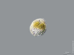
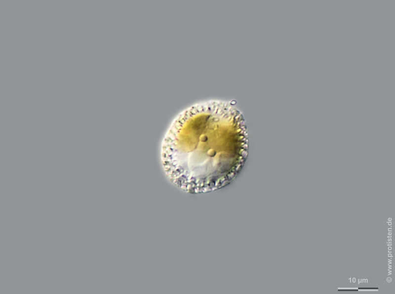
Hymenomonas roseola Scale bar indicates 10 µm. The specimen was gathered in a pond in the forest of Altenholz-Stift near Kiel. Sampling date 5/2017. The image was built up using several photomicrographic frames with manual stacking technique. Images were taken using Zeiss Axioskop with Olympus OM-D M5 MKII. Image under Creative Commons License V 3.0 (CC BY-NC-SA). Place name: Pond in the forest of Altenholz-Stift (Schleswig-Holstein, Germany) Latitude: 54.384913 Longitude: 10.125691 Der Messbalken markiert eine Länge von 10 µm. Die Probe wurde in einem Waldteich bei Altenholz-Stift (nahe Kiel) im Mai 2017 gesammelt. Mikrotechnik: Zeiss Axioplan, Kamera: Olympus OM-D M5 MKII. Creative Commons License V 3.0 (CC BY-NC-SA). For permission to use of (high-resolution) images please contact postmaster@protisten.de.
-
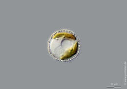
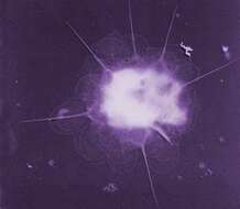
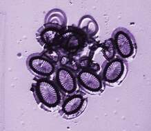
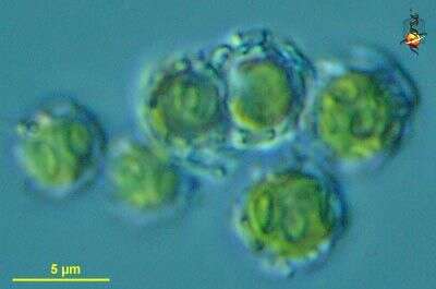
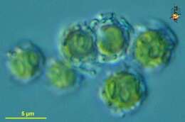
Emiliania (emil-ee-ann-ee-a) huxleyi, a coccolithophorid haptophyte. It can exist in several different forms, and these are the non-motile coccospheres, in which the cells, with golden plastids, are enclosed in layers of small calcareous scales. Differential interference microscopy.
" data on this strain.
-
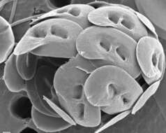
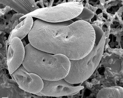
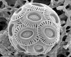
SEM of coccosphere - E. huxleyi type A
-
This image was made from samples taken during a scientific cruise in the Pacific. Water was filtered to concentrate the organisms that were present, then dried onto a thin sheet of plastic and then shadowed with a fine layer of metal to provide contrast. The preparation was then observed with an electron-microscope. This technique has been used to document the diversity of marine microbes, especially, protists in the oceans.
-
This image was made from samples taken during a scientific cruise in the Pacific. Water was filtered to concentrate the organisms that were present, then dried onto a thin sheet of plastic and then shadowed with a fine layer of metal to provide contrast. The preparation was then observed with an electron-microscope. This technique has been used to document the diversity of marine microbes, especially, protists in the oceans. According to Jeremy Young, this is a fragment of a coccosphere of Umbilicosphaera sibogae.
-
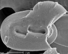
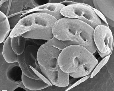
SEM of coccosphere. Flagellar opening is toward top right. Note considerable variation in coccolith size and wing development
-
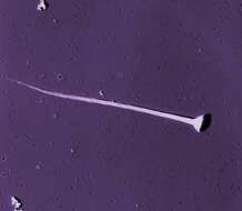
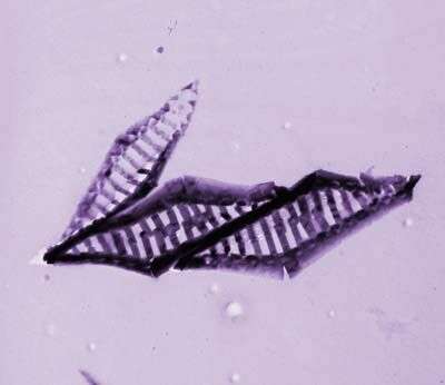
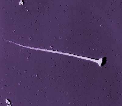
Broken coccolith of H. wallichii in distal view, breakage shows the proximal shield.
-
Helicosphaera wallichii (Lohmann 1902) Okada & McIntyre 1977 [Coccolithophora] Like H. carteri but: central-area with oblique twisted slits; bridge typically better developed; and liths perhaps slightly larger. NB Slits obliquity: In distal view the slits are rotated about 10-20° clockwise (and so away from the wing), this is the ânormalâ sense of obliquity in Helicosphaera, as shown by many fossil species. The Pleistocene species H. inversa is similar but shows the opposite sense of obliquity. HOL phase - unknown but H. wallichii often co-occurs with Syracolithus dalmaticus in our samples (Geisen et al., 2004).
-
This image was made from samples taken during a scientific cruise in the Pacific. Water was filtered to concentrate the organisms that were present, then dried onto a thin sheet of plastic and then shadowed with a fine layer of metal to provide contrast. The preparation was then observed with an electron-microscope. This technique has been used to document the diversity of marine microbes, especially, protists in the oceans.
-
This image was made from samples taken during a scientific cruise in the Pacific. Water was filtered to concentrate the organisms that were present, then dried onto a thin sheet of plastic and then shadowed with a fine layer of metal to provide contrast. The preparation was then observed with an electron-microscope. This technique has been used to document the diversity of marine microbes, especially, protists in the oceans.