-
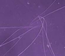
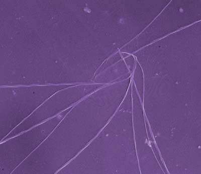
This image was made from samples taken during a scientific cruise in the Pacific. Water was filtered to concentrate the organisms that were present, then dried onto a thin sheet of plastic and then shadowed with a fine layer of metal to provide contrast. The preparation was then observed with an electron-microscope. This technique has been used to document the diversity of marine microbes, especially, protists in the oceans.
-
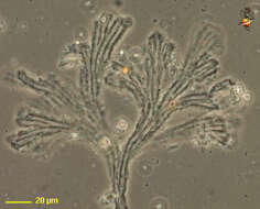
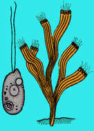
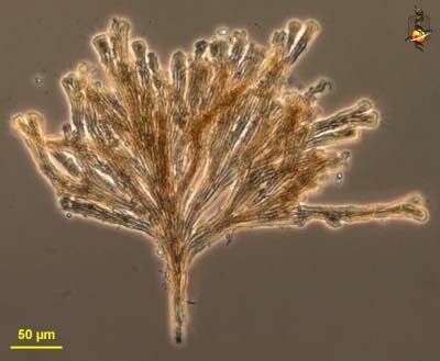
Rhipidodendron (rip-ee-doe-dend-ron, is a colonial spongomonad flagellate, in which the cells are located at the end of a branching (aborescent) colony. The matrix of the colony is formed from adhering small globules of mucilage. The branches are flat, with several channels in each blade. One cell is located at the end of each channel (many of the cells were dislodged from this preparation). Phase contrast.
-
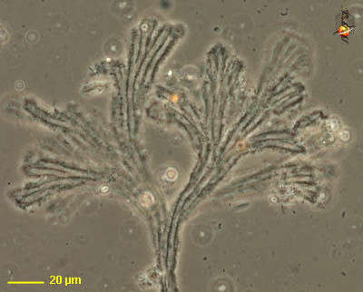
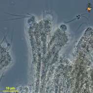
Rhipidodendron (rip-ee-doe-dend-ron, is a colonial spongomonad flagellate, in which the cells are located at the end of a branching (aborescent) colony. The matrix of the colony is formed from adhering small globules of mucilage. The branches are flat, with several channels in each blade. One cell is located at the end of each channel and the cells have two flagella. Phase contrast.
-
-
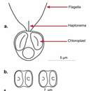
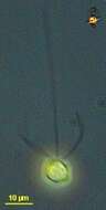
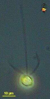
Chrysochromulina (cry-so-crumb-you-line-a) ericina a single-celled haptophyte, with two similar flagella, long anterior haptonema and a golden colour from two yellow-brown chloroplasts. Small scales lie on the surface of the cell but these are not evident in this image. Phase contrast microscopy.
data on this strain.