-
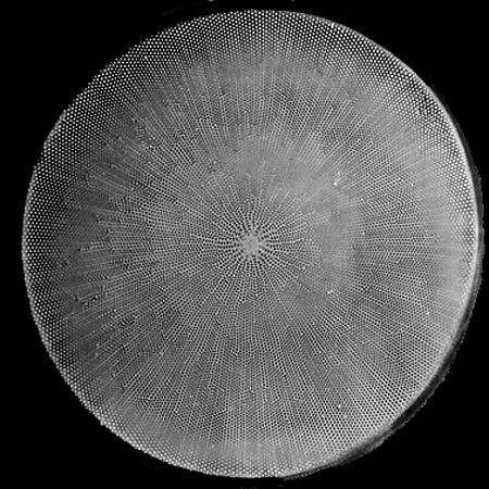
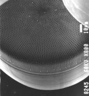
Fig 2: Coscinodiscus wailesii Light micrograph of valve face of a live cell
-
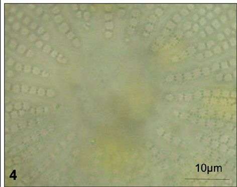
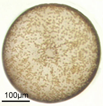
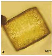
Fig 4: Coscinodiscus wailesii Light micrograph central hyaline area of a live cell
-
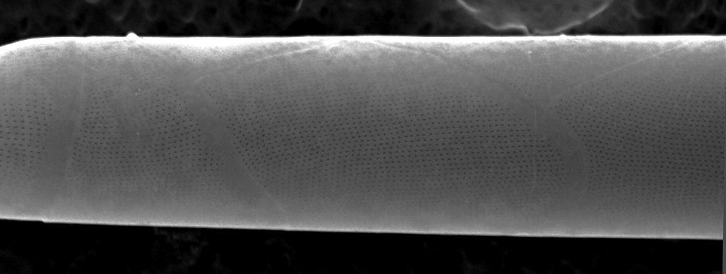
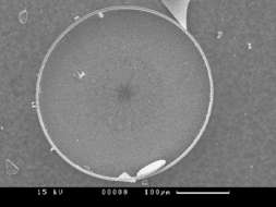
Fig 5: Scanned electron micrograph image of C.wailesii in the valve view.
-
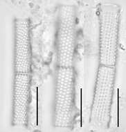
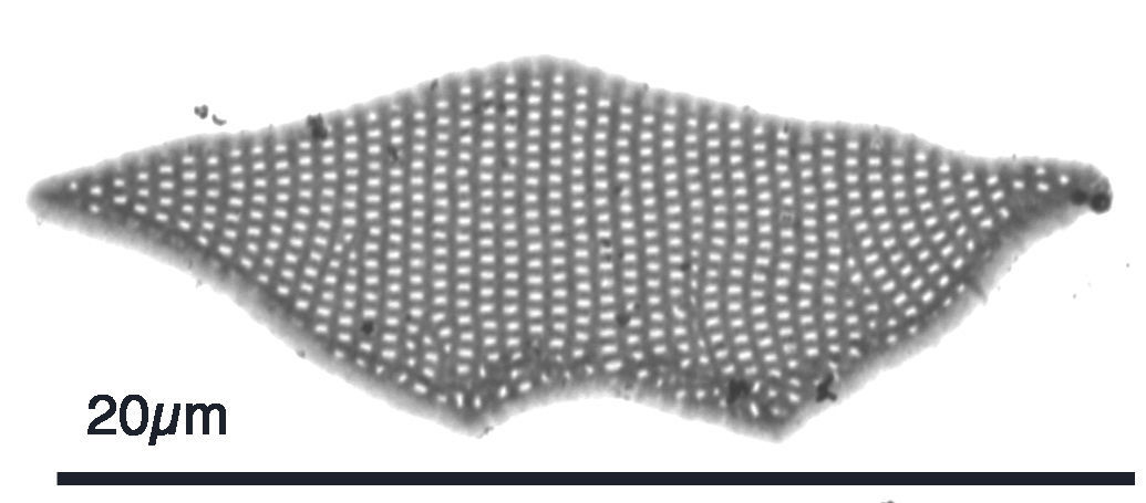
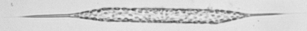
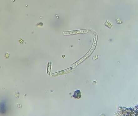
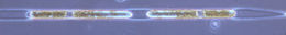
Fig 3: Coscinodiscus wailesii Light micrograph of a Lugol's preserved cell in girdle view
-
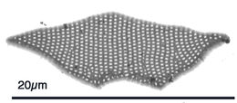
Coscinodiscus wailesii. oblique SEM . Visible are: girdle bands, two regular rows of rimoportulae (2-3 areolae from margin & at valve face/mantle junction), one irregular row of rimoportulae near center, irregular hyaline central area. scale bar is 10µm.
-
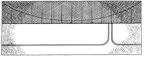
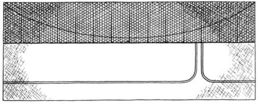
Coscinodiscus wailesii,schematic girdle view of one theca (modified from Gran & Angst, Fig. 26)
-
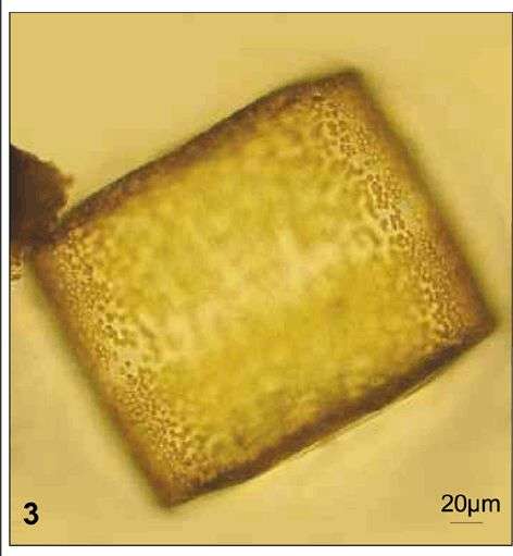
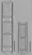
Coscinodiscus wailesii, cleaned valve, light microscope
-
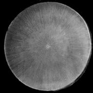
Coscinodiscus wailesii, living cell in valve view
-
R. setigera: valve apex, transitional to var. pungens
-
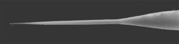
R. setigera: valve apex of var. setigera (SEM)
-
R. setigera: individual girdle band (TEM)
-
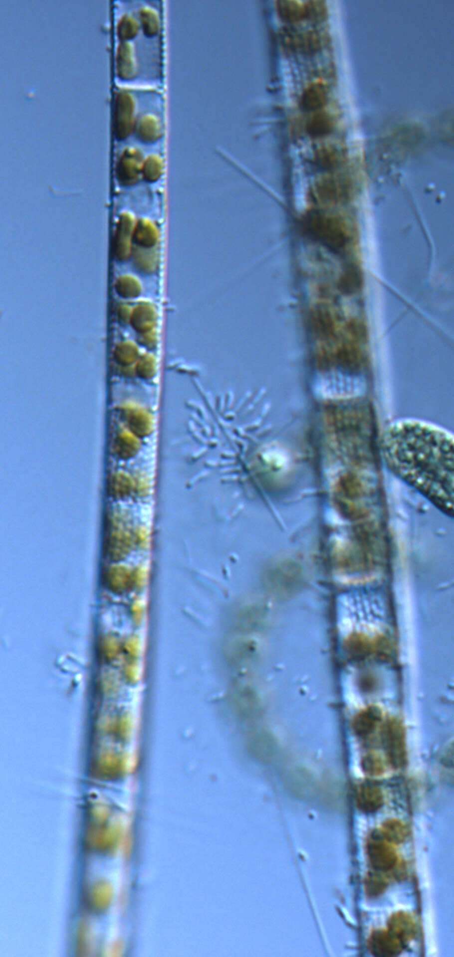
R. setigera: girdle bands in two dorsiventral columns
-
R. setigera: one spine typical of var. setigera; other spine transitional to var. pungens
-
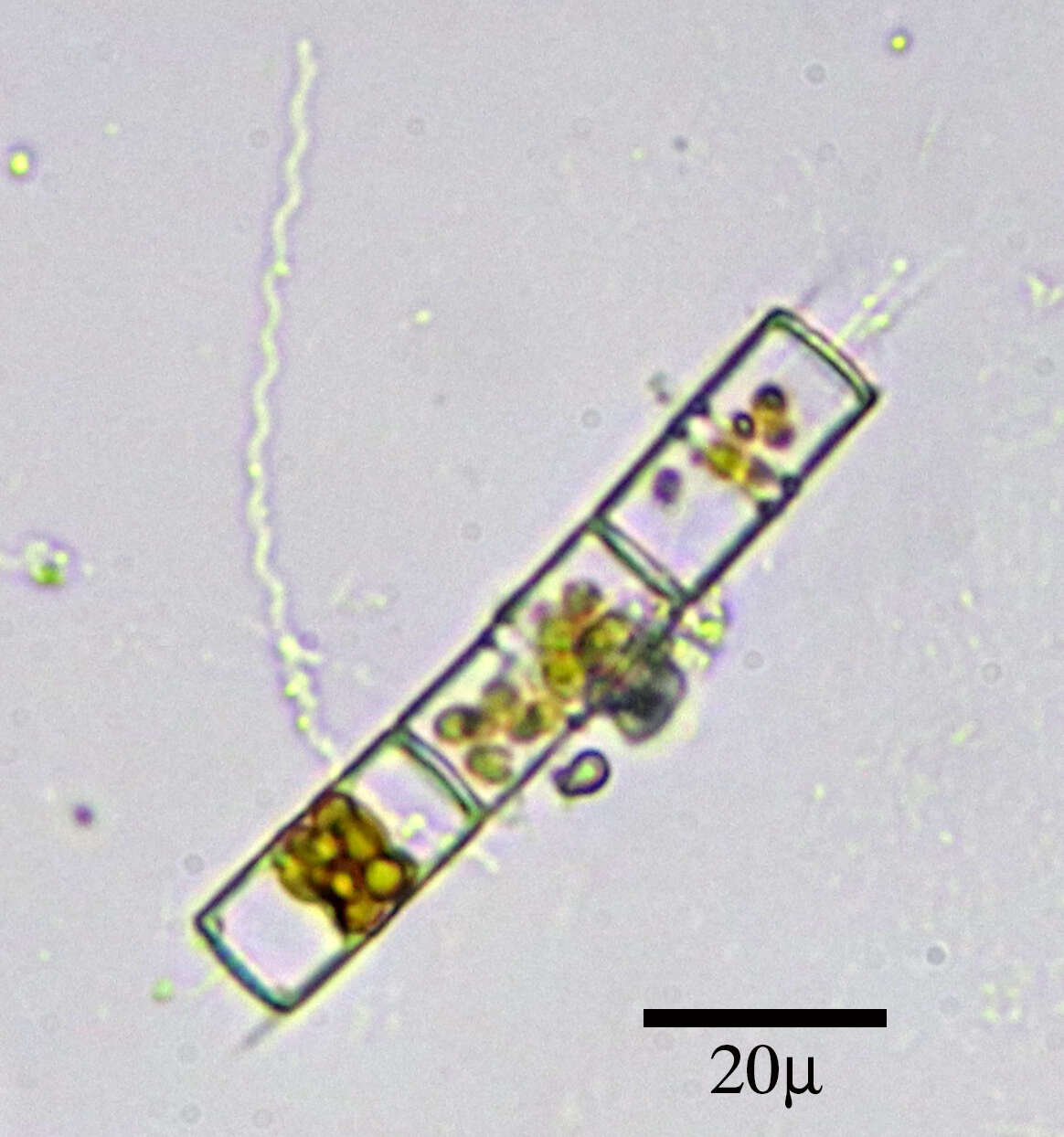
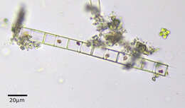
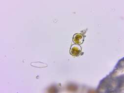
R. setigera fully-formed resting spore pair in parent cell
-
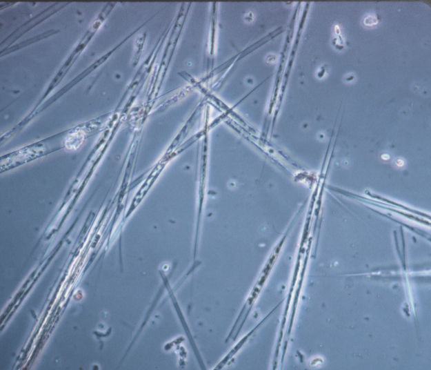
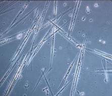
R. setigera bloom: var setigera + var. pungens; diameters 6-9µm
-
-
-
-
-
-
-
-
-