Comprehensive Description
provided by Smithsonian Contributions to Zoology
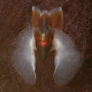
Tetronychoteuthis massyae Pfeffer, 1912
The mantle of this species is covered with many solid, stellate, minute, tightly-packed, papillose tubercles (Figures 14, 15). Each tubercle is roughly mushroom-shape in profile with a slightly concave central disc and a thick base. The periphery of the tubercle contains several (mostly 7 to 10) 2-, 3-, or 4-prong, conical papillae (Figures 14–16). The discs on a specimen of 100 mm ML are about 0.3 mm in diameter and 0.15 mm in height. The tubercles consist of very densely packed material with chondrocytes, reminiscent of elastic cartilage, regularly distributed throughout the dense connective tissue matrix of the disc and the base (Figures 17–22). SEM examination shows that the dense tissue is interspersed throughout with small vacuoles of varied sizes and shapes (Figures 18, 19). The walls of the vacuoles and the matrix are relatively thick and fibrous. The small vacuoles are mixed with large tubule-like spaces that give an overall dense spongy structure.
Histologically the following features occur through the tubercles and mantle (Figures 20–22). A delicate acellular cuticle envelops the surface of the tubercles and the papillae. The surface of the mantle between the tubercles is covered with normal epidermis that is fused with the bases of the tubercles. The base of each tubercle is continuous with and has the same chondrofibrous composition as the outer layer of the dermis, the foundation layer that envelops the mantle superior to the muscle layers. Pigment sacs (chromatophores) that provide the background coloration occur in the boundary zone between epidermis and dermis. Along the inferior boundary of the foundation layer occurs a layer of large, closely packed chondrocytes that immediately overlie the dense, thin layer of longitudinal dense connective tissue fibers of the mantle (Figure 20). Inferior to the connective tissue fibers is another thin layer of connective tissue regularly penetrated by oblique and circular muscle fibers that extend from the underlying mantle musculature. Inferior to this boundary zone lie several alternating layers of compact circular and oblique muscles that comprise most of the thick wall of the mantle (not shown in figure sections). The inferior-most layer of the mantle musculature consists of interspersed circular and oblique muscles which insert into the inferior mantle epithelium. No inferior layer of longitudinal muscle exists.
- bibliographic citation
- Roper, Clyde F. E. and Lu, C. C. 1990. "Comparative morphology and function of dermal structures in oceanic squids (Cephalopoda)." Smithsonian Contributions to Zoology. 1-40. https://doi.org/10.5479/si.00810282.493