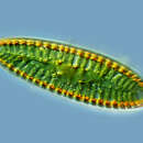
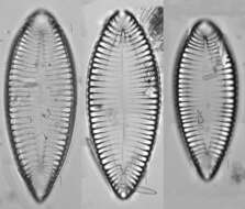
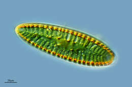
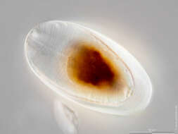
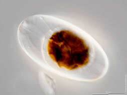
Scale bars indicate 50 µm (1) and 100 µm (2, 3).Three images.First:Girdleband view, focus is on epivalve.Second:The same specimen in the Petri dish, viewed with a stereo microscope.Third:The same specimen in the Petri dish turned to valvar view, viewed with a stereo microscope.Please click on < or > on the image edges or on the dots at the bottom edge of the images to browse through the slides!Place name: Baltic Sea, Kieler Förde, Kiel Fjord (Germany) Latitude: 54.3894126 Longitude: 10.1749055a) Microscope Zeiss Axioplan, camera Olympus OM-D M5 MKII. DOF images.b) Stereo microscope Olympus SZX16, camera Olympus OM-D M5 MKII. DOF images.© Wolfgang Bettighofer,images under Creative Commons License V 3.0 (CC BY-NC-SA).For permission to use of (high resolution) images please contact
postmaster@protisten.de.For further information about the image, please click here:
Link to protisten.de page