-
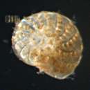
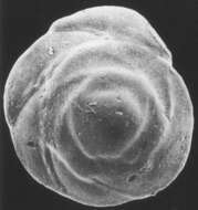
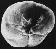
Individual collected in Saanich Inlet, Vancouver Island, British Columbia. This species was most common in deep water in the center of the inlet. Image courtesy of R. Timothy Patterson, Carleton University. This image first appeared in J. Foram. Res. 28:201-219 and is used with permission.
-
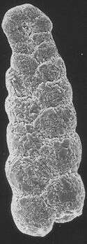
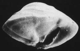
Fossil (Badenian) specimen, from Nussdorff, Austria. Image courtesy of Stefan Revets. This image first appeared in Hansen and Revets, J. Foram. Res. 22:166-180 (1992) and is used with permission.
-
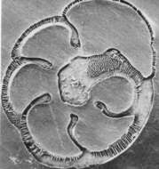
Polished and etched horizontal section through test. Image courtesy of Stefan Revets. This image first appeared in Hansen and Revets, J. Foram. Res. 22:166-180 (1992) and is used with permission.
-
Image courtesy of Stefan Revets. This image first appeared in Hansen and Revets, J. Foram. Res. 22:166-180 (1992) and is used with permission.
-
Image courtesy of Stefan Revets. This image first appeared in Hansen and Revets, J. Foram. Res. 22:166-180 (1992) and is used with permission.
-
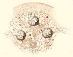
Section of cell with central capsule, associated black pigment, central nucleus, and calymma. Inset is an endoplasmic vacuole.
-
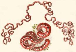
The central capsules are worm-like. With oil droplets, nuclei, small pigment spots and yellow symbiotic algae.
-
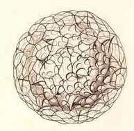
Living coenobium, with serpentine central capsules. Numerous yellow algal cells are scattered among the radial pseudopodia.
-
Large colonial coenobium or jelly colony, and a single isolated amoeboid central capsule with oil droplet.
-
-
Each shell contains numerous large granules
-
-
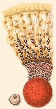
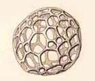
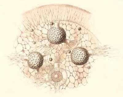
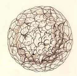
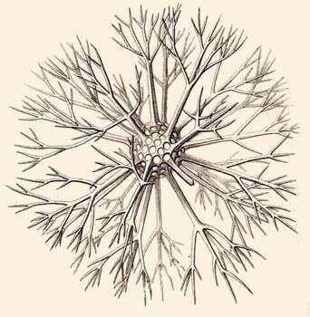
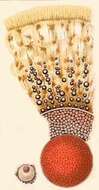
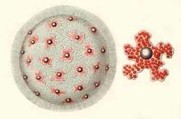
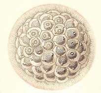
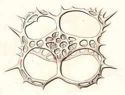
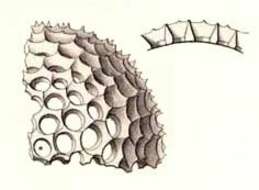
Haeckels fiugure legend reads .... a small piece of the surface of a living coenobium, seen from the surface. Only four individuals are visible, the central capsule of which contains numerous small nuclei and a central oil-globule. The including spherical lattice-shell is provided with a few (one to four) larger apertures, which are prolonged into short cylindrical tubules. Through these latter radiate bundles of fine pseudopodia, branching and anastomosing, and forming a fine sarcode network between the alveoles of the calymma. On the surface of the alveolated jelly-sphere the pseudopodia form a dense radiating zone. Xanthella or yellow cells are everywhere scattered.
-
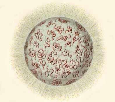
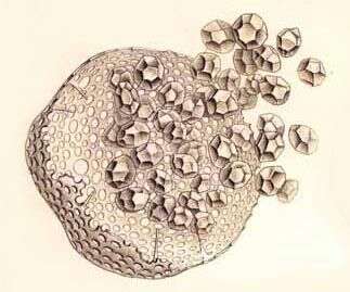
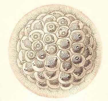
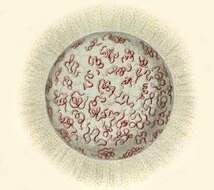
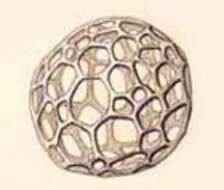
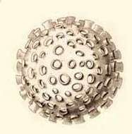
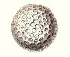
Haeckels figure legend reads: A small coenobium or colony in the state of alveolation, forming a jelly-sphere, composed of a great number of capsulated individuals, densely aggregated. Each central capsule contains an oil-globule, and is enclosed by a spherical lattice-shell, which bears a few (one to four) short cylindrical tubules. Each shell is again enveloped by a membranous polyhedral alveole and separated from it by structureless jelly. The thick cortical jelly-envelope, which surrounds the whole spherical colony, exhibits a fine radial striation, produced by radiating pseudopodia; many xanthella or yellow cells are scattered in the calymma.
-
-
-
-
Dorsal view.
-
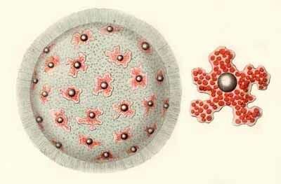
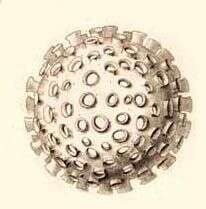
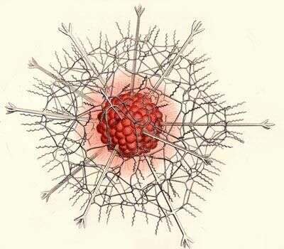
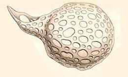
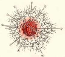
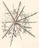
Haeckel says: Entire shell and central capsule. Numerous club-shaped radial apophyses or coecal sacs arise from the pink central capsule and are protruded through the pores of the medullary shell, which is completely hidden by them. The sarcomatrix in the calymma, surrounding the central capsule, exhibits a fine radial striation. Numerous retracted pseudopodia, bearing red granules, arise from the sarcomatrix and piece the calymma radially. the interval between the two concentric shells is filled up by the hyaline calymma.
-
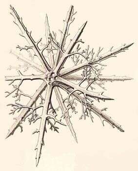
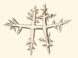
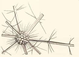
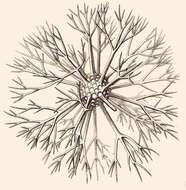
Haechel says: Medullary shell and the basal parts of the radial spines arising from it.
-
-
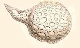
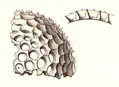
Shell with vertical section through the wall.
-
-