-
-
-
John D. Taylor, Emily A. Glover
Zookeys
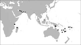
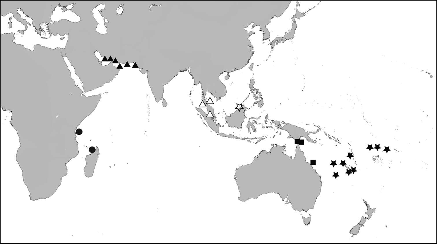
Figure 4.Distribution map - Scabrilucina victorialis, solid triangles; Scabrilucina vitrea, open triangles; Scabrilucina melvilli, solid squares; Gonimyrtea ferruginea, solid stars; Myrtina reflexa, solid circles, Ferrocina brunei open star.
-
-
-
-
-
John D. Taylor, Emily A. Glover
Zookeys
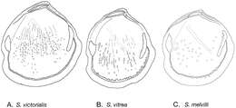
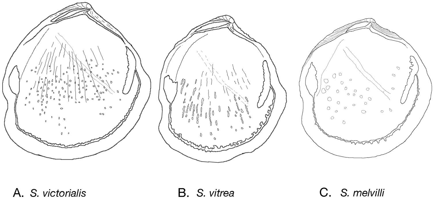
Figure 3.Internal drawings Scabrilucina species. A S. victorialis B Scabrilucina vitrea C Scabrilucina melvilli.
-
-
-
-
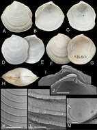
John D. Taylor, Emily A. Glover
Zookeys
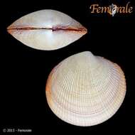
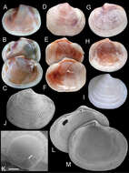
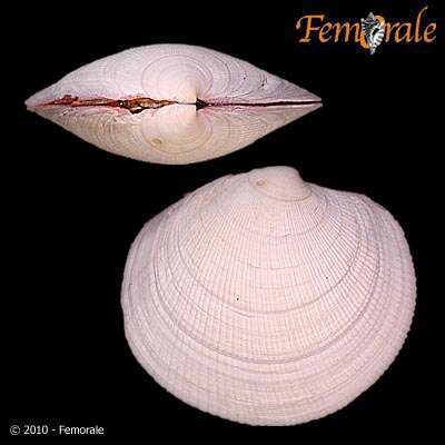
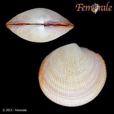
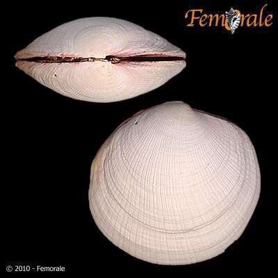
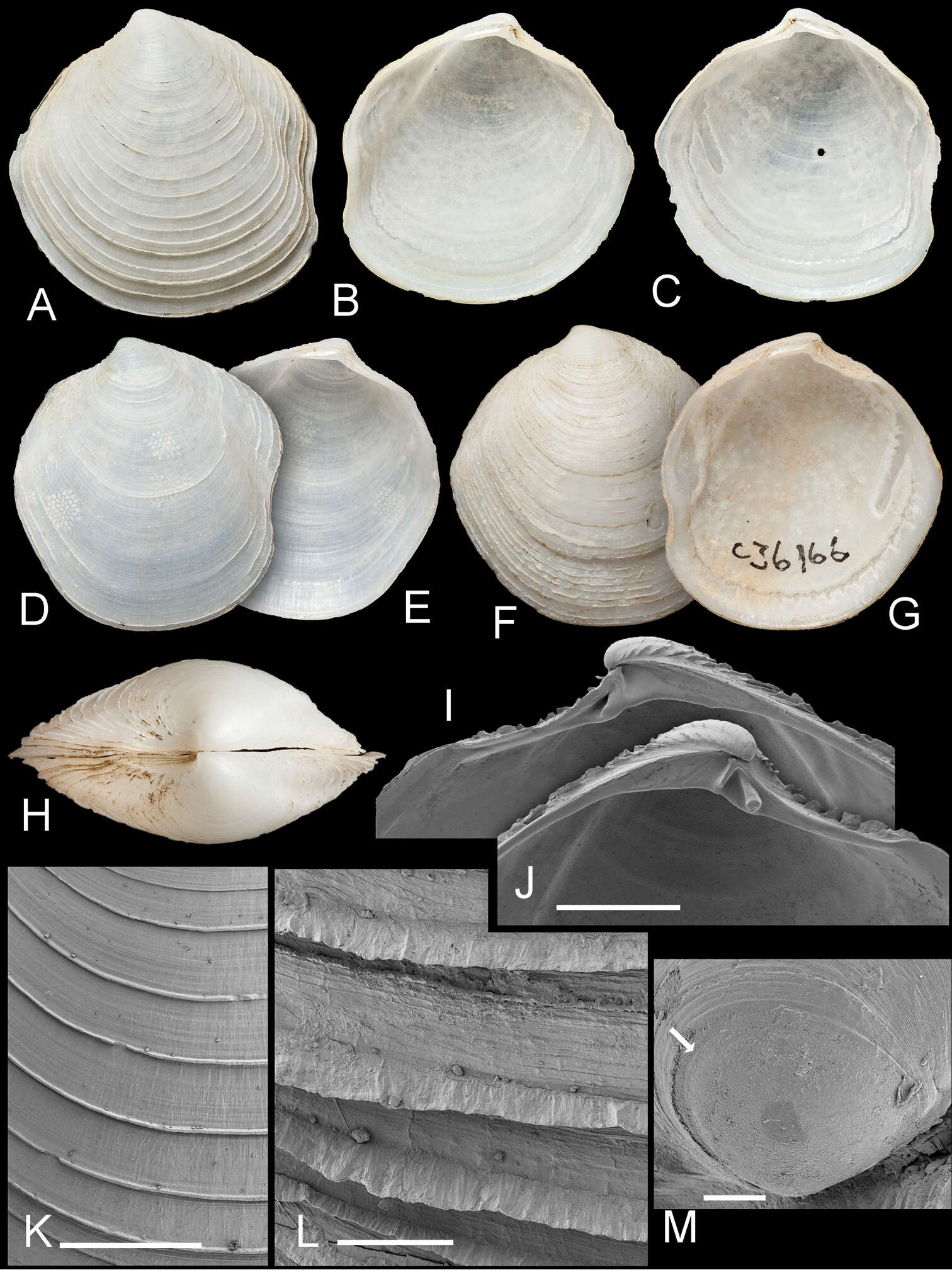
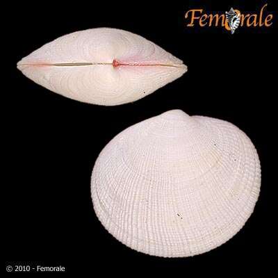
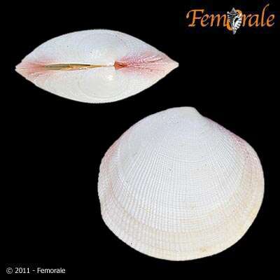
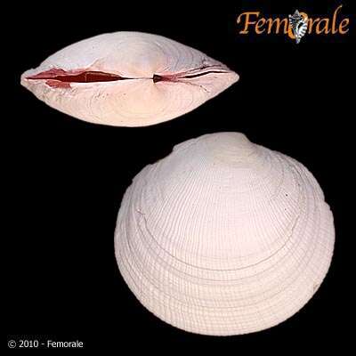
Figure 6.Scabrilucina melvilli sp. n. A–C Holotype AMS C. 360708 exterior of left valve and interior of left and right valves, L = 21 mm D–E paratype AMS C. 360708 exterior and interior of left valve, L= 18.3 mm F–G paratype AMS 036166 exterior and interior of left valve, L= 23.1 mm H Dorsal view of holotype I–J Detail of hinge teeth of right and left valves of juvenile shells. Scale bar = 1 mm K External sculpture. Scale bar = 1 mm L Detail of commarginal lamellae. Scale bar = 200 µm M Protoconch. Arrow marks boundary between PI and PII. Scale bar = 50 µm.
-
-
-
-
John D. Taylor, Emily A. Glover
Zookeys
Figure 4.Distribution map - Scabrilucina victorialis, solid triangles; Scabrilucina vitrea, open triangles; Scabrilucina melvilli, solid squares; Gonimyrtea ferruginea, solid stars; Myrtina reflexa, solid circles, Ferrocina brunei open star.
-
2016 University of California Museum of Paleontology
CalPhotos
-
-
-
John D. Taylor, Emily A. Glover
Zookeys
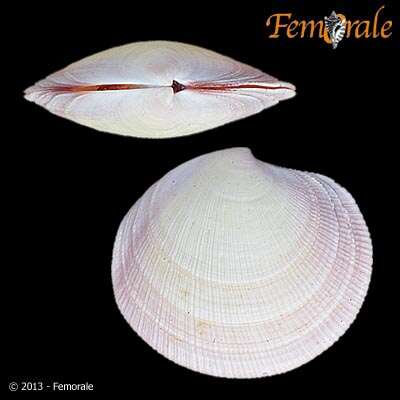
Figure 7.Ferrocina brunei sp. n. A–C Holotype NHMUK 20130122 Exterior of left valve and interior of right and left valves, L = 8.2 mm D–F Paratype NHMUK 20130123 exterior of right valve and interior of left and right valves, L = 8.4 mm. pbv trace of pallial blood vessel G–H Paratype NHMUK 20130123 exterior and interior of left valve, L = 8.9 mm I Exterior of right valve of white form NHMUK 20130123, L = 7.9 mm J SEM of right valve L = 6.0 mm K Protoconch, arrow at PI /PII junction. Scale bar = 50 µm L–M Interior of right and left valves L = 5.2 mm.
-
2017 University of California Museum of Paleontology
CalPhotos
-
-
-
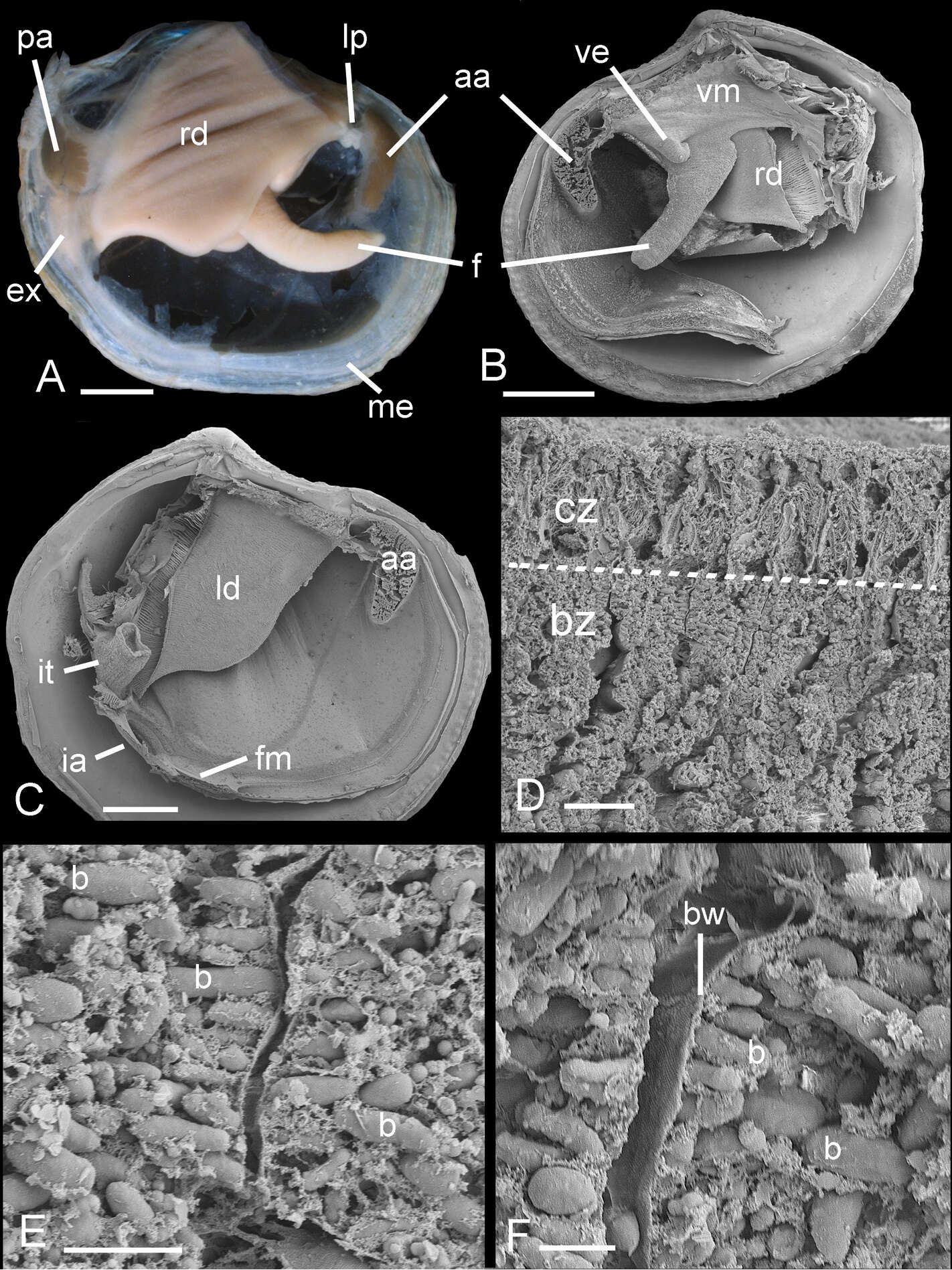
John D. Taylor, Emily A. Glover
Zookeys
Figure 8.Ferrocina brunei sp. n. A Body from right side. Scale bar = 1 mm B Body from left side, mantle and left demibranch removed showing foot and visceral extension. Scale bar = 1 mm C Body from right side with visceral mass, right demibranch and mantle removed. Scale bar = 1 mm D Section through part of demibranch showing ciliated and bacteriocyte zones. Scale bar = 20 µm E Part of ctenidial filament in bacteriocyte zone showing bacteria. Scale bar = 5 µm F Detail of bacteriocytes and bacteria aligned normal to the apical cell wall. Scale bar = 2 µm. aa, anterior adductor muscle. b, bacteria. bw, bacteriocyte apical wall. bz, bacteriocyte zone. cz, ciliated zone. ex, exhalant aperture. f, foot. fm, fused mantle. ia, inhalant aperture. it, inverted tube of posterior exhalant aperture. ld, left demibranch. lp, labial palps. me, mantle edge. pa, posterior adductor muscle. rd, right demibranch. ve, visceral extension. vm, visceral mass.