-
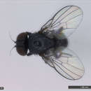
All Biocode files are based on field identifications to the best of the researcher’s ability at the time.
-
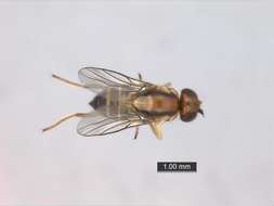
All Biocode files are based on field identifications to the best of the researcher’s ability at the time.
-
Wayne N. Mathis, Alessandra Rung, Marion Kotrba
Zookeys
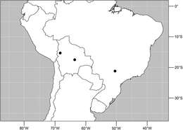
Figure 34.Distribution of Planinasus miradorus sp. n. (square) and Planinasus atriclypeus (dots).
-
Stéphanie Boucher, Kenji Nishida
Zookeys
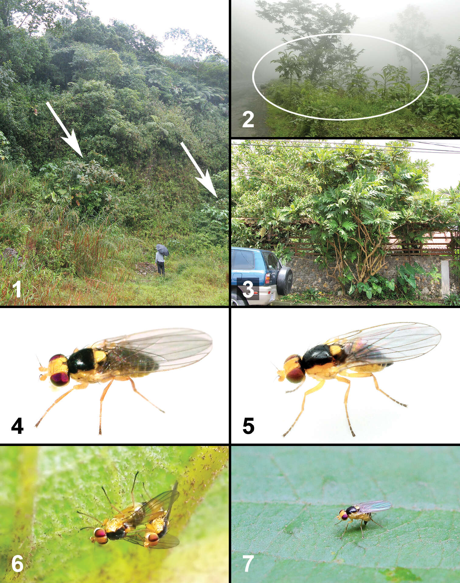
Figures 1–7.Life history of two new species of Liriomyza. 1–3 Habitats 1 Open area in a valley near Reserva Biológica Manuel Alberto Brenes in San Ramón (site 2). Arrows indicate Bocconia frutescens trees 2 Bocconia frutescens saplings (in circle) growing along the road after land slides caused by 2009 earthquake in Cinchona-Vara Blanca area (site 10) 3 Ornamental Bocconia frutescens tree (in middle) in urban area of San Isidro de Coronado (site 15) 4 Liriomyza mystica female (from site 5) 5 Liriomyza prompta female (from site 13) 6 Mating couple of Liriomyza prompta on the underside of Bocconia frutescens leaf at 7:00 am (30.v.2009, site 13) 7 Liriomyza prompta ovipositing on the upper side of Bocconia frutescens leaf blade at 3:00 pm (17.vi.2011, site 2).
-
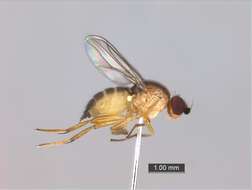
Viviane Rodrigues de Sousa, Márcia Souto Couri
Zookeys
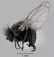
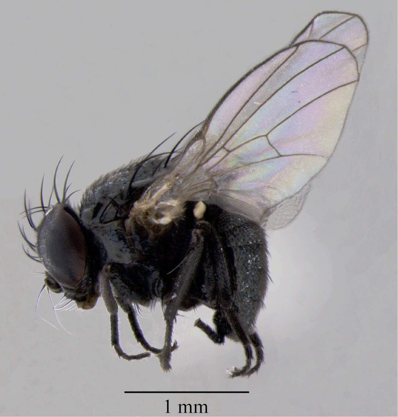
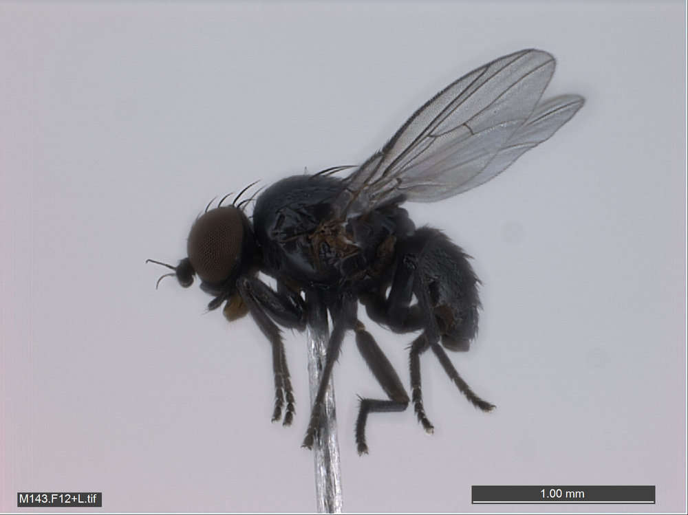
Figure 2.Japanagromyza inferna Spencer, male, in lateral view.
-
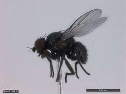
All Biocode files are based on field identifications to the best of the researcher’s ability at the time.
-
All Biocode files are based on field identifications to the best of the researcher’s ability at the time.
-
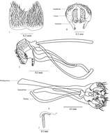
Wayne N. Mathis, Alessandra Rung, Marion Kotrba
Zookeys
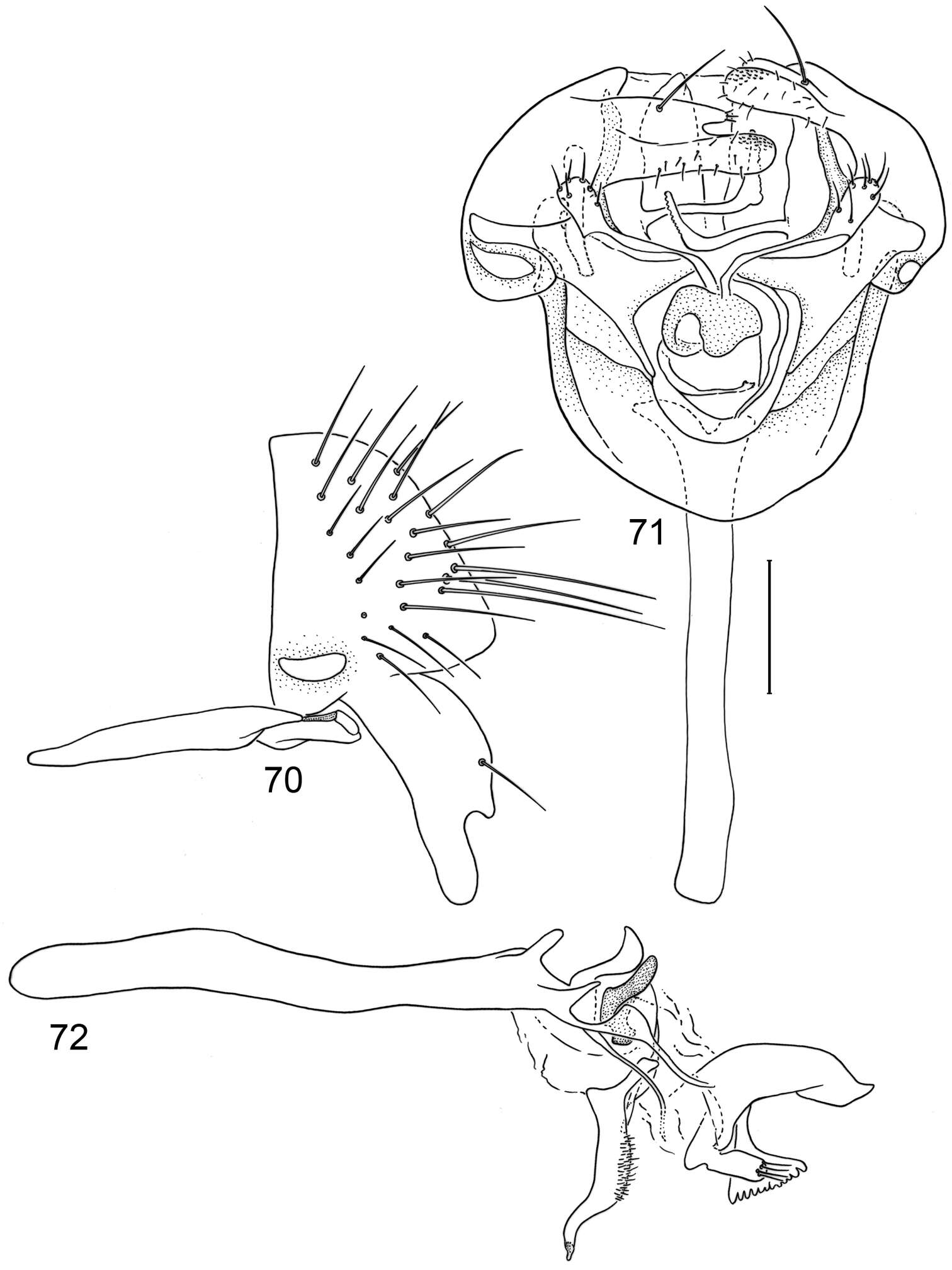
Figures 70–72.Illustrations of Planinasus atriclypeus sp. n. (male). 70 epandrium, surstylus, hypandrium, and pregonite, lateral view 71 structures of internal male terminalia, ventral view 72 internal structures of male terminalia, lateral view, lateral view. Scale bar = 0.1 mm.
-
Stéphanie Boucher, Kenji Nishida
Zookeys
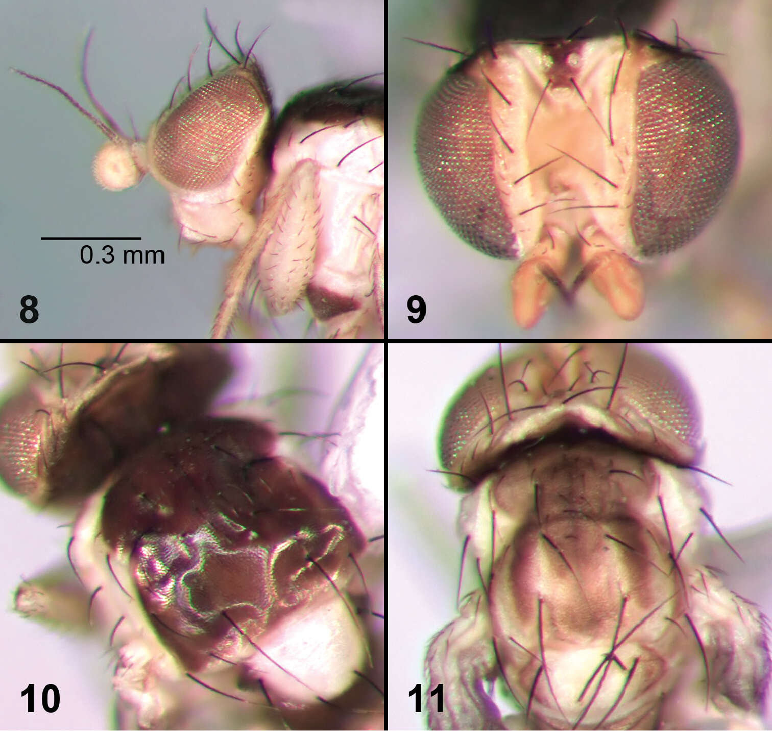
Figures 8–11.External morphology of adult Liriomyza mystica. 8 Head, lateral 9 Head, dorso-frontal 10 Thorax, dorsal 11 Thorax, dorsal (teneral specimen).
-
Viviane Rodrigues de Sousa, Márcia Souto Couri
Zookeys
Figures 3–7.Male terminalia of Japanagromyza inferna Spencer 3 sternite 5 4 epandrium, cercal plate and surstylus 5 hypandrium 6 phallapodeme, hypandrium, phallus 7 ejaculatory apodeme.
-
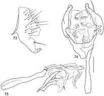
Wayne N. Mathis, Alessandra Rung, Marion Kotrba
Zookeys
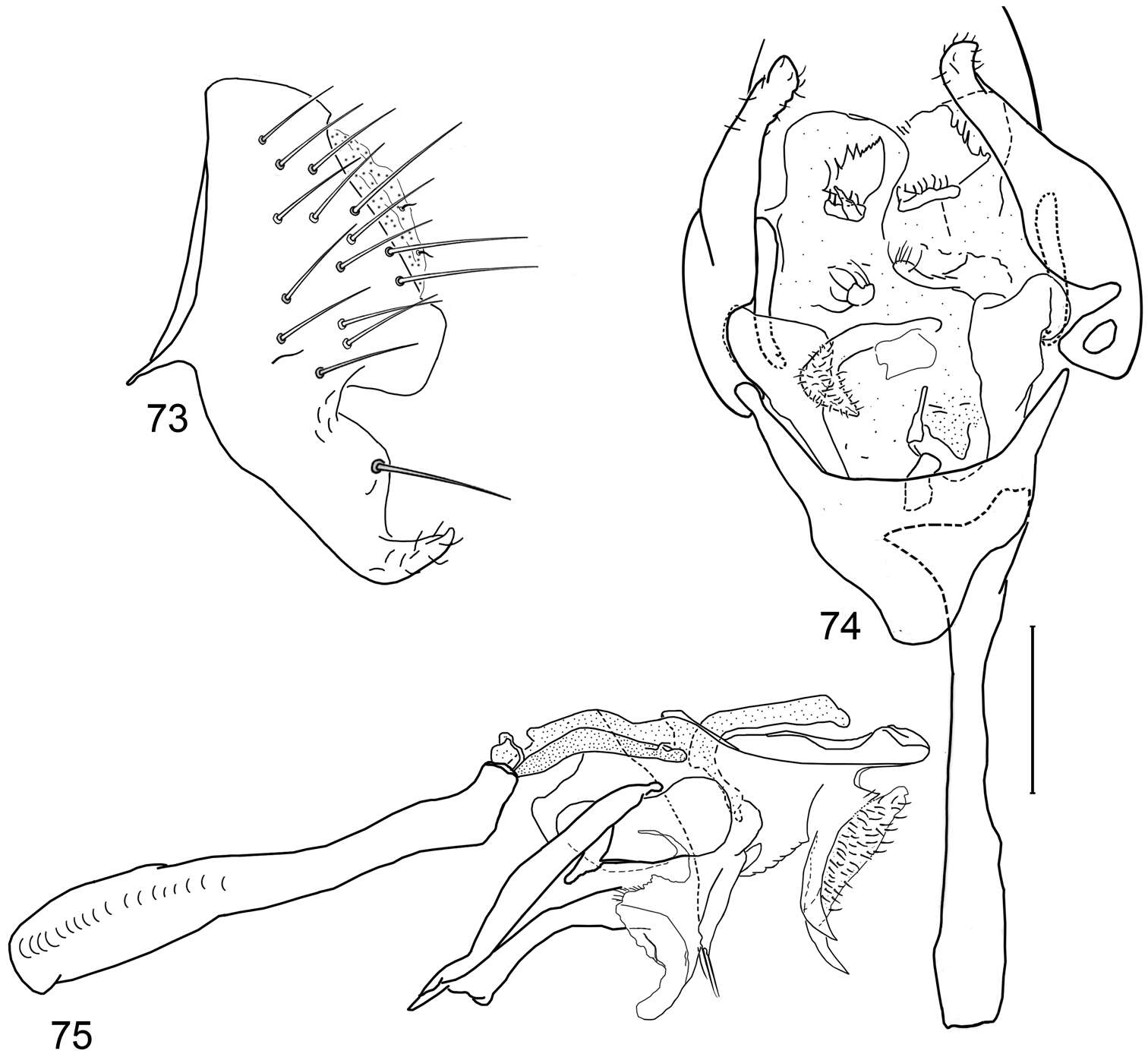
Figures 73–75.Illustrations of Planinasus atrifrons sp. n. (male). 73 epandrium and surstylus, lateral view 74 structures of internal male terminalia, ventral view 75 internal structures of male terminalia, lateral view. Scale bar = 0.1 mm.
-
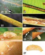
Stéphanie Boucher, Kenji Nishida
Zookeys
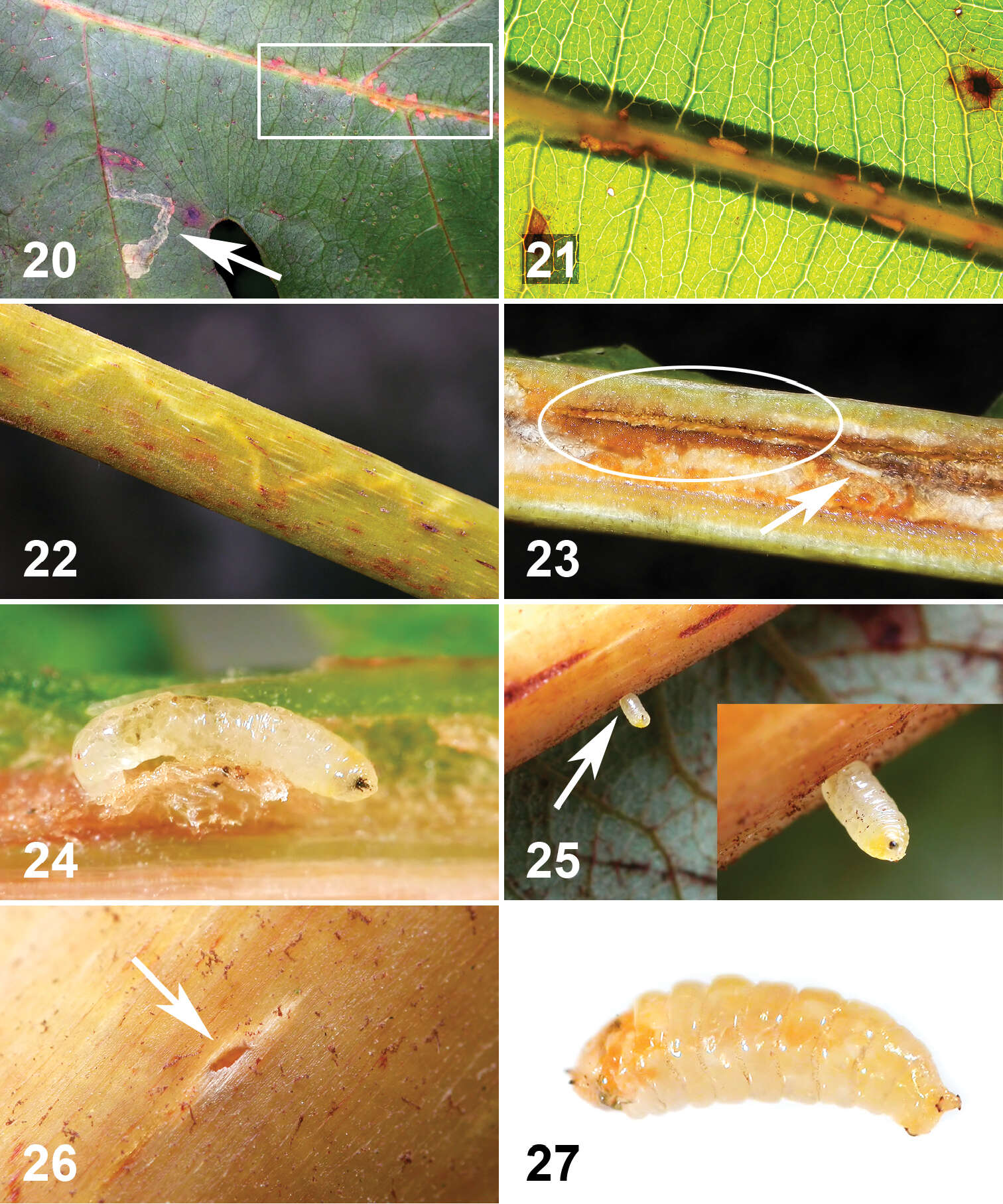
Figures 20–27.Life history of Liriomyza mystica larvae on Bocconia frutescens. 20–22 External evidence caused by internal larval feeding on vein and petiole 20 Brown to reddish brown spots (ca. 1–2 mm long) on upperside along primary vein, marked by rectangular line. Arrow indicates Liriomyza prompta mine 21 Pale brown linear spots along the primary vein seen through strong sunlight from the back. Note that lower part of vein (underside) is thicker and shown as shadow 22 Mine in pale colour zigzag, approximately 30 mm long 23 Longitudinally opened primary vein with linear mine (circle) and late instar larva (arrow) 24 Late instar larva in situ, ventral view. Cephalopharyngeal skeleton on right. Notice orange spot at head 25 Mature larva exiting from underside of vein (arrow). Close-up view, lower right. Notice orange spot at head 26 Exit hole (ca. 1 mm wide) on underside of primary vein 27 Mature larva in pre-puparial stage. Posterior on right.
-
Viviane Rodrigues de Sousa, Márcia Souto Couri
Zookeys
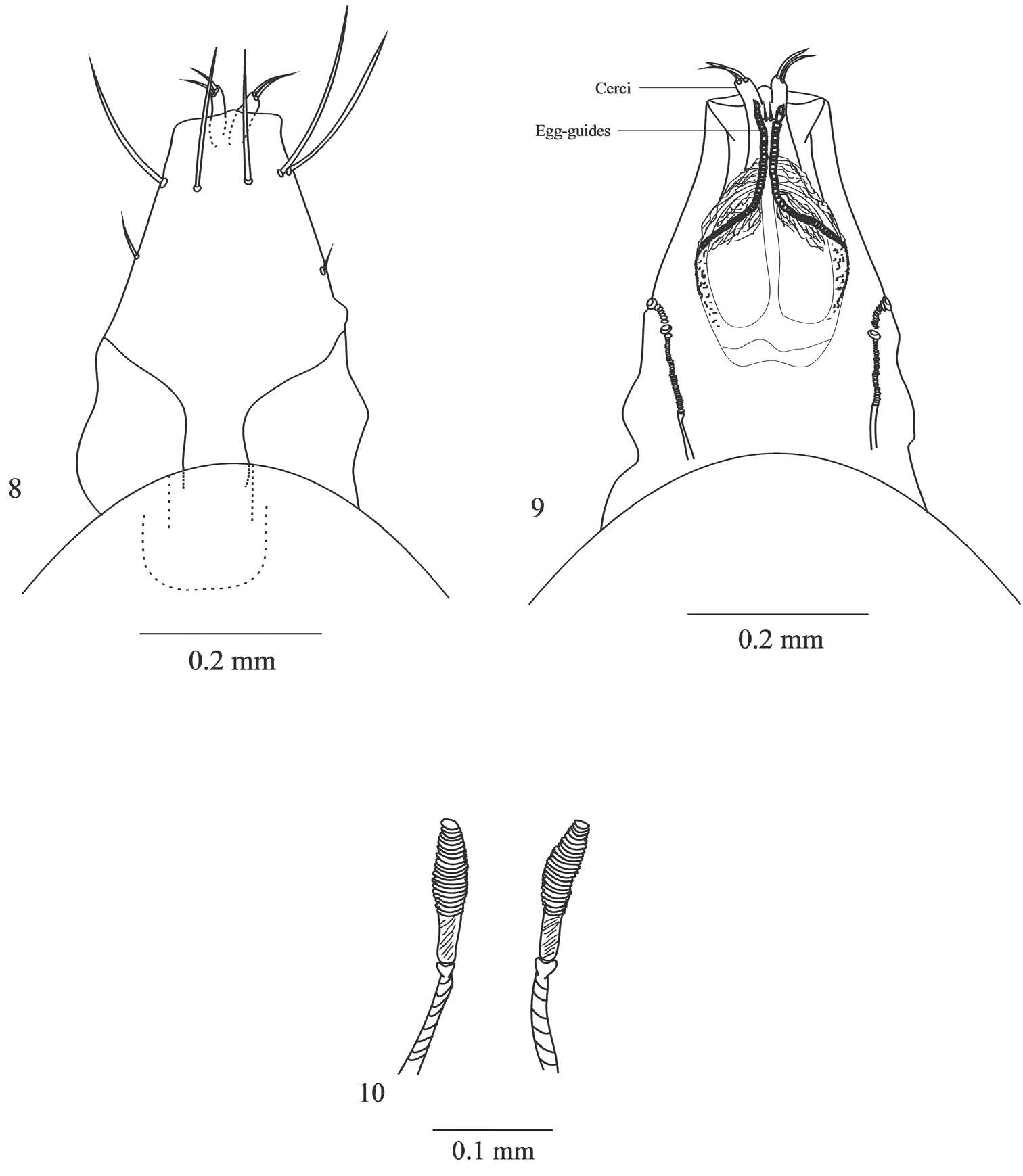
Figures 8–10.Female ovipositor of Japanagromyza inferna Spencer 8 dorsal view 9 ventral view 10 spermathecae.
-
Wayne N. Mathis, Alessandra Rung, Marion Kotrba
Zookeys
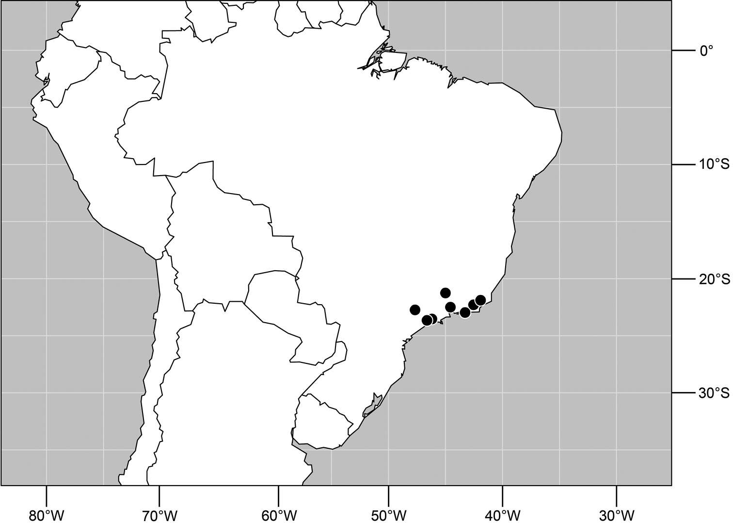
Figure 76.Distribution of Planinasus atrifrons sp. n.
-
Stéphanie Boucher, Kenji Nishida
Zookeys
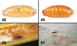
Figures 48–51.Life history of two new species of Liriomyza. 48 Puparium of Liriomyza mystica, in situ 49 Puparium of Liriomyza prompta, in situ 50 Pupa of Pteromaline parasitoid wasp (sp. 01) inside Bocconia frutescens leaf vein parenchyma 51 Braconid parasitoid wasp, most likely Opius sp., attempting to oviposit in mature Liriomyza prompta larva at site 13.
-
Viviane Rodrigues de Sousa, Márcia Souto Couri
Zookeys
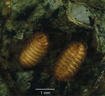
Figure 11.Pupae of Japanagromyza inferna Spencer in gall of the Centrosema virginianum L. (Fabaceae).
-
Wayne N. Mathis, Alessandra Rung, Marion Kotrba
Zookeys
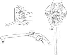
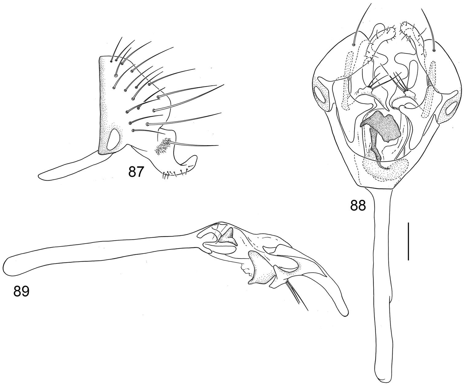
Figures 87–89.Illustrations of Planinasus nigrifacies sp. n. (male). 87 epandrium, surstylus, hypandrium, lateral view 88 structures of internal male terminalia, ventral view 89 internal structures of male terminalia, lateral view. Scale bar = 0.1 mm.
-
Stéphanie Boucher, Kenji Nishida
Zookeys
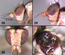
Figures 28–31.External morphology of adult Liriomyza prompta. 28 Head, lateral 29 Head, lateral (variation of eye colour) 30 Head, dorsal 31 Thorax, dorsal.
-
Viviane Rodrigues de Sousa, Márcia Souto Couri
Zookeys
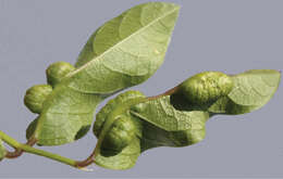
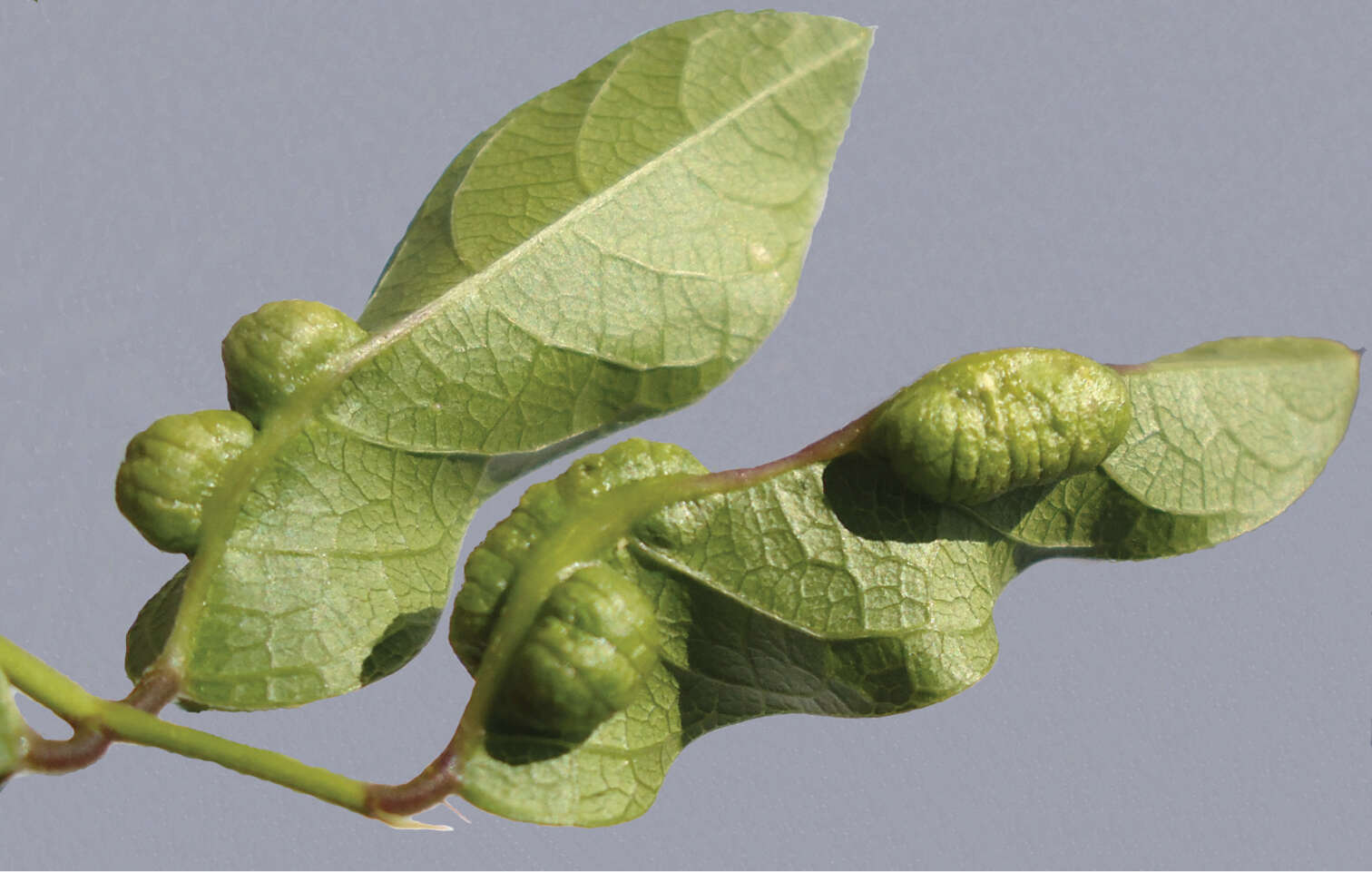
Figure 12.Gall of Japanagromyza inferna in Centrosema virginianum L. (Fabaceae).
-
Wayne N. Mathis, Alessandra Rung, Marion Kotrba
Zookeys
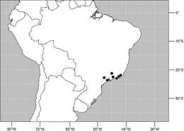
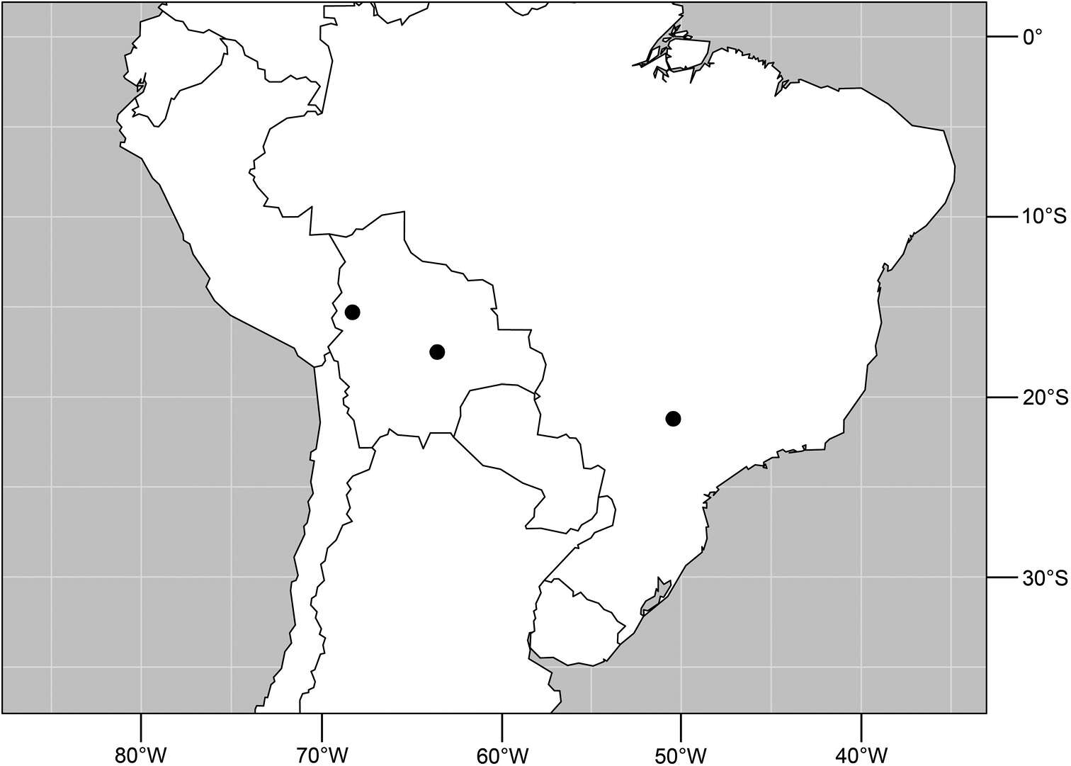
Figure 90.Distribution of Planinasus nigrifacies sp. n.
-
Stéphanie Boucher, Kenji Nishida
Zookeys
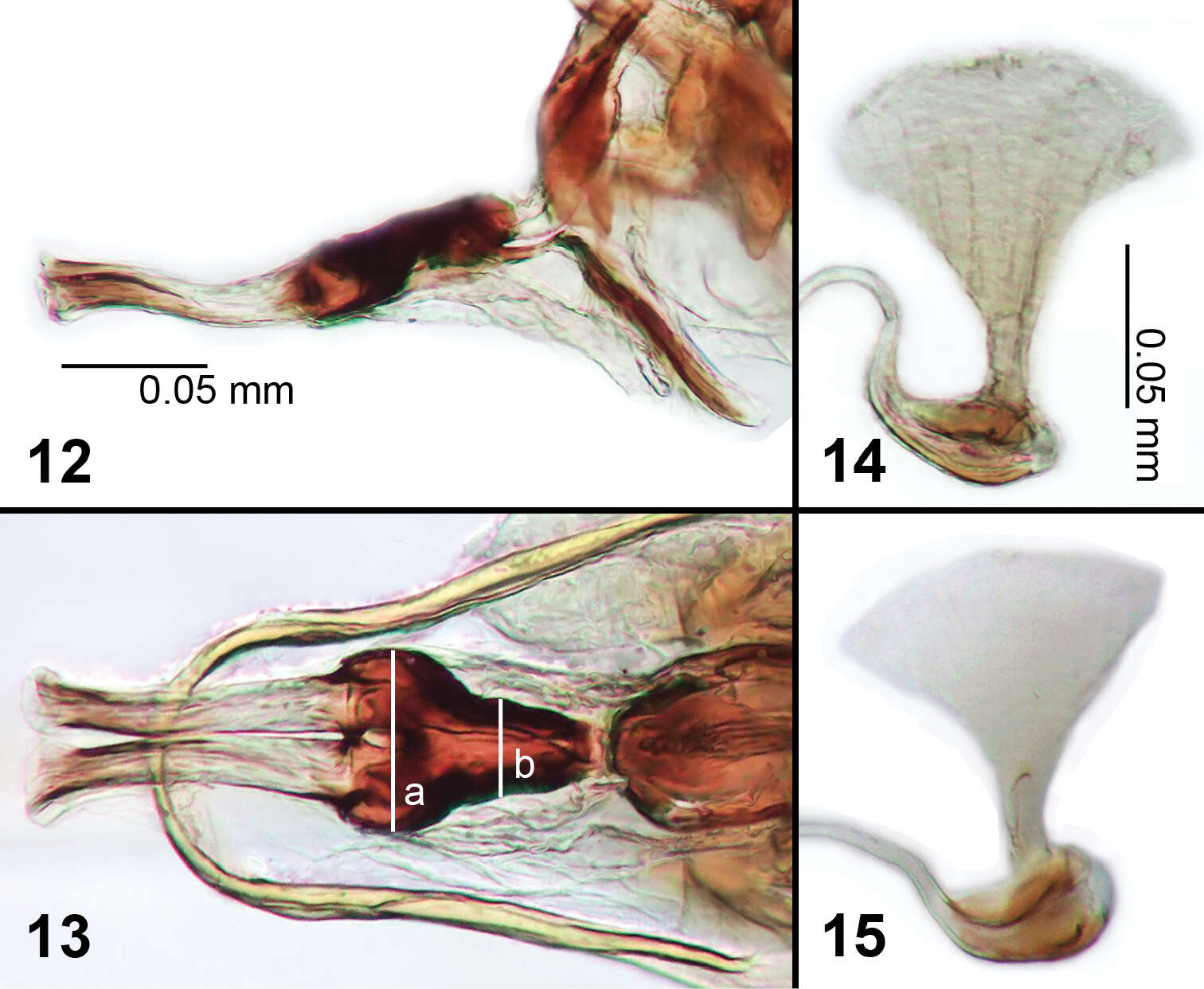
Figures 12–15.Male genitalia of Liriomyza mystica. 12 Phallus, lateral 13 Phallus, ventral (see text for lines ‘a’ and ‘b’) 14, 15 Ejaculatory apodeme.
-
Wayne N. Mathis, Alessandra Rung, Marion Kotrba
Zookeys
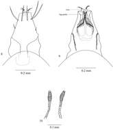
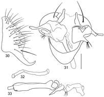
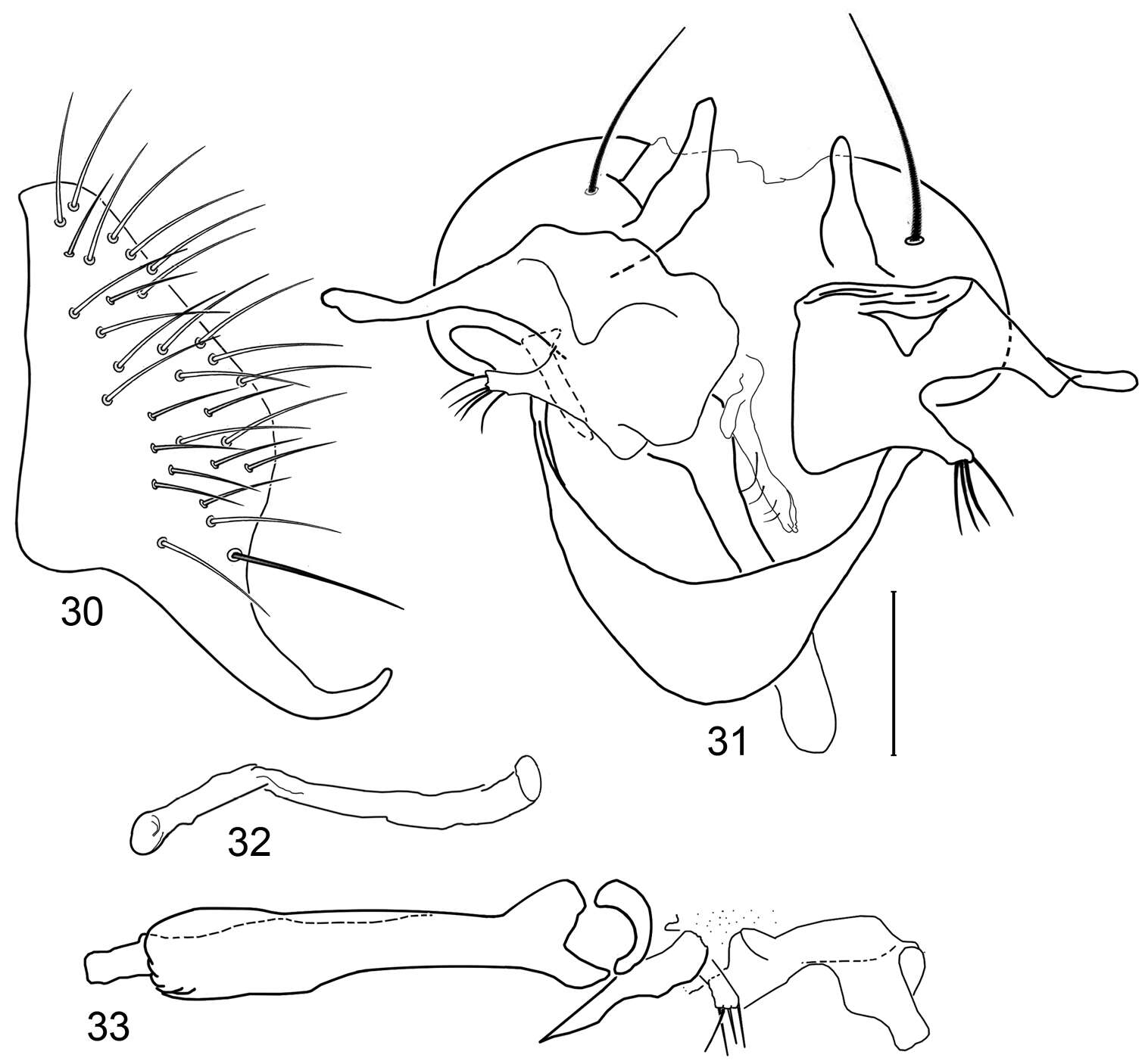
Figures 30–33.Illustrations of Planinasus miradorus sp. n. (male). 30 epandrium, surstylus, , lateral view 31 epandrium, hypandrium, and internal structures of male terminalia, ventral view 32 ejaculatory apodeme, lateral view 33 internal structures of male terminalia, lateral view. Scale bar = 0.1 mm.
-
Stéphanie Boucher, Kenji Nishida
Zookeys
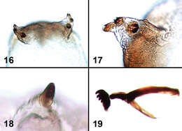
Figures 16–19.Larval characters of Liriomyza mystica. 16 Posterior spiracles 17 Posterior spiracle (close-up) 18 Anterior spiracle (note angle of view is different from Fig. 38) 19 Cephalopharyngeal skeleton.
-
Wayne N. Mathis, Alessandra Rung, Marion Kotrba
Zookeys
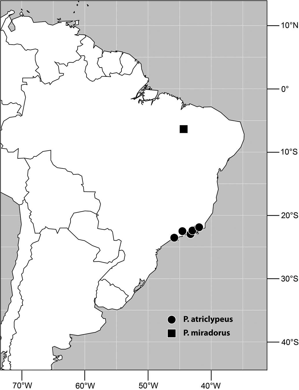
Figure 34.Distribution of Planinasus miradorus sp. n. (square) and Planinasus atriclypeus (dots).