Anna Halász, Catherine S. McFadden, Dafna Aharonovich, Robert Toonen, Yehuda Benayahu
Zookeys
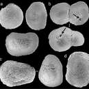
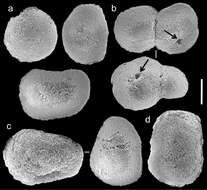
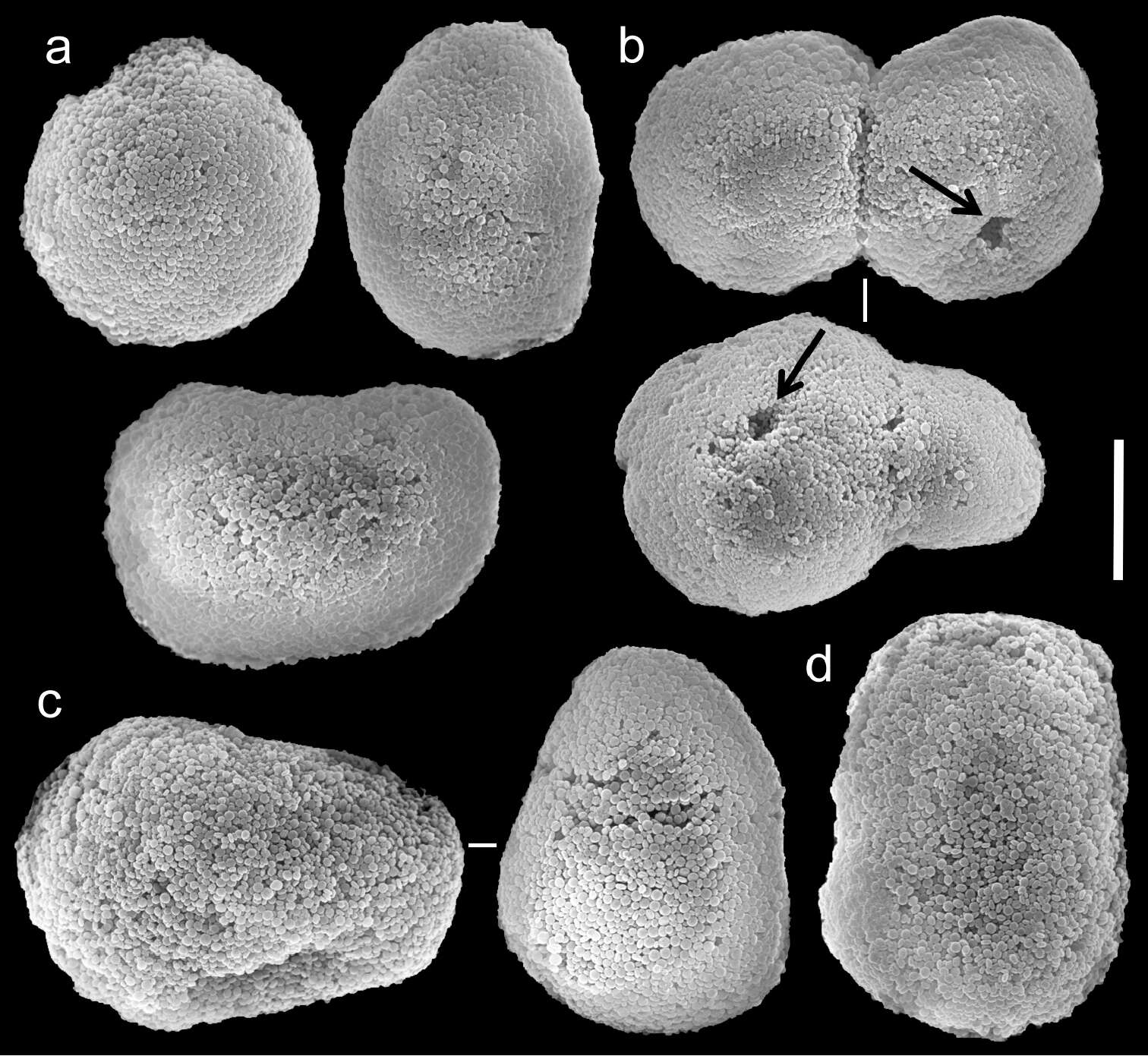
Figure 10.Scanning electron micrographs of polyp sclerites of Ovabunda crenata (Reinicke, 1997) (RMNH Coel. 23517). a Regular sclerites b Fused sclerites c Egg-shaped sclerites d Rectangular sclerite. Arrows indicate surface dents. Scale bar 10 µm.
Anna Halász, Catherine S. McFadden, Dafna Aharonovich, Robert Toonen, Yehuda Benayahu
Zookeys
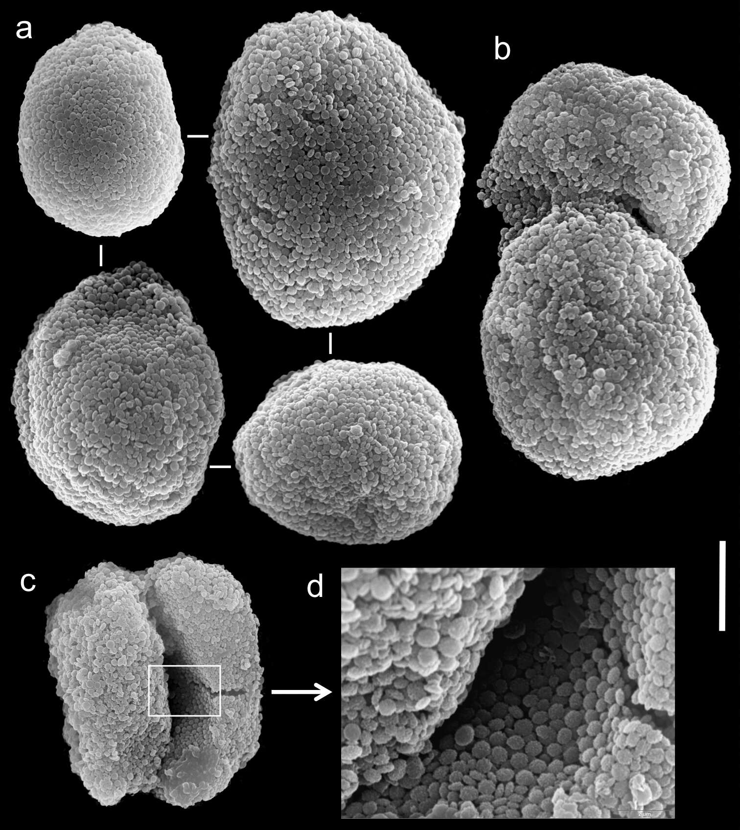
Figure 5.Scanning electron micrographs of polyp sclerites of Xenia crista Reinicke, 1997 holotype (RMNH 18677). a Regular sclerites b–c Fused sclerites d White rectangle in c indicates magnified area. Scale bar 10 µm.