Diagnostic Description
provided by Fishbase
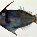
Blackish brown or bluish black head and anterior portion of body, rest of the body orange. A black blotch surrounding the gill opening. Caudal fin orange; soft dorsal and anal fins yellow; first dorsal spine blackish brown. Pelvic rudiment large not broadly attached to posterior margin of ventral flap. Scale ridge rugosities of male usually develop at about 65 mm SL. Scale spinulation on midbody not closely packed.
- Recorder
- Estelita Emily Capuli
Comprehensive Description
provided by Smithsonian Contributions to Zoology
Pervagor melanocephalus (Bleeker)
MUSCLES OF THE CREER
ADDUCTOR MANDIBULAE (Figures 87b, 91, 92; see also Figure 101 of Stephanolepis auratus: A 1α, A 1β, A 1γ, A 2α, A 2β, A 3).—The A 1 section of the adductor mandibulae has become considerably modified. A 1α and A 1α′ arise from the lateral face of the ethmoid (and the ventral surface of the lateral flange) as well as from the prefrontal and the infraorbital ligament (between the prefrontal and hyomandibular). A 1α′ (Figure 91) grades into an aponeurotic sheet which fades into the tissues of the upper lip. It is a fairly distinct subsection, although there is some intermingling of fibers posteriorly. The main mass of A 1 is formed by A 1α, which inserts on the posteroventrolateral face of the maxilla by a broad aponeurosis. Medial to this are two other sections, arising from the infraorbital ligament. The dorsal of these, A 1γ (see Figure 101) soon grades into a flat aponeurosis which attaches to the lateral process of the ethmoid supporting the palatine, and the palatine itself. Its function is obscure. Beneath this muscle is A 1β′ (Figure 101), a smallish, somewhat flattened muscle which develops a tendon anteriorly joining that of A 1α laterally and that of A 1β medially. A 1β originates from the lateral faces of the mesopterygoid and metapterygoid and inserts on the medial face of the maxilla. A 2 consists of two subdivisions. A 2α originates from the prefrontal, infraorbital ligament, and the hyomandibular. A 2β is well developed and is separated from A 2α by ramus mandibularis V. It arises from the anterior hyomandibular, the preopercle, symplectic, quadrate, and posterior metapterygoid. Some of the anteroventral fibers originate from the extreme ventral flange of the preopercle, but do not form a distinct section as in the balistids. A 2α and A 2β insert by a common tendon on the medial face of the dentary. A 3 is completely separate from A 2 except for the shared tendinous insertion on the dentary. It originates from the lateral faces of the metapterygoid and quadrate.
LEVATOR ARCUS PALATINI.—The muscle is poorly developed, and no longer visible in superficial lateral view. It originates from the ventromedial face of the orbital process of the sphenotic and the anterolateral prootic. The fibers fan out to insert on the posterodorsal and posterolateral faces of the hyomandibular, the two regions being separated by a thin ridge.
DILATATOR OPERCULI (Figure 91: D.O.).—Origin is from the ventrolateral face of the orbital process of the sphenotic and the pterotic, the fibers inserting on the anterolateral face and dorsal rim of the opercle.
LEVATOR OPERCULI (Figure 91: L.O.).—The muscle originates from the anterodorsal supracleithrum and anterolateral posttemporal and inserts on the posterodorsal ridge of the opercle.
ADDUCTOR ARCUS PALATINI (Figure 92: A.A.P.).—Fibers arise from the ventrolateral faces of the vomer and parasphenoid and insert on the dorsomedial hyomandibular and metapterygoid and dorsal mesopterygoid. The anterior region of the pterygoids underlies the parasphenoid, with which it is in contact dorsally. The muscle is not developed in the posterior half of the floor of the orbit.
RETRACTOR ARCUS PALATINI (Figures 91, 92: R.A.P.).—The muscle has completely separated from the adductor arcus palatini, and the division of the fibers into anterior and posterior sections as in balistids is not nearly as striking (the ventral aponeurosis of the anterior portion being absent). A small anterolateral bundle arises from the lateral process of the ethmoid. Fibers from this region insert mainly on the cartilagenous plug between the ectopterygoid, maxilla, and vomer, the rest of the muscle inserting predominantly on the ectopterygoid. Origin is entirely from the lateral and ventral surfaces of the dorsal flange of the ethmoid.
ADDUCTOR OPERCULI (Figure 95: AD.OP.).—This is a well-developed muscle originating from the lateral prootic and pterotic, and passing lateroventrally in front of the ventral process of the pterotic. It inserts on the dorsomedial margin of the opercle posterior to that bone’s articulation with the hyomandibular.
MUSCLES OF THE HYOID REGION
INTERMANDIBULARIS.—The muscle crosses transversely between the halves of the lower jaw just behind the symphysis. It lies completely dorsal to the protractor hyoidei.
PROTRACTOR HYOIDEI (Figures 91, 92: PR.HY.).—The fibers arise from the posteroventrolateral face of the anterohyal. The dorsal fibers pass anteriorly to the ventromedial face of the dentary. The more ventral fibers originate at an increasingly transverse angle and meet their antimeres in the midventral line. There is an obvious myocomma which passes anteriorly as it proceeds toward the midline in the dorsal region.
HYOHYOIDEUS INFERIORIS (Figures 91, 95: HY.IN.).—Origin is from the entire ventral surface of the anterohyal, the fibers passing ventrally to a midventral raphe and attachment on the ventrolateral face of the urohyal.
HYOHYOIDEI ABDUCTORES (Figure 91: H.AB.).—Only the three ventral rays possess these muscles. The more dorsal two are small, passing from the medial faces of the rays to the fascia on the medial face of the anterohyal. The ventralmost muscle is broad and flat, arising anteriorly in the region of the urohyal and ventrohyal and inserting along the dorsomedial border of the first ray.
HYOHYOIDEI ADDUCTORES (Figures 91, 92: H.AD.).—The fibers arise from the fascia overlying the dorsal surface of the sternohyoideus, and pass posterodorsally, medial to the branchiostegal rays and the preopercle, to fade out in the tissue beneath the opercle and subopercle.
STERNOHYOIDEUS (Figures 91, 95: STH.).—The fibers arise mainly from the posterior myocomma separating the muscle from the obliquus inferioris, with only a few medial fibers from the anteroventral cleithrum. There are two myocommata, the fibers inserting anterodorsally by a tendon to the posteroventral face of the ventrohyal and anteroventrally on the posterolateral face of the urohyal. Ventromedially, a small bundle of fibers arises from the anteroventral tip of the cleithrum and inserts on the posteroventral tip of the urohyal. It is very similar to the vertical bundle found in the same position in balistids, and is probably a homologous structure.
STERNOBRANCHIALIS (Figures 94, 95: STB.).—Origin is from the anteroventromedial cleithrum. The fibers pass vertically upward, meeting briefly with their antimeres. The tendon divides into two sections, the anterior of which inserts broadly on the posterodorsal urohyal while the posterior part inserts on the ventral face of hypobranchial 3.
RETRACTOR INTEROPERCULI (Figure 93: R.I.).—Origin is from the medial face of the posteroventral flange of the preopercle. The fibers converge to insert on the anterior part of the interopercleopercle ligament. Just in front of this, the interopercle attaches to the posterohyal and the medial preopercle. The muscle is probably a derivative of the hyohyoidei adductores.
VENTRAL BRANCHIAL MUSCLES
PHARYNGOCLAVICULARIS EXTERNUS (Figures 94, 95: PHC.E.).—The fibers arise from the anterior face of the cleithrum, and diverge as they pass dorsally. The anterior section inserts on the extreme anterolateral tip of ceratobranchial 5, while the tendon of the posterior part joins the dorsal tendon of the coracobranchialis internus to insert on the anteroventral face of ceratobranchial 5.
PHARYNGOCLAVICULARIS INTERNUS (Figures 94, 95: PHC.I.).—The muscle originates from the anteromedial face of the dorsal cleithrum, and passes anteriorly. It grades into a tendon which divides into a dorsal section to ceratobranchial 5 (joining the posterior tendon of the pharyngoclavicularis externus) and a main anterior tendon which inserts on the anteroventromedial faces of the fourth ceratobranchials.
OBLIQUI VENTRALES I, III (Figure 94: OBL.V.).—Both muscles are small, spanning the joints between the hypobranchials and ceratobranchials of their respective arches. Obliquus II has apparently been modified into a section of rectus II.
TRANSVERSI VENTRALES III–V (Figure 94: TR.V.).—The anteriormost of this series connects the ventral faces of the third ceratobranchials, and is single. In the cases of transversi IV and V, certain changes have occurred. Both these muscles consist of two bundles of fibers, crossing each other in the midline at some 30 degrees. The anterior muscles both connect the anteromedial faces of the fourth ceratobranchials across the midline, the posterior muscles connecting the fifth ceratobranchials in a like manner. In both pairs, the fibers passing from anterior left to posterior right lie ventral to those passing in the opposite direction.
RECTI VENTRALES I, II, IV (Figures 94, 95: RECT.V.).—Rectus I connects the ventrolateral surface of basibranchial 1 (with some fibers to the posterodorsal face of the dorsohyal) to the anterolateral face of ceratobranchial I. The main mass of rectus II lies between the anteroventral face of ceratobranchial 2 and the dorsolateral face of the urohyal. A very small lateral section attaches to the posteroventromedial face of ceratobranchial I. The major section of rectus IV passes between the ventromedial face of hypobranchial 3 and the anterolateral tip of ceratobranchial 4. The posteroventral section found in triacanthoids and balistids is presumably represented by a slip of fibers diverging from the posterior face of transversus ventralis III, and attaching to the dorsal surface of the tendon of the rectus communis.
RECTUS COMMUNIS (Figures 94, 95: R.COMM.).—The muscle originates from the posterodorsal urohyal and grades into a tendon which inserts on the anterolateral face of ceratobranchial 5. As described above, the tendon receives a small bundle of fibers from the posterior face of the transversus ventralis III.
DORSAL BRANCHIAL MUSCLES
LEVATORES EXTERNI (Figure 95: L.EXT.).—The four muscles originate from the anteroventral face of the prootic, the origins becoming more lateral posteriorly. Levator III inserts on epibranchials 3 and 4, the other three levators inserting on the epibranchials of their respective arches. The third levator is partially fused to the fourth, and a small aponeurosis connects the latter muscle to the dorsal tip of ceratobranchial 5.
LEVATORES INTERNI II–III (Figure 96: L.INT.).—Origin is from the prootic posterior to the hyomandibular foramen. Levator II passes anteroventrally to insert on the medial face of infrapharyngobranchial 2, while levator III inserts on the dorsolateral face of infrapharyngobranchial 3.
OBLIQUUS DORSALIS III (Figure 96: OBL.D.).—There are two sections, the anterior of which passes between the anterodorsal face of infrapharyngobranchial 2 and the anterodorsal face of epibranchial 3, lying beneath transversus dorsalis II. The smaller, posterior portion connects the third infrapharyngobranchial to epibranchial 3.
OBLIQUUS POSTERIOR (Figure 96: OBL.P.).—A small muscle, which passes from the anterodorsal tip of ceratobranchial 5 to the posterodorsal face of epibranchial 4.
TRANSVERSI DORSALES II–III (Figure 96: TR.D.).—The anterior muscle originates from the lateral parasphenoid and passes out laterally to insert on the posterodorsal face of epibranchial 2. Transversus III crosses the dorsal midline between the dorsal faces of the third epibranchials, with a few of the more anterior fibers arising from the parasphenoid.
RETRACTOR DORSALIS (Figure 96: D.RETR.).—Origin is from the posterodorsolateral surface of the basioccipital, the fibers coursing anteroventrally to insert on the dorsomedial face of the infrapharyngobranchial 3.
ADDUCTORES IV–V (Figure 96: AD.).—Adductor IV passes across the angle between the medial faces of the fourth epibranchial and ceratobranchial. Adductor V connects the anterodorsal face of ceratobranchial 5 to the posterodorsal face of ceratobranchial 4.
SPHINCTER OESOPHAGI (Figures 94, 95, 96: S.O.).—The muscle encircles the esophagus, attaching to ceratobranchial 5, epibranchial 4, and infrapharyngobranchial 3. A fairly large section passes anterodorsal to the retractor dorsalis.
MUSCLES OF THE PECTORAL REGION
ABDUCTOR SUPERFICIALIS (Figure 91: ABD.S.).—Origin is from the ventrolateral and posteroventral cleithrum. The fibers insert on the anterolateral bases of each of the principal fin rays.
ABDUCTOR PROFUNDUS (Figure 95: ABD.P.).—The fibers arise from the anterolateral face of the coracoid and insert on the bases of the principal fin rays in the region of the posteroventral flange.
ARRECTOR VENTRALIS (Figure 91: ARR.V.).—The muscle originates from the lateral cleithrum behind the ala laminaris and above the abductor superficialis. The fibers insert on the lateral base of the vestigial fin ray, of which only the medial half remains.
ADDUCTOR SUPERFICIALIS (Figure 97: ADD.S.).—Origin is from the dorsomedial face of the cleithrum, the fibers inserting on the dorsomedial faces of the principal rays, a little distal to their bases. The more dorsal fibers serve the more ventral rays, and lie more medially.
ADDUCTOR PROFUNDUS (Figure 97: ADD.P.).—The fibers arise from the medial faces of the cleithrum and coracoid, and insert on the posteroventromedial flanges of the principal fin rays.
ARRECTOR DORSALIS (Figure 97: ARR.D.).—The muscle originates from the medial face of the cleithrurn and passes posterodorsally to insert on the posteromedial flange of the vestigial fin ray.
CORACORADIALIS (Figure 97: COR.R.).—This small musicle arises from the anterior face of the medial flange of the coracoid. It passes laterally to insert on the medial face of the ventral (fourth) radial.
PROTRACTOR PECTORALIS (Figure 95: P.P.).—The muscle connects the posteroventral tip of the pterotic to the anterodorsal face of the cleithrum.
LEVATOR PECTORALIS (Figure 95: TR.).—A somewhat smaller muscle, arising from the ventral process of the pterotic and inserting on the extreme anterodorsal tip of the cleithrum.
MUSCLES OF THE PELVIC GIRDLE ARRECTOR VENTRALIS PELVICUS (Figure 98: A.V.P.).—The fibers arise from the midventral groove in the pelvis, the structure becoming somewhat bipinnate posteriorly. A tendon develops (and may be split into left and right components), which inserts on the anteroventral tip of the cartilagenous plug. There did not appear to be a ventral fin ray element, but the specimens were not alizarin stained.
ADDUCTOR SUPERFICIALIS PELVICUS (Figure 98: A.S.P.).—The muscle consists of left and right halves, and originates in the trough on the posterodorsal face of the pelvis in front of the dorsal lobe. The tendons pass posteriorly through a bony canal and insert on the fin ray element, the right tendon also attaching partly to the cartilagenous plug.
MUSCLES OF THE DORSAL FIN
INCLINATORES DORSALES (Figure 91: INC.).—There muscles serve the soft dorsal fin only. They arise in the fascia between the lateral processes of the distal ends of the pterygiophores and insert on the lateral bases of the fin rays, decreasing in size posteriorly.
ERECTORES DORSALES (Figures 91, 92: EREC.).—The erector to the first spine is large, and originates from the anterolateral face of the pterygiophore, the frontal, and the supraoccipital. It inserts on the anterolateral base of the spine. The erector to the reduced second spine arises from the frontal and inserts on the lateral surface of the main body of the spine. The erectors to the fin rays originate from the posterolateral halves of the pterygiophores and the anterolateral faces of the neural spines. They insert on the anterolateral bases of the rays.
DEPRESSORES DORSALES (Figure 91: DEPR.).—The first depressor passes from the frontal to the posterolateral base of the spine, while that to the second spine arises from the frontal and lateral face of the pterygiophore and inserts on the distal tip of the ventrolateral process. The muscles to the fin rays originate from the anterolateral halves of the pterygiophores and posterolateral faces of the neural spines and insert on the posterolateral bases of the rays.
SUPRACARINALIS MEDIUS (Figure 91: S.MED.).—The muscle passes from the posterior tip of the first basal pterygiophore to attach in a long, vertical groove in the anterior face of the first pterygiophore of the soft dorsal fin. Some of the more medial fibers attach to the tips of the neural spines of the second and third vertebrae.
SUPRACARINALIS POSTERIOR (Figure 99: S.POST.).—The muscle connects the posterodorsal face of the last pterygiophore of the dorsal fin with the anterodorsal tip of the last neural spine. The medial part of the muscle attaches to the dorsolateral tips of the neural spines of the antipenultimate and penultimate vertebrae.
MUSCLES OF THE ANAL FIN
INCLINATORES ANALES (Figure 91: INC.).—The enlarged, residual first inclinator passes from the fascia overlying the obliquus inferioris to attach to the anteroventral face of the first anal pterygiophore. All the other inclinators, one of which inserts on the lateral base of each ray, arise in the fascia between the lateral pterygiophore processes and the hypaxial body muscles.
ERECTORES ANALES.—The erectors originate from the posterior halves of the pterygiophores and insert tendinously on the anterolateral base of each fin ray.
DEPRESSORES ANALES.—The fibers arise from the anterolateral halves of the pterygiophores and insert on the posterolateral bases of the fin rays.
INFRACARINALIS ANTERIOR (Figures 91, 95: INF.A.).—The muscle originates from the ventromedial coracoid and posteroventral cleithrum. Insertion is in the large ventrolateral groove in the pelvis, the fibers almost reaching the posterior margin of the bone.
INFRACARINALIS MEDIUS (Figure 91: INF.M.).—This is a thin, sheetlike muscle extending posteriorly from the lateral face of the pelvis. It joins the ventral fibers of the obliquus inferioris, the more dorsal fibers reaching the posteroventral tip of the postcleithrum, while the posteroventral fibers attach to the ventrolateral face of the first anal pterygiophore.
INFRACARINALIS POSTERIOR (Figure 99: INF.P.).—Fibers arise from the posteroventral tip of the last anal pterygiophore. They attach to the tips of the next two haemal spines before ending on the anteroventral tip of the last haemal spine.
MUSCLES OF THE CAUDAL FIN
INTERRADIALIS (Figure 99a: INT.).—The muscle is made up of slips which pass from the lateral faces of the rays toward the midline. They usually attach to the bases of the rays lying nearer the midline, but cross over each other in the case of the fibers from rays V 1 and D 1.
HYPOCHORDAL LONGITUDINALIS (Figure 99a: H.L.).—The fibers arise from the ventrolateral face of the hypural plate and divide into three tendons, which insert on the ventrolateral bases of rays D 1–3.
FLEXOR DORSALIS (Figure 99b: F.D.).—Origin is from the dorsolateral faces of the 18th and 19th centra, the neural spine of the former, the urostyle, and the epurals. The fibers divide into six tendons, inserting on the bases of rays D 1–6.
FLEXOR DORSALIS SUPERIOR (Figure 99: F.D.S.).—Origin is from the dorsolateral faces of the third and fourth last vertebrae and their neural spines, the fibers inserting on the base of ray D 6.
FLEXOR VENTRALIS (Figure 99b: F.V.).—The fibers arise from the ventrolateral faces of the antipenultimate and penultimate centra, the haemal spine of the forrner and the hypural plate. The muscle inserts on the bases of rays V 1–6.
FLEXOR VENTRALIS INFERIOR (Figure 99: F.V.T.).—The muscle originates from the ventrolateral faces of the centra of the third and fourth last vertebrae and their haemal spines and inserts on the base of ray V 6.
TRANSVERSUS CAUDALIS (Figure 99b: TR.C.).—Origin is from the dorsolateral face of the hypural plate. The fibers pass posteroventrally to insert on the dorsal base of ray V 1.
LATERAL BODY MUSCLES
EPAXIALIS (Figures 91, 95, 99a: EPAX.).—The fibers attach anteriorly to the epiotic, exoccipital, and supraoccipital. Posteriorly, the angle of the myocommata increases to a sharp V, whose apex lies posteriorly. Only the section closest to the midline contributes fibers to the insertion tendons serving rays D 1–5. The dorsal portion fades into a fascia covering the whole region, joining with a similar fascia from the obliquus inferioris. A small slip of the ventral section attaches to epural 2.
OBLIQUUS SUPERIORIS (Figures 91, 99a: OBL.S.).—The anterior region does not reach the skull, arising from the lateral surface of the vertebral column. The size of the muscle increases posteriorly, the last myomere inserting on the bases of rays D 1 and V 1–5, with a small ventral slip to the posterolateral face of the first hypural.
OBLIQUUS INFERIORIS (Figures 91, 95, 99a: OBL.I.).—The anteroventral portion of the muscle arises from the fascia overlying the lateral coracoid-cleithrum region, and is anteriorly continuous with the sternohyoideus. Some fibers arise from the posterolateral face of the coracoid. Most of the fibers from this region attach to the anteroventral face of the postcleithrum. A number of myomeres arise from the lateral midline, the fibers passing anteroventrally to insert aponeurotically on the posterodorsal face of the postcleithrum. Fiber direction gradually becomes horizontal posteriorly, and the muscle grades into a vague aponeurosis which joins that from the dorsal portion of the epaxialis to cover the caudal peduncular region.
SPINALIS.—The muscle originates from the posterior face of the epiotic and the first dorsal pterygiophore and inserts on the dorsolateral tips of the neural spines of the second and third vertebrae.
CUTANEOUS MUSCLES
TRANSVERSUS CUTANEOUS (Figure 91: TR.CUT.).—This is a small muscle arising from the middle of the anteroventral face of the postcleithrum and passing downward to insert on the ventrolateral face of the pelvis.
- bibliographic citation
- Winterbottom, Richard. 1974. "The familial phylogeny of the Tetraodontiformes (Acanthopterygii: Pisces) as evidenced by their comparative myology." Smithsonian Contributions to Zoology. 1-201. https://doi.org/10.5479/si.00810282.155