Comprehensive Description
provided by Smithsonian Contributions to Zoology
Triacanthodes anomalus (Schlegel)
MUSCLES OF THE CHEEK
ADDUCTOR MANDIBULAE (Figure 49: A 1, A 2α, A 2β; see also Figure 68a of Hollardia).—The adductor mandibulae is well separated into two portions by the path of ramus mandibularis V. The posterodorsal section (A 2α) originates from the anterodorsal face of the hyomandibular and from a sheetlike aponeurosis covering the dorsal surface of the section. Beneath the eye, A 1 begins to develop, increasing in size as it passes anteriorly. The fibers from A 1 grade into a tendon which receives contributions from A 2β and A 3 ventrally. This composite tendon then divides, the dorsal part passing anterodorsally to insert near the medial face of the cranial condyle of the maxilla, beneath its articulation with the palatine, while the ventral part inserts on the anteromedial face of the dentary in the Meckelian fossa. The lateral portion of the ventral fibers contributing to the tendon mentioned above forms A 2β, which originates from the dorsal preopercle and quadrate and the anterior face of the hyomandibular. The ventromedial section, A 3, originates from the lateral faces of the quadrate, symplectic, and metapterygoid.
LEVATOR ARCUS PALATINI (Figure 49: L.A.P).—The muscle originates from the ventral face of the postorbital process of the sphenotic, and inserts on the dorsal and anterodorsal faces of the hyomandibular. It is a pyramidal muscle, whose apex lies dorsally.
DILATATOR OPERCULI (Figure 49: D.O.).—Origin is from the ventrolateral sphenotic and pterotic and the dorsal tip of the hyomandibular. The fibers pass ventrolaterally to insert on the tip of the dorsal process of the opercle. It is a conical muscle, with the apex pointing ventrally.
LEVATOR OPERCULI (Figure 49: L.O.).—The muscle consists of two portions. The anterior part originates from the ventrolateral face of the pterotic and inserts on the dorsomedial face of the posterior flange of the opercle. The posterior section arises from the lateral pterotic and anteromedial supracleithrum and passes ventrally to insert on the posteromedial flange of the opercle just behind the insertion of the anterior part.
ADDUCTOR ARCUS PALATINI (Figure 49: A.A.P.).—This parallel fibered muscle originates from the ventrolateral surface of the parasphenoid and the lateral prootic. It inserts on the dorsomedial faces of the metapterygoid, mesopterygoid, and hyomandibular.
ADDUCTOR OPERCULI (Figure 50: AD.OP.).—Origin is from the ventral face of the pterotic, the fibers inserting on the dorsomedial face of the opercle.
MUSCLES OF THE HYOID REGION
INTERMANDIBULARIS (see Figure 65 of Hollardia, IMD.).—This small muscle connects the halves of the lower jaw, attaching to the dentaries just below the Meckelian fossae.
PROTRACTOR HYOIDEI (Figure 49: PR.HY.).—Origin is from the inner face of the dentary, both dorsal and ventral to the intermandibularis. The fibers pass posteriorly and split into left and right halves. Each section inserts on the lateral face of the anterohyal as far back as the third branchiostegal ray.
HYOHYOIDEUS INFERIORIS (Figures 49, 50: HY.IN.).—The muscle is relatively well developed, and passes from a median ventral raphe to the antero lateral and ventral suriaces of the anterohyal. The muscle attaches ventrally to the ventrohyals by a pair of ligaments passing anterolaterally from the anteromedial border of the fibers. These are similar, although somewhat weaker, than those figured for Hollardia (Figure 65).
HYOHYOIDEI ABDUCTORES (Figure 49: H.AB.).—There are four sections of this muscle, one to each of the ventral four branchiostegal rays. The section to the first ray originates as a long tendon from the posteroventral face of the ventrohyal, that to the next ray from the base of the first ray. The two following sections arise from the base of the second ray. All insert on the medial faces of the rays distal to their bases.
HYOHYOIDEI ADDUCTORES (Figure 49: H.AD.).—The muscle passes between the dorsalmost four branchiostegal rays and continues above them as a thin layer of fibers in the membrane lining the opercular cavity, fading out beneath the opercle and subopercle. There is a fairly well-developed section along the posterior border of the opercle in the tissues of the opercular valve. Fibers also continue inward ventrally along the floor of the opercular cavity for a short distance.
STERNOHYOIDEUS (Figures 49, 50: STH.).—A well-developed muscle, consisting of three bundles of fibers separated by myocommata. The posterolateral region is continuous with the anterolateral fibers of the obliquus inferioris. Origin is from the anteroventral cleithrum and insertion mainly on the posterior and lateral faces of the urohyal. The dorsomedial fibers tend to pass dorsally rather than anterodorsally. The more anterior of these fibers attach to the fascia separating the halves of the sternohyoideus, while the more posterior ones are to a greater or lesser extent continuous with the fibers of the sternobranchialis.
STERNOBRANCHIALIS (Figure 50: STB.).—The muscle originates from the anteroventromedial face of the cleithrum. It passes dorsally, the anterior fibers intermingling with those of the sternohyoideus, and grades into a broad, flat aponeurosis which fuses with that from the other side. The two sheets then separate and divide into anterior and posterior components. The latter passes dorsally, lateral to the rectus communis, to insert on the ventral faces of the third hypobranchial and fourth ceratobranchial. From the posteromedial face of the anterior portion a tendon arises, fusing briefly with its antimere before diverging to insert on the ventral surface of the third hypobranchial. It passes medial to the rectus communis. The rest of the anterior sheet continues upward, lateral to the rectus communis. to insert on the ventral surfaces of the first three hypobranchials.
VENTRAL BRANCHIAL MUSCLES
PHARYNGOCLAVICULARIS EXTERNUS (Figures 50, 51: PHC.E.).—Origin is from the anterodorsal face of the medial region of the cleithrum, the fibers passing dorsomedially to insert on the anteroventral face of ceratobranchial 5.
PHARYNGOCLAVICULARIS INTERNUS (Figures 50, 51: PHC.I.).—The fibers arise from the anteromedial face of the cleithrum and pass forward to insert on the ventral border of ceratobranchial 5. The insertion is medial to that of the pharyngoclavicularis externus.
OBLIQUI VENTRALES I–III (Figure 51: OBL.V.).—These muscles pass between the ceratobranchials and hypobranchials of the first three arches. The first is small, the second being somewhat better developed. The third muscle attaches to the ventral process of the third hypobranchial. The more posterior fibers of this section attach to the arch-shaped ligament between the left and right hypobranchial processes.
TRANSVERSI VENTRALES IV–V (Figure 51: TR.V.).—The muscles pass transversely across the midline, connecting the ventromedial faces of the fourth and fifth ceratobranchials, respectively.
RECTI VENTRALES I AND IV (Figure 51: RECT.V.).—The first of these muscles connects the anterolateral face of hypobranchial 1 to the posterolateral face of the dorsohyal. Rectus IV arises from the ventral face of ceratobranchial 4 and passes anteromedially to attach to the posterior surface of the arch-shaped ligament between the third hypobranchials.
RECTUS COMMUNIS (Figures 50, 51: R.COMM.).—Origin is from the posterodorsolateral face of the urohyal. The fibers pass posteriorly, the muscle becoming aponeurotic about the level of the third hypobranchial. It inserts on the ventrolateral face of ceratobranchial 5.
DORSAL BRANCHIAL MUSCLES
LEVATORES EXTERNI I–IV (Figure 50: L.EXT.).—These four cylindrical muscles insert aponeurotically on the posterodorsal faces of the four epibranchials. They originate from the lateral prootic, and a small part of the pterotic, increasing in size while attaining a more lateral origin as one proceeds posteriorly in the series.
LEVATORES INTERNI II–III (Figure 52: L.INT.).—Both muscles originate from the prootic. Levator II inserts on the dorsomedial face of the infrapharyngobranchial 2, while levator III (which originates more dorsolaterally on the prootic) inserts on the anterodorsolateral face of the infrapharyngobranchial 3.
OBLIQUUS DORSALIS III (Figure 52: OBL.D.).—The muscle passes from the anterodorsal face of epibranchial 2 to the posterodorsal face of infrapharyngobranchial 3.
OBLIQUUS POSTERIOR (Figure 52: OBL.P.).—The muscle connects the anterodorsal face of ceratobranchial 5 to the posterodorsal face of ceratobranchial 4.
TRANSVERSI DORSALES II–III (Figures 50, 52: TR.D.).—The anterior muscle originates partly from the ventrolateral surface of the parasphenoid and partly from a raphe with its antimere behind this. It inserts on the dorsal face of epibranchial 2. The posterior muscle arises from a raphe in the dorsal midline and courses laterally to insert on the posterodorsal face of epibranchial 3.
RETRACTOR DORSALIS (Figure 52: D.RETR.).—This is a well-developed muscle originating from the ventrolateral surfaces of the basioccipital and the first vertebra. lt passes anteroventrally to insert on the dorsal face of infrapharyngobranchial 3.
ADDUCTORS IV–V (Figures 50, 52: AD.).—Adductor IV is a small muscle connecting the medial faces of the fourth epi- and ceratobranchials. Adductor V is also small, and passes from the anterodorsal tip of ceratobranchial 5 to the distal tip of epibranchial 4. It lies lateral to the obliquus posterior.
SPHINCTER OESOPHAGI (Figures 51, 52: S.O.).—The fibers of the muscle surround the esophagus, attaching mainly to the posteromedial faces of the fifth ceratobranchials. In the ventral midline, it lies dorsal to transversus ventralis V, and develops a raphe at its extreme anterior margin. It is continuous across the dorsal midline, and is attached anteriorly to epibranchial 4 by connective tissue.
MUSCLES OF THE PECTORAL GIRDLE
ABDUCTOR SUPERFICIALIS (Figure 49: ABD.S.).—The fibers originate from the posteroventral face of the cleithrum, and pass posterodorsally to insert by a series of tendons on the dorsolateral bases of the 12 principal fin rays.
ABDUCTOR PROFUNDUS (Figures 49, 50, 53: ABD.P.).—Origin is from the posterior face of the cleithrum and the anterior face of the coracoid. The fibers pass dorsally to insert tendinously on the lateral faces of the posteroventral flanges of the 12 principal fin rays and the lateral half of the base of the vestigial fin ray.
ARRECTOR VENTRALIS (Figure 49: ARR.V.).—The muscle originates just dorsal to the abductor superficialis on the cleithrum. It courses posterodorsally to insert tendinously on the base of the medial half of the vestigial fin ray.
ADDUCTOR SUPERFICIALIS (Figure 53: ADD.S.).—Origin is from the posterior face of the cleithrum and the ventromedial scapula. The fibers insert by a series of 12 tendons on the dorsomedial faces of the principal fin rays. The more medial and dorsal portion of the muscle serves the more ventral fin rays, attaching at right angles to the longitudinal axes of the rays. The more ventral fibers attach to the more dorsal rays with an increasingly parallel orientation. The muscle has thus effectively “folded under” itself.
ADDUCTOR PROFUNDUS (Figure 53: ADD.P.).—Origin is from the medial cleithrum and coracoid, the muscle inserting tendinously on the ventromedial flanges of the bases of the 12 principal fin rays.
ARRECTOR DORSALIS (Figure 53: ARR.D.).—The muscle originates from the medial face of the scapula and cleithrum, and passes posterodorsally to insert on the medial half of the vestigial fin ray.
CORACORADIALIS (Figure 53: COR.R.).—A small muscle, arising from the dorsal face of the posterior process of the coracoid, and inserting on the ventromedial face of the last (ventralmost) radial.
ADDUCTOR RADIALIS (see Figure 62 of Parahollardia, ADD.R.).—The fibers arise from the ventromedial faces of the middle two radials and insert on the bases of the ventralmost two fin rays. It courses at right angles to the coracoradialis, and lies lateral to the adductor profundus.
PROTRACTOR PECTORALIS (Figure 50: P.P.).—Origin is from the ventrolateral pterotic just behind the hyomandibular fossa, the fibers passing ventrally to insert on the anterodorsal face of the cleithrum.
LEVATOR PECTORALIS (Figure 50: TR.).—The muscle arises from the pterotic just behind the protractor pectoralis. The fibers fan out to insert on the medial face of the supracleithrum.
MUSCLES OF THE PELVIC GIRDLE
ARRECTOR DORSALIS PELVICUS (Figures 50, 53: A.D.P.).—Origin is from the anterolateral face of the anterior part of the pelvis, and is bordered ventrally by the lateral flange of the bone. It inserts on the anterolateral aspect of the pelvic spine, dorsal to the articulation with the pelvis.
ARRECTOR VENTRALIS PELVICUS (Figure 53: A.V.P.).—The muscle arises from the anteroventral surface of the anterior part of the pelvis. It inserts on the anterolateral aspect of the pelvic spine, ventral to the articulation with the pelvis.
ADDUCTOR SUPERFICIALIS PELVICUS (Figures 50, 53: A.A.P.).—Originating on the posteromedial face of the anterior part of the pelvis, the fibers pass posterolaterally to insert on the dorsal surface of the dorsal flange of the pelvic spine.
ADDUCTOR PROFUNDUS PELVICUS (Figure 53: A.P.P.).—There are two slips of this muscle, which originate from the dorsomedial face of the pelvis posterior to the adductor superficialis pelvicus. Each slip inserts on the dorsomedial surface of a pelvic ray, the larger and more anterior section inserting on the more anterior of the two rays.
MUSCLES OF THE DORSAL FIN
INCLINATORES DORSALES (Figures 49, 55a: INC.).—The second to fourth dorsal spines and their pterygiophores receive a sheet of muscle fibers arising from the fascia overlying the epaxialis. The inclinator to the sixth spine and those to the soft dorsal fin rays are separate, inserting only on the bases of the spine or ray. The muscles to the rays are well developed, particularly anteriorly.
ERECTORES DORSALES (Figures 49, 54, 55a: EREC.).—All spines and fin rays receive an erector. The fibers arise from the lateral faces of the pterygiophores and associated neural spines, and insert on the anterolateral bases of the spines or rays. The first erector is extremely powerful and is bipinnate. The size of these muscles in both the spiny and the soft dorsal fins decreases posteriorly.
DEPRESSORES DORSALES (Figures 49, 54, 55a: DEPR.).—All spines and rays receive a depressor, which originates from the lateral pterygiophores and neural spines in between the erectors. The muscles are smaller than the erectors, and insert on the posterolateral bases of the spines and rays. They also decrease in size posteriorly in both fins.
SUPRACARINALIS ANTERIOR (Figures 49, 54: S.ANT.).—Origin is from the lateral and dorsal faces of the supraoccipital, the fibers inserting on the anterodorsolateral face of the first pterygiophore of the dorsal fin.
SUPRACARINALIS POSTERIOR (Figure 56: S.POST.).—Fibers arise from the lateral surface of the last pterygiophore of the dorsal fin, and course posteriorly. The muscle attaches to the dorsolateral tips of the neural spines of the caudal vertebrae, terminating on the neural spine of caudal vertebra 10.
MUSCLES OF THE ANAL FIN
INCLINATORES ANALES (Figure 55b: INC.).—Each anal fin ray receives an inclinator on the lateral face of the fin base, which originates in the fascia overlying the hypaxialis. The anterior muscles are better developed than the posterior ones. There is an additional, residual inclinator muscle originating in front of the first normal inclinator and inserting on the ventrolateral face of the first anal fin pterygiophore.
ERECTORES ANALES (Figure 55b: EREC.).—Each fin ray possesses an erector, originating from the lateral faces of the pterygiophores and haemal spines and inserting on the anterodorsal base of the ray.
DEPRESSORES ANALES (Figure 55b: DEPR.).—These muscles originate just posterior to the erectors and insert on the posterolateral bases of the fin rays.
INFRACARINALIS ANTERIOR (Figures 49, 50, 53: INF.A.).—Fibers arise from the posteroventral extremity of the cleithrum and fan out to insert on the anterolateral face of the pelvis.
INFRACARINALIS MEDIUS (Figure 49: INF.M.).—The muscle originates from the anteroventral margin of the first anal pterygiophore and inserts on the dorsal face of the posterior tip of the pelvis. It consists of two bundles of fibers separated by a myocomma, just posterior to which is a small knot of fibers passing dorsally to join the ventral surface of the obliquus inferioris.
INFRACARINALIS POSTERIOR (Figure 56: INF.P.).—Fibers arise from the posteroventral face of the last anal pterygiophore and attach to the ventrolateral tip of the haemal spine of caudal vertebra 10. The medial fibers of the muscle attach to the lateral tips of the haemal spines of the two intervening vertebrae (8 and 9).
MUSCLES OF THE CAUDAL FIN
INTERRADIALIS (Figure 56a,b: INT.).—The muscle interconnects the caudal fin rays, with a section inserting on the ventrolateral face of each of the six dorsal rays and on the dorsolateral face of each of the six ventral rays. The two strips of muscle thus formed mingle between the rays on either side of the midline.
HYPOCHORDAL LONGITUDINALIS (Figures 56a,b,: H.L.).—Origin is from the lateral face of hypurals 1 and 2, the fibers coursing posterodorsally to insert on the lateral bases of the second to fourth (D 2–4) dorsal fin rays.
FLEXOR DORSALIS (Figure 56b: F.D.).—The muscle originates from the lateral surfaces of the neural spines and dorsolateral surfaces of the centra of the ninth to eleventh caudal vertebrae, and the dorsolateral face of the urostyle. The fibers pass posterodorsally to insert on the lateral bases of rays D 2–5.
FLEXOR DORSALIS SUPERIOR (Figure 56: F.D.S.).—Origin is from the neural spines of the tenth and eleventh caudal vertebrae, the fibers passing posteriorly to insert on the lateral base of D 6.
FLEXOR VENTRALIS (Figure 56b: F.V.).—Fibers arise from the posterolateral faces of the haemal spines and ventrolateral surfaces of the centra of the ninth to eleventh caudal vertebrae, and the ventrolateral face of the urostyle. Fibers pass posteroventrally to insert on the bases of rays V 1–6.
FLEXOR VENTRALIS EXTERNUS (Figure 56b: F.V.E.).—The muscle originates from the posterolateral faces of the haemal spines of caudal vertebrae 9–10, and passes posteriorly to insert on the lateral bases of rays V 1–2. It lies directly beneath the hypaxial body musculature.
FLEXOR VENTRALIS INFERIOR (Figure 56: F.V.I.).—Origin is from the posterolateral tips of the haemal spines of caudal vertebrae 10–11. Insertion is on the base of ray V 6.
TRANSVERSUS CAUDALIS (Figure 56c: TR.C.).—This fan-shaped muscle originates from the dorsolateral face of hypural 5 and the posterolateral surface of the urostyle. It courses posteroventrally beneath the hypochordal longitudinalis to insert on the dorsal base of ray V 1.
LATERAL BODY MUSCLES
EPAXIALIS (Figures 49, 56a: EPAX.).—The epaxial musculature consists of nineteen myomeres, all vertebrae except the urostyle thus being represented. There are two major directions of the myocommata, and a third direction is present anterodorsally. The first two merge in the posterior region of the body, where they insert by a series of broad aponeuroses on the lateral bases of rays D 1–6. The aponeuroses to rays D 1–3 pass medial to the hypochordal longitudinalis. Anteriorly, the mass of the muscle attaches to the posterior surface of the skull.
OBLIQUUS SUPERIORIS (Figures 49, 50, 56a: OBL.S.).—This section forms the main mass of the hypaxialis, and consists of eighteen myomeres. It is poorly developed anteriorly, where it attaches to the extreme posterodorsal tip of the cleithrum. (The anterior section probably contains part of the lateralis superficialis, which usually forms a thin sheet covering both epaxial and hypaxial musculature near the anterior midline. The fibers apparently always pass anteroposteriorly, with little dorsal or ventral angulation. Owing to the extreme difficulty of observing the nature of this subdivision in preserved (or even fresh) material, it will not be considered further. Suffice it to say that a detailed, accurate description of the lateral body muscles in general, and that of the lateralis superficialis in particular, would require serial sectioning of the material. Greene (1913), in his diagnosis of the lateralis superficialis, relies on color and histological criteria.) The obliquus superioris broadens out posteriorly, forming the dorsal and posterior borders of the abdominal cavity. It contributes fibers to the compound section of the hypaxialis inserting on the lateral bases of caudal rays V 1–6.
OBLIQUUS INFERIORIS (Figures 49, 56a: OBL.I.).—The anteroventral portion of this section arises from the ventrolateral face of the cleithrum and the posterolateral pelvis. The anterior fibers are continuous with the sternohyoideus, most of the fibers behind this attaching to the anteroventral margin of the postcleithrum. The anterodorsal fibers arise from the fascia over the midline and epipleurals and course anteroventrally to the posterodorsal surface of the postcleithrum. The fibers become increasingly horizontal posteriorly, and the section separates somewhat from the obliquus superioris before finally fusing with it in the peduncular region. The fibers from this part attach to the bases of caudal fin rays V 1–6. The muscle is made up of sixteen myomeres, the more anterior of which are parallel with the muscle fiber direction.
- bibliographic citation
- Winterbottom, Richard. 1974. "The familial phylogeny of the Tetraodontiformes (Acanthopterygii: Pisces) as evidenced by their comparative myology." Smithsonian Contributions to Zoology. 1-201. https://doi.org/10.5479/si.00810282.155
描述
provided by The Fish Database of Taiwan
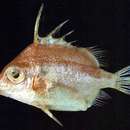
體長橢圓形,側扁而高;尾柄短小。吻不突出,吻長等於眼眶長,眶間隔凸出。眼稍大,眼徑等於或略小於吻長。
口端位,唇薄;上下頜齒同型,齒圓錐狀,除外緣齒列外,內側另有一疏齒列,約1-6顆(通常2顆)。鰓裂長;擬鰓長,
延伸至胸鰭基下方;為32-40片。體被細鱗,每一鱗片的表面有1-4直立的棘突排列成行,大型魚可達6棘突,棘多細尖,大型魚有分叉的棘突。背鰭兩個,硬棘VI,發育良好,各棘長度由前向後漸減;腹鰭軟條2,明顯可見,通常向下超過胸鰭基2/3處,甚或更低,腹鰭骨扁平中寬。
體淡紅色,有兩條明顯黃帶,一從眼上方延伸至背鰭末端,一從眼後延伸至臀鰭基前緣,但上述之條紋會予春天時消失。