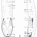
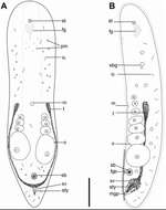
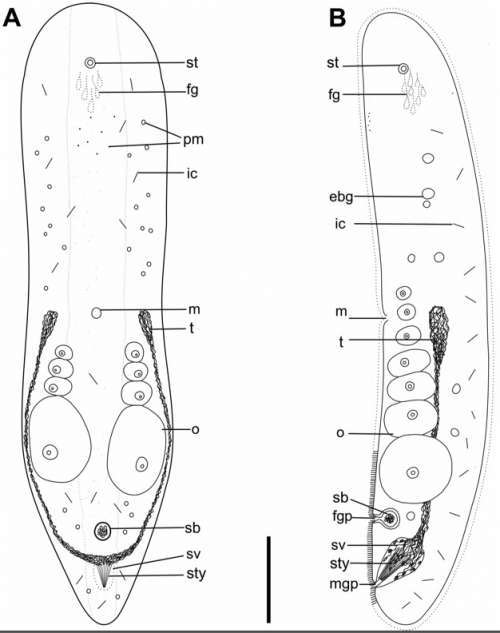
Childia aculifera sp.nov. Reconstructions show arrangement of organs. A. Dorsal reconstruction of whole living animal. B. Sagittal reconstruction of the whole animal. Scale bar: 220 μm.
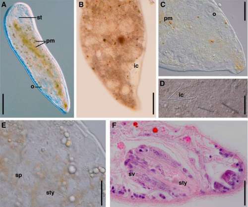
Childia aculifera sp.nov. ?E photomicrographs of living specimens. A. Dorsal view of whole specimen. Scale bar: 250 μm. B. View of posterior part of body. Scale bar: 100 μm. C. View of posterior end. Scale bar: 75 ?m. D. Inclusions (arrowhead). Scale bar: 30 μm. E. View of posterior part of body. Scale bar: 40 μm. F. Photomicrograph of sagittal histological sections stained with hematoxylin-eosin. Scale bar: 30 μm.