-
All Biocode files are based on field identifications to the best of the researcher’s ability at the time.
-
All Biocode files are based on field identifications to the best of the researcher’s ability at the time.
-
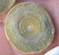
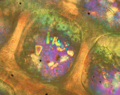
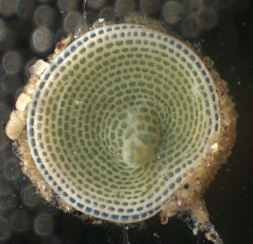
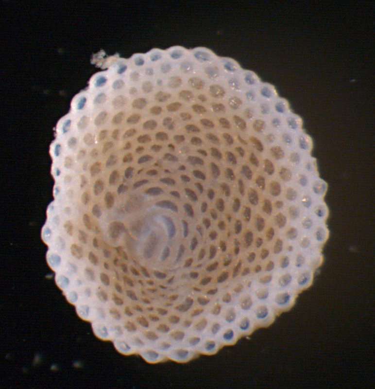
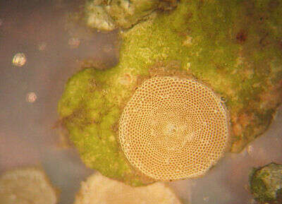
This polarized-light image shows some of the "windows" that cover the surface of the test. Image courtesy of Samuel S. Bowser, Wadsworth Center.
-
Orbiculina adunca.
-
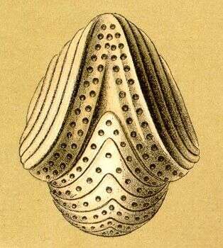
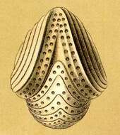
Orbitolites tenuissimus, Carpenter MSS..
-
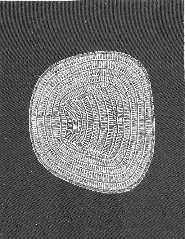
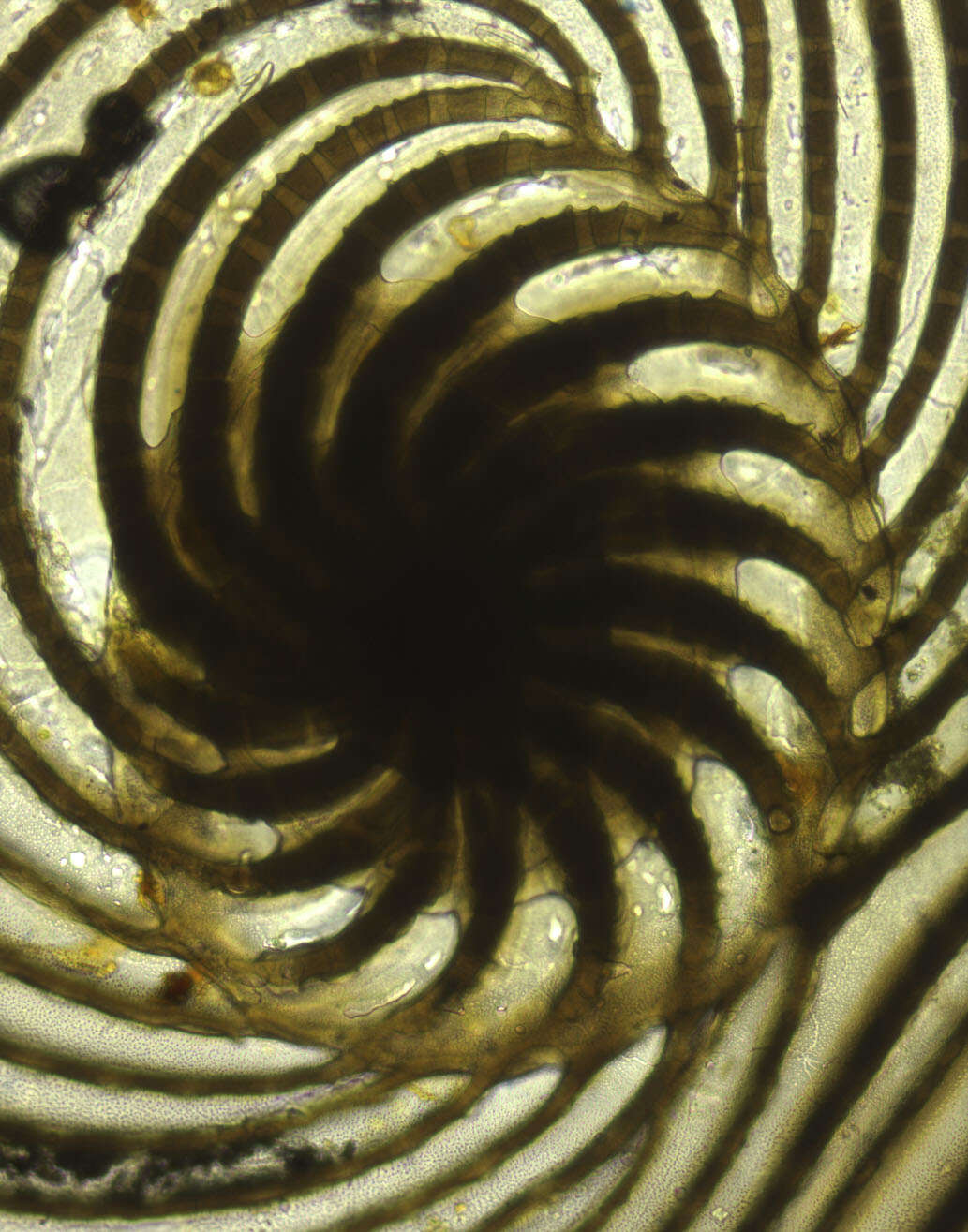
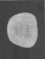
Cycloputeolina discoidea (Flint, 1899). Photomicrograph of early coil in specimen figured in Flint, 1899, Plate 49, fig. 2 (left image). Uncatalogued slide F-4446 in Cushman Collection of Foraminifera, National Museum of Natural History, Smithsonian Institution, Washington, DC.
-
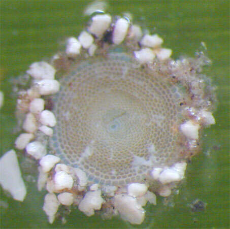
Live individual of the foraminiferan Sorites dominicensis attached to a blade of turtle grass (Thalassia testudinum) from Belize
-
Live individuals of the foraminiferans Archaias angulatus and one specimen of Coscinospira (=Peneroplis) antillarum from Belize
-
All Biocode files are based on field identifications to the best of the researcher’s ability at the time.
-
All Biocode files are based on field identifications to the best of the researcher’s ability at the time.
-
The green color of this calcareous foraminiferan is due to the presence of symbionts. Notice the very large size of the test; this species can be well over 1 cm across. Image (and hand) courtesy of Samuel S. Bowser, Wadsworth Center.
-
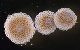
Live individual of Sorites dominicensis undergoing reproduction by multiple fission
-
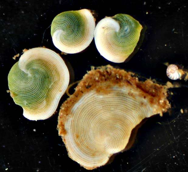
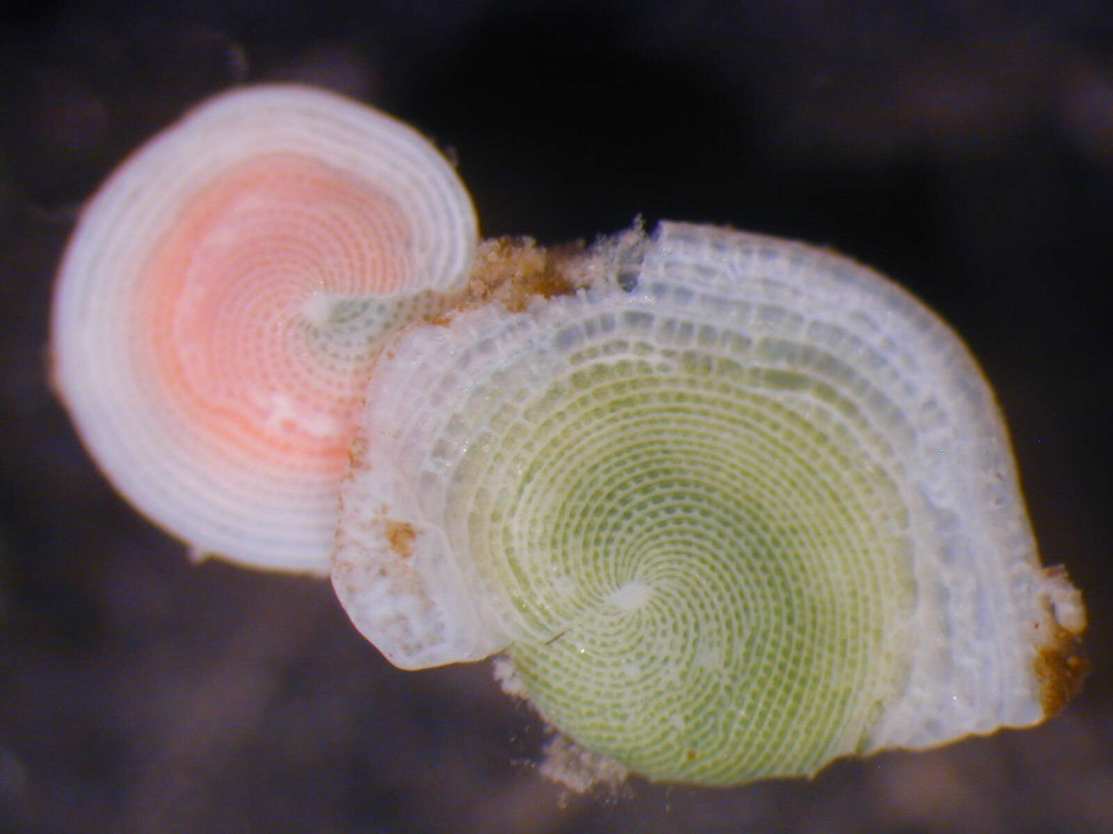
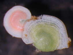
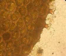
""Watermelon forams"--These foraminiferans possess chlorophyte (green algal) endosymbionts which give to the living cell a characteristic "grass green" coloration (specimen on right). Under high light (high UV?) conditions, the symbionts produce carotenoid pigments (salmon pink specimen on left)."
-
All Biocode files are based on field identifications to the best of the researcher’s ability at the time.
-
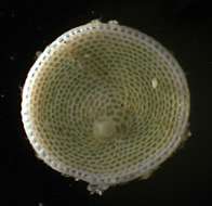
Live individual collected in Florida, USA. Phase-contrast photomicrograph by Scott Fay, 2004.
-
-
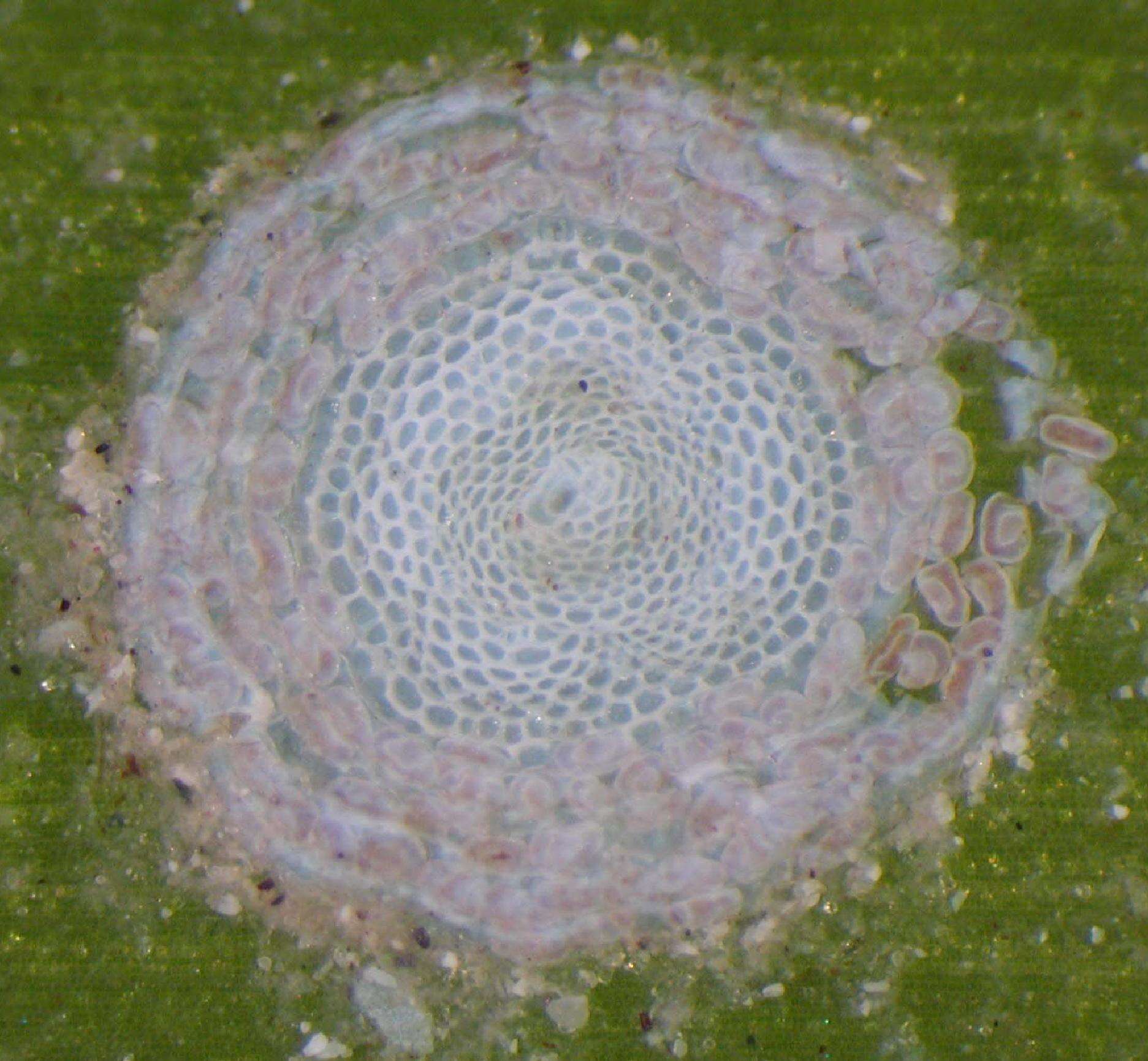
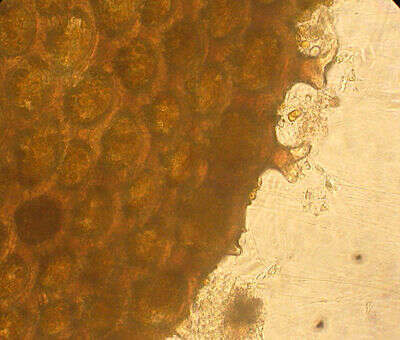
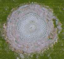
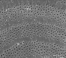
Close up of the pseudopores (pits) on the chamber surface of the foraminiferan Archaias angulatus from Belize, CA
-
All Biocode files are based on field identifications to the best of the researcher’s ability at the time.
-
Notice the reticulopodia protruding from pores in the edge of the test. Specimen collected in Florida, USA. Phase-contrast photomicrograph by Scott Fay, 2004.
-
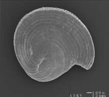
Scanning electron micrograph of a shell of the foraminiferan Archaias angulatus from Belize
-
All Biocode files are based on field identifications to the best of the researcher’s ability at the time.
-
An individual in natural surroundings. The test's chambers are particularly distinct in this photograph. The genus name
Sorites means "a heap" in Greek, and is also the name of a philosophical problem (the "
sorites paradox"). This paradox deals with the process of adding individual objects to a group: at what point do, say, individual sand grains added to a pile become "a heap of sand"? In this case, even though the foram adds its chambers one by one, it only takes one chamber to make a
Sorites. Problem solved. This individual was collected from Cook's Bay, Moorea, French Polynesia. Light micrograph by Scott Fay, 2005.
-
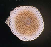
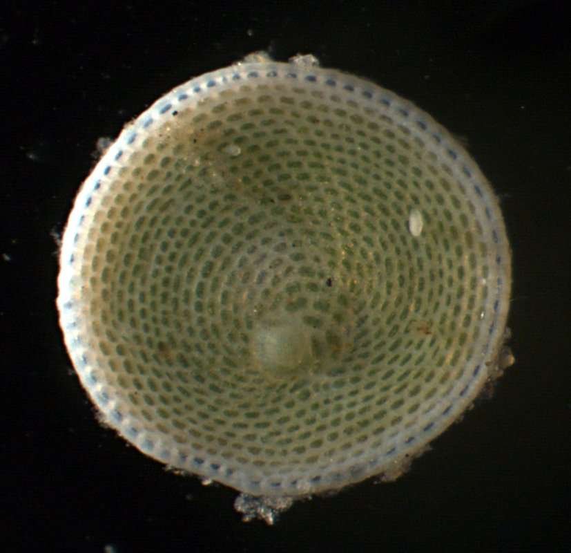
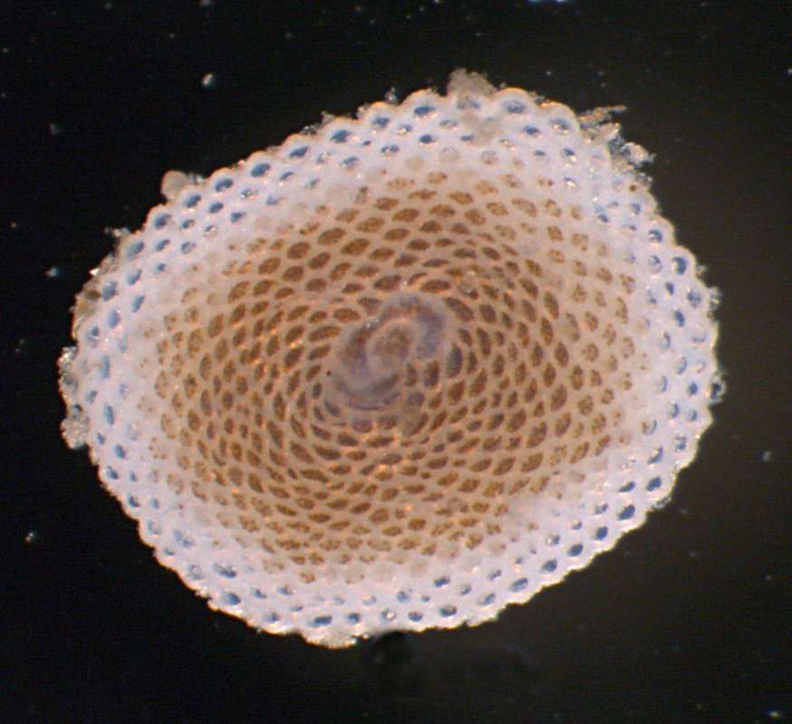
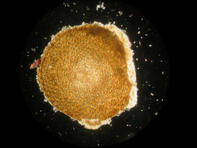
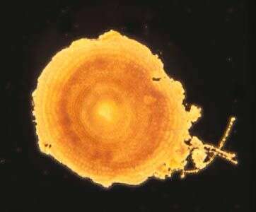
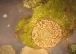
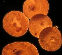
Large disc-shaped soritid foraminifera. The test is calcareous, flattened and disc-like, cells may be several millimetres in diameter from Bahamas. This is an image of a dead test only, dark ground image by Dave Caron. This organism is found in benthic habitats.
-
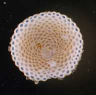
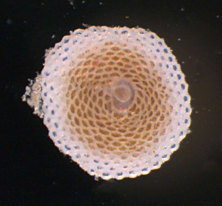
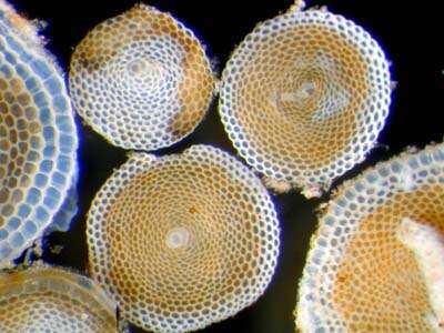
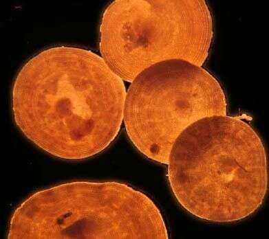
Large disc-shaped soritid foraminifera. The test is calcareous, flattened and disc-like, cells may be several millimetres in diameter from Bahamas. This organism is found in benthic habitats. This is an image of tests only, dark ground image by Dave Caron.