-
Muelas del Pan, Castille and Leon, Spain
-
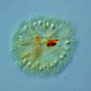
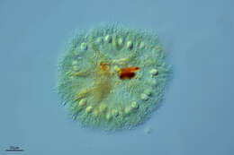
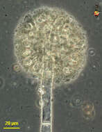
Spongomonas (spong-owe-moan-ass), is a solitary or colonial spongomonad flagellate, in which the cells are located within a more or less globular matrix formed from adhering small globules of mucilage. Many cells were dislodged while this sample was being prepared. Phase contrast.
-
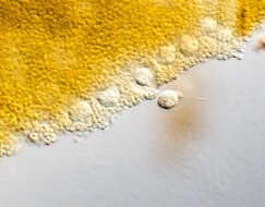
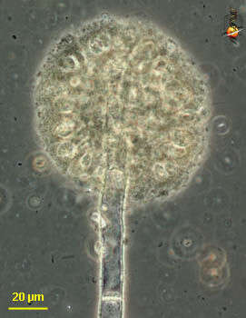
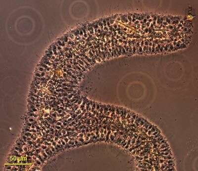
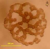
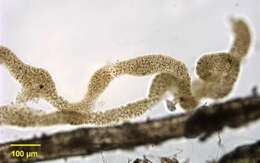
Spongomonas (spong-owe-moan-ass), is a solitary or colonial spongomonad flagellate, in which the cells are located within a more or less globular matrix formed from adhering small globules of mucilage. In this case the colony has formed at the end of some extraneous fibre. Phase contrast.
-
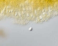
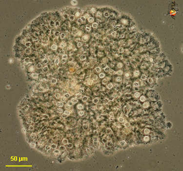
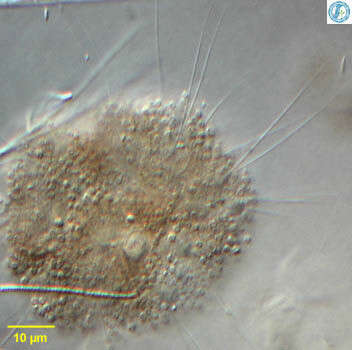
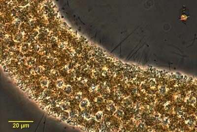
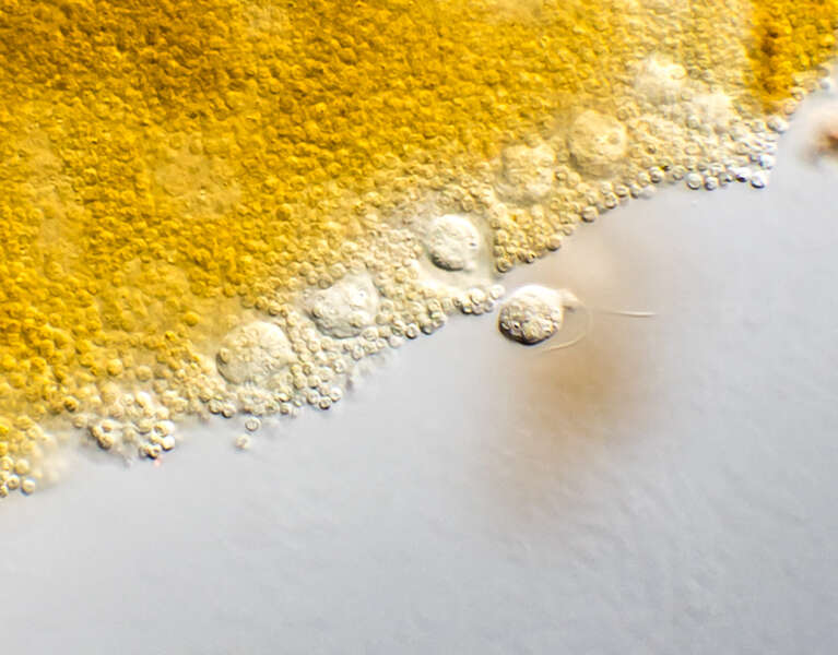
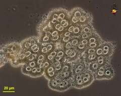
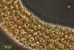
Spongomonas (spong-owe-moan-ass), is a solitary or colonial spongomonad flagellate, in which the cells are located within a more or less globular matrix formed from adhering small globules of mucilage. This is an image of a cluster of cells picked from the surface of the pond. Phase contrast.
-
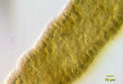
Spongomonas (spong-owe-moan-ass), is a solitary or colonial spongomonad flagellate, in which the cells are located within a more or less globular matrix formed from adhering small globules of mucilage. This is an image of a thin cluster of cells. Phase contrast.
-
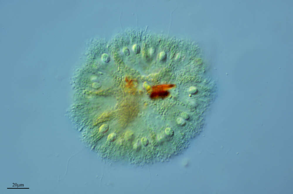
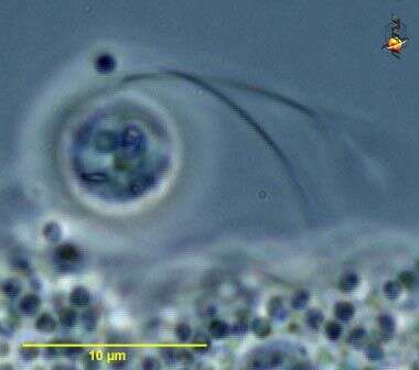
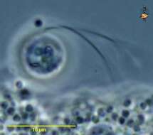
Spongomonas (spong-owe-moan-ass), is a solitary or colonial spongomonad flagellate, in which the cells are located within a more or less globular matrix formed from adhering small globules of mucilage. Detail of single cell showing the two flagella Phase contrast.
-
-
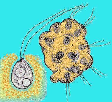
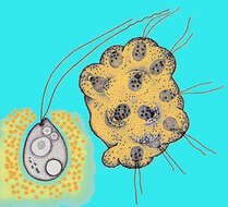
Spoongomonas, detail of several cells from within a globular colony. Each cell is more or less spherical and gives rise to two long flagella that are slightly different in length. The matrix of the colony is made up up of brown globular mucoid balls. From Lake Donghu, China. Differential interference contrast micrograph.
-
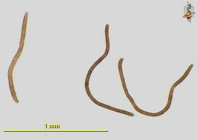
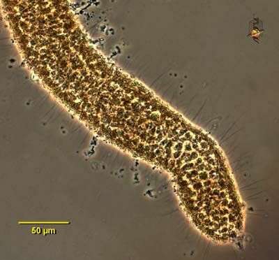
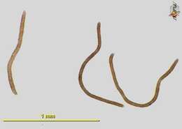
Spongomonas - a heterotrophic flagellate. The cells are spherical and give rise to two flagella that are slightly different in length. The cells form colonies, being embedded in a common mucous matrix that is made up of small globules of orange or brown mucus. This species forms sausage-shaped colonies that are up to a millimetre in length. Bright field illumination.
-
Spongomonas - a heterotrophic flagellate. The cells are spherical and give rise to two flagella that are slightly different in length. The cells form colonies, being embedded in a common mucous matrix that is made up of small globules of orange or brown mucus. This species forms sausage-shaped colonies that are up to a millimetre in length. Phase contrast micrograph.
-
Portrait of Spongomonas intestinum colony. Colonies are composed of small spherical colorless individual flagellates each with two equal flagella embedded in a serpiginous gelatinous matrix. Flagella are about 3 times cell length. Some species form spherical colonies. Gelatinous matrix may be brownish-green as seen here or nearly colorless. From freshwater pond near Boise, Idaho. Brightfield.
-
Detail of Spongomonas colony showing individual cells embedded in gelatinous matrix. Each cell has two equal flagella about three times the cell length. From fresh water pond near Boise, Idaho. Oblique illumination.
-
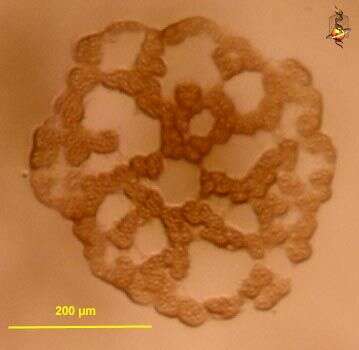
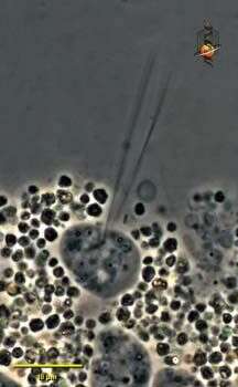
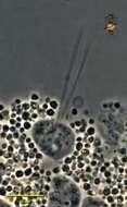
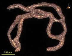
Low power image of a colony - thousands of cells in a common gelatinous matrix makes this branching spongy colony. Dark ground illumination.
-
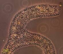
Detail of a small section of a colony showing the numerous individual cells within the mucus matrix. Phase contrast optics.
-
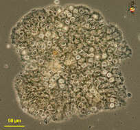
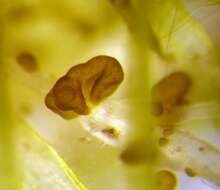
Sampling date 05/2007. Larger colony of S. uvella growing mushroom-shaped on Sphagnum leaflets. The diameter of the colony in the center of the picture is about 2 mm.Place name: Bog Dosenmoor near Neumuenster (Schleswig-Holstein, Germany) Latitude: 54.136219 Longitude: 10.026433Dissecting microscope Zeiss SV6, camera Olympus C7070WZ.© Wolfgang Bettighofer,images under Creative Commons License V 3.0 (CC BY-NC-SA).For permission to use of (high resolution) images please contact
postmaster@protisten.de.For further information about the image, please click here:
Link to protisten.de page
-
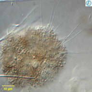
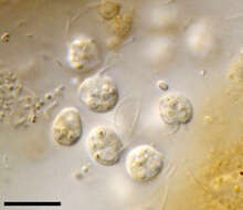
Scale bar indicates 10 µm.Cells out of the mucilaginous colony lump. The two flagella per cell are clearly depicted.Place name: Bog Dosenmoor near Neumuenster (Schleswig-Holstein, Germany) Latitude: 54.136219 Longitude: 10.026433Microscope Zeiss Universal, camera Olympus C7070WZ.© Wolfgang Bettighofer,images under Creative Commons License V 3.0 (CC BY-NC-SA).For permission to use of (high resolution) images please contact
postmaster@protisten.de.For further information about the image, please click here:
Link to protisten.de page
-
Place name: Pond Birkensee near Rödelsee (Lower Franconia, Germany) Latitude: 49.71819841 Longitude: 10.27807474Microscope Zeiss Axioplan, camera Canon DSLR.Copyright Dr. Rainer Meisch, Würzburg, Germany.© Wolfgang Bettighofer,images under Creative Commons License V 3.0 (CC BY-NC-SA).For permission to use of (high resolution) images please contact
postmaster@protisten.de.For further information about the image, please click here:
Link to protisten.de page