-
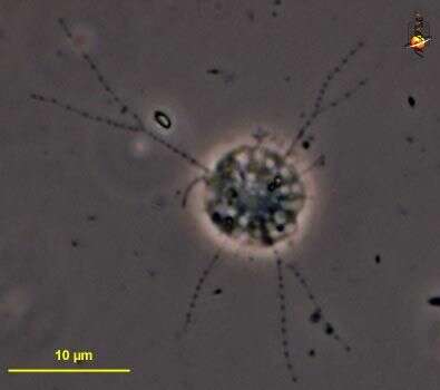
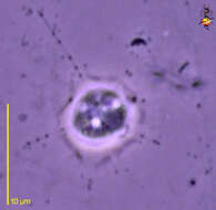
Microcometes (mike-row-come-eat-ees) are flagellates that are usually treated as amoebae. Cell usually located within an irregular organic lorica, the walls of which vary from delicate to thick, from colourless to dark brown. There are multiple apertures from which thin arms bearing extrusomes emerge. The nucleus is typically eccentric, and the arms end on a dark element. The organization is very similar to the centrohelids, except Microcometes has a flagellum (4 o clock). Phase contrast.
-
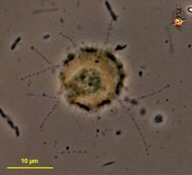
Microcometes (mike-row-come-eat-ees) are flagellates that are usually treated as amoebae. Cell usually located within an irregular organic lorica, the walls of which vary from delicate to thick, from colourless to dark brown. there are multiple apertures from which thin arms bearing extrusomes emerge. Phase contrast.
-
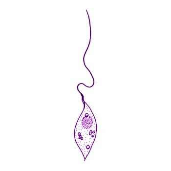
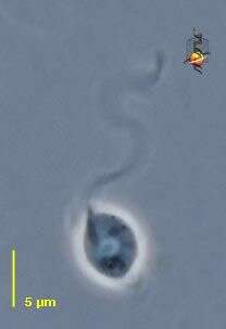
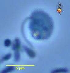
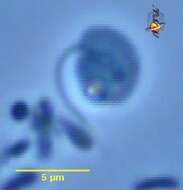
Microcometes (mike-row-come-eat-ees) are flagellates that are usually treated as amoebae. The flagella are very short and inactive and very hard to see - particularly because the cells are usually enclosed in a thick walled organic test. However, this cell must have recently settled and has yet to form the lorica, and one flagellum is quite obvious. Phase contrast.
-
Microcometes (mike-row-come-eat-ees) are flagellates that are usually treated as amoebae. Cell usually located within an irregular organic lorica, the walls of which vary from delicate to thick, from colourless to dark brown. there are multiple apertures from which thin arms bearing extrusomes emerge. Phase contrast.
-
-
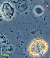
Microcometes, a flagellate living in a lorica that adheres to the substrate. The lorica has a number of ports through which fine pseudopodia that bear granules extend. The pseudopodia usually adhere to the substrate. Flagella are inactive and often hard to see. The lorica seems to be transparent when first formed, with age it becomes brown. From Lake Donghu, China. Differential interference contrast micrograph.
-
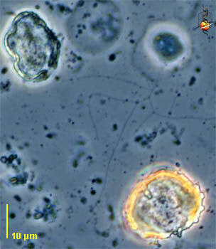
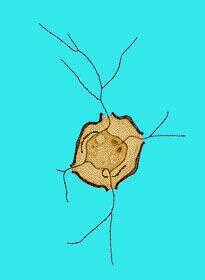
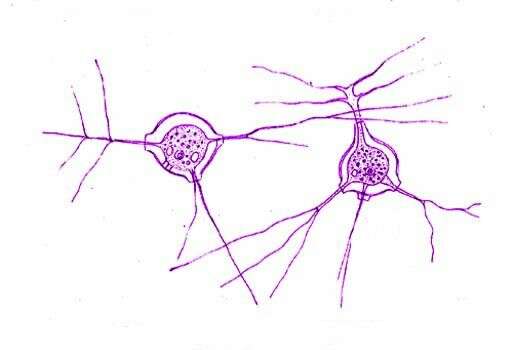
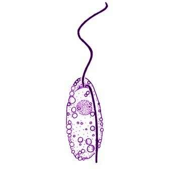
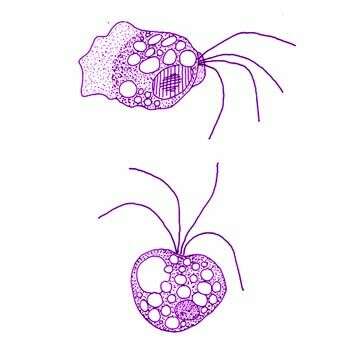
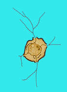
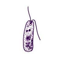
Microcometes paludosa Cienkowski 1876. Flattened amoeboid cell with thin branching pseudopodia adpressed to the substrate, pseudopodia with sparse granules, usually within a lorica which is transparent and then when first formed, but becomes brown and thickened with age, pseudopodia emerge from slightly tubular aperture, this image shows the understanding by Cienkowski of this species.
-
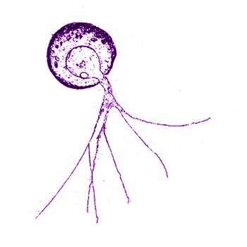
Microcometes paludosa Cienkowski 1876. Flattened amoeboid cell with thin branching pseudopodia adpressed to the substrate, pseudopodia with sparse granules, usually within a lorica which is transparent and then when first formed, but becomes brown and thickened with age, pseudopodia emerge from slightly tubular apertures, with two short inactive flagella (not usually reported), 5-15 microns diameter. This image shows the current understanding of this species.
-
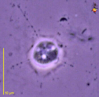
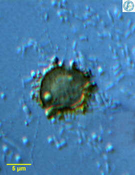
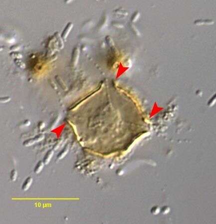
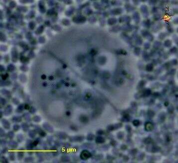
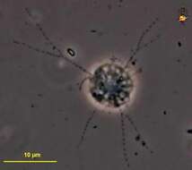
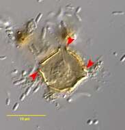
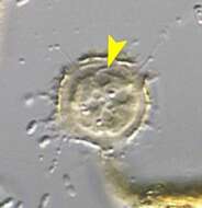
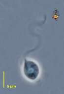
Microcometes paludosa (Cienkowski, 1876). Red arrowheads mark the short -necked test apertures through which thin pseudopodia with extrusomes protrude. From the margins of a slow-moving freshwater stream near Boise, Idaho. April 2007. DIC.
-
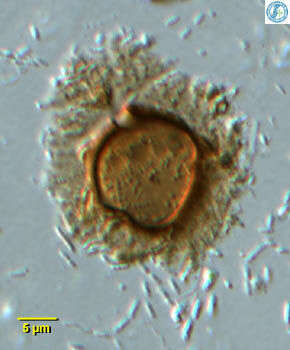
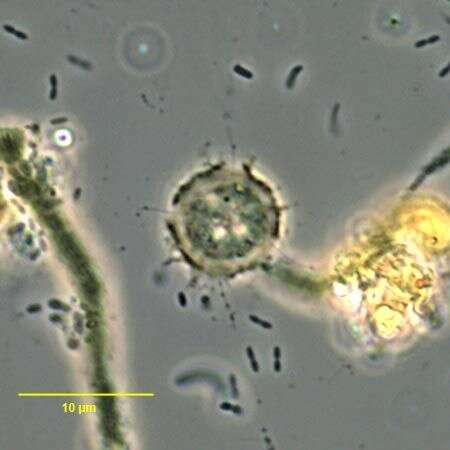
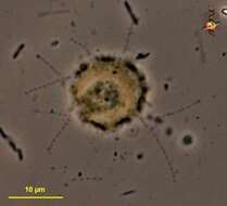
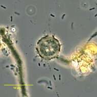
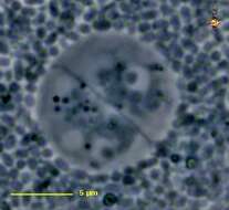
Microcometes paludosa (Cienkowski, 1876). there are multiple contractile vacuoles. From the margins of a slow-moving freshwater stream near Boise, Idaho. April 2007. Phase contrast.
-
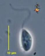
Microcometes paludosa (Cienkowski, 1876).The yellow arrowhead marks a short flagellum. There are usually two and they are easily overlooked. there are multiple contractile vacuoles.From the margins of a slow-moving freshwater stream near Boise, Idaho. April 2007. DIC
-
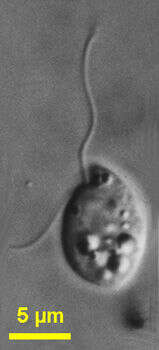
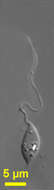
Phyllomitus (file-owe-mite-us) is a swimming phagotrophic flagellate - believed to be related to the stramenopiles (because of the sine wave pattern of beating of the anterior flagellum and because the flagellum draws the cell forward suggesting the presence of flagellar hairs). There is a second trailing flagellum. This is not a particular familiar genus, but is one of the more voracious heterotrophic flagellates. They may ingest particles of food many times bigger than themselves - and is not unknown to see swimming diatoms which after carefully scrutiny can be seen to be a large diatom enclosed by a Phyllomitus that has become stretched very thinly. Phase contrast.
-
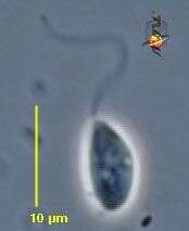
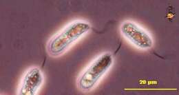
Phyllomitus (file-owe-mite-us) undulans Stein, 1878. Cells are about 11 - 12 microns long and have two flagella that adhere to each other. The cells are pointed anteriorly and posteriorly. The flagella arise at the anterior end of the cell. The flagella appear to be similar in length, about 3 times longer than the cell, and undulate. The pellicle is smooth and the nucleus is located on the mid-anterior part of the cell. Cytoplasmic strands may be seen from the posterior end. The cells may attach to the substrate by the posterior end of the cell. Rarely observed.
-
Phyllomitus undulans Stein, 1878. Cells are about 11 - 12 microns long and have two flagella that adhere to each other. The cells are pointed anteriorly and posteriorly. The flagella arise at the anterior end of the cell. The flagella appear to be similar in length, about 3 times longer than the cell, and undulate. The pellicle is smooth and the nucleus is located on the mid-anterior part of the cell. Cytoplasmic strands may be seen from the posterior end. The cells may attach to the substrate by the posterior end of the cell.
-
Phyllomitus (file-owe-mite-us) granulatus Larsen and Patterson, 1990. Cell outline is sac-shaped. Cells are flexible, 7 to 21 microns long and slightly flattened. The anterior flagellum beats with a sine-wave, is about 1.0 to 1.5 times the length of the cell and is directed to the front and slightly to the right during swimming. The posterior flagellum inserts to the left of the anterior flagellum, varies in length from 0.5 to 2.5 times the length of the cell. Cytoplasm is drawn out at the posterior end. Refractile granules underlie the cell surface. The nucleus is located below the anterior pocket, near the centre of the cell and is roundish. The cells contained ingested eukaryotic algae. Sometimes very common (late culture).
-
Phyllomitus granulatus Larsen and Patterson, 1990. Cell outline is sac-shaped. Cells are flexible, 7 to 21 microns long and slightly flattened. The anterior flagellum beats with a sine-wave, is about 1.0 to 1.5 times the length of the cell and is directed to the front and slightly to the right during swimming. The posterior flagellum inserts to the left of the anterior flagellum, varies in length from 0.5 to 2.5 times the length of the cell. Cytoplasm is drawn out at the posterior end. Refractile granules underlie the cell surface. The nucleus is located below the anterior pocket, near the centre of the cell and is roundish. The cells contained ingested eukaryotic algae.
-
Phyllomitus salinus Lackey, 1940. Cells are about 12 microns long and 5 microns wide. This small fonn is very active. Typically elongate, cylindrical, metabolic, with 2 flagella emerging from a slight anterior depression. The trailing flagellum is about two-thirds body length, the anterior is about one and one half body length. Nucleus median, cytoplasm clear, granular, a few small spheres sometimes present. Nutrition and reproduction not reported. It also has shorter flagella and a less pronounced mouth region than P. undulans.
-
Proleptomonas (pro-lep-toe-moan-ass) a ellipsoidal heterotrophic flagellate of uncertain affinities, one apical flagellum emerging from the front of the cell, nucleus usually located anteriorly. Reported usually from soils. Phase contrast.
-
Proleptomonas (pro-lep-toe-moan-ass) a ellipsoidal heterotrophic flagellate of uncertain affinities, one apical flagellum emerging from the front of the cell, nucleus usually located anteriorly. Reported usually from soils. Phase contrast.
-
Proleptomonas (pro-lep-toe-moan-ass) a ellipsoidal heterotrophic flagellate of uncertain affinities, one apical flagellum emerging from the front of the cell, nucleus usually located anteriorly. Reported usually from soils. Bilaterally symmetrical cyst. Phase contrast.
-
Quadricilia rotundata (Skuja, 1948) V+rs, 1992. Cells are 5-9 x 10-15 microns, globular cells 10-20 microns in diameter. Cell globular, or nearly so, with 4 (-8) unequal, smooth, acronematic flagella inserted anteriorly in a shallow depression. The flagella are 1-3 times the diameter of the cell body. Many thin, branched pseudopodia may be produced from any point of the cell surface. Cytoplasm sometimes highly vesiculate, nucleus central or in the cell anterior.
-
Sainouron (sigh-noo-ron) is a small gliding flagellate from soils. Body elliptical, one trailing flagellum, rarely with an anterior flagellum. This image shows a subapical insertion of the flagellum. The cell shape is somewhat distorted (swollen). Phase contrast.