-
-
-
-
-
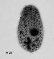
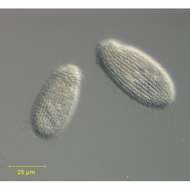
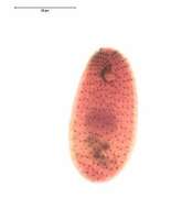
Detail view of Dexiotricha granulosa (Kent, 1881) Foissner, 1994 showing the prominent ring-shaped cytoplasmic glycogen granules. From freshwater pond near Boise, Idaho. DIC.
-
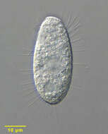
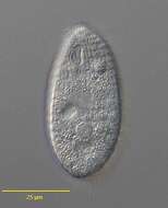
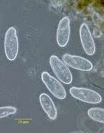
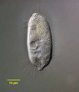
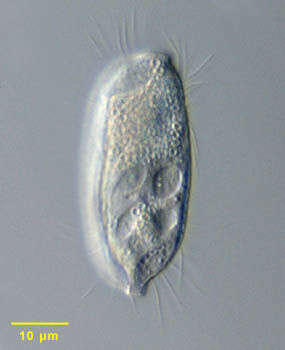
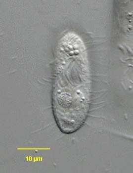
Portrait of the hymenostome ciliate, Dexiotricha granulosa (Kent, 1881) Foissner, 1994, synonymous with Loxocephulus granulosa. The cell is ovoid, broadly rounded posteriorly and truncate anteriorly. Regular longitudinal kineties terminate at a subapical band of circumferential kineties demarcating a cilia-free truncate apical area or frontal plate. There is a single long caudal cilium. The oral aperture is small and difficult to visualize in vivo. It is located in the anterior quarter with an undulating membrane on the right (seen faintly here) and 3 membranelles (not seen here). The macronucleus is spheroid and located in the mid-cell. The contractile vacuole is seen here to the left of the macronucleus. The spherical micronucleus is not seen here. The cytoplasm contains many small refractile ring-shaped glycogen granules, which are diagnostic for the species (see detail images). Dexitricha is bactiverous. From freshwater pond near Boise, Idaho. Differential interference contrast.
-
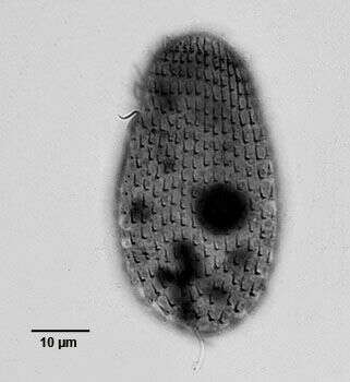
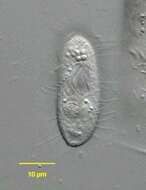
Ventrolateral view of the infraciliature of the hymenostome ciliate, Dexiotricha granulosa (Kent, 1881) Foissner, 1994. Synonym of Loxocephulus granulosa. The cell is ovoid, broadly rounded posteriorly and truncate anteriorly. Regular longitudinal kineties terminate at a subapical band of circumferential kineties demarcating a cilia-free truncate apical area or frontal plate. Fibrils radiate anteriorly from the kinetids of the anteriormost paratene (seen here). There is a single long caudal cilium. The oral aperture is small and difficult to visualize in vivo. It is located in the anterior quarter with an undulating membrane on the right (seen here) and 3 membranelles (the posterior most seen here). The macronucleus is spheroid and located in the mid-cell. Single contractile vacuole. From freshwater pond near Boise, Idaho. Silver carbonate stain (see Foissner, W. Europ. J. Protistol., 27:313-330;1991).Brightfield. Black and white.
-
Dorsal view of the infraciliature of the hymenostome ciliate, Dexiotricha granulosa (Kent, 1881) Foissner, 1994. Synonym of Loxocephulus granulosa. The cell is ovoid, broadly rounded posteriorly and truncate anteriorly. Regular longitudinal kineties terminate at a subapical band of circumferential kineties demarcating a cilia-free truncate apical area or frontal plate. Fibrils radiate anteriorly from the kinetids of the anteriormost paratene (seen here). There is a single long caudal cilium. The oral aperture is small and difficult to visualize in vivo. It is located in the anterior quarter with an undulating membrane on the right (seen here) and 3 membranelles (the posterior most seen here). The macronucleus is spheroid and located in the mid-cell. Single contractile vacuole. From freshwater pond near Boise, Idaho. Silver carbonate stain (see Foissner, W. Europ. J. Protistol., 27:313-330;1991).Brightfield. Black and white.
-
Ventral view of the infraciliature of late division of the hymenostome ciliate, Dexiotricha granulosa (Kent, 1881) Foissner, 1994. Synonym of Loxocephulus granulosa. The cell is ovoid, broadly rounded posteriorly and truncate anteriorly. Regular longitudinal kineties terminate at a subapical band of circumferential kineties demarcating a cilia-free truncate apical area or frontal plate. Fibrils radiate anteriorly from the kinetids of the anteriormost paratene (seen here). There is a single long caudal cilium. The oral aperture is small and difficult to visualize in vivo. It is located in the anterior quarter with an undulating membrane on the right (seen here) and 3 membranelles ( seen most clearly in the proter). The macronucleus is spheroid and located in the mid-cell. Single contractile vacuole. From freshwater pond near Boise, Idaho. Silver carbonate stain (see Foissner, W. Europ. J. Protistol., 27:313-330;1991).Brightfield. Black and white.
-
Portrait of the hymenostome ciliate, Dexiotricha granulosa (KENT,1881) FOISSNER, 1994, synonymous with Loxocephulus granulosus. The cell is ovoid, broadly rounded posteriorly and truncate anteriorly. Regular longitudinal kineties terminate at a subapical band of circumferential kineties demarcating a cilia-free truncate apical area or frontal plate. There is a single long caudal cilium. The oral aperture is small and difficult to visualize in vivo. It is located in the anterior quarter with an undulating membrane on the right (seen faintly here) and 3 membranelles (not seen here). The macronucleus is spheroid and located in the mid-cell. The contractile vacuole is seen here to the left of the macronucleus. The spherical micronucleus is not seen here. The cytoplasm contains many small refractile ring-shaped glycogen granules, which are diagnostic for the species (see detail images). Dexitricha is bactiverous. From freshwater pond near Boise, Idaho. Differential interference contrast.
-

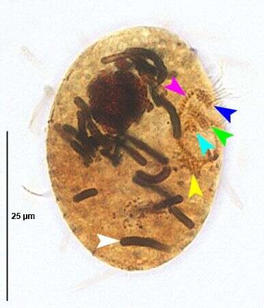
Ventrolateral view of the infraciliature of the hymenostome ciliate, Dexiotricha granulosa (Kent, 1881) Foissner, 1994. Synonym of Loxocephulus granulosa. The cell is ovoid, broadly rounded posteriorly and truncate anteriorly. Regular longitudinal kineties terminate at a subapical band of circumferential kineties demarcating a cilia-free truncate apical area or frontal plate. Fibrils radiate anteriorly from the kinetids of the anteriormost paratene. There is a single long caudal cilium (green line). The oral aperture is small and difficult to visualize in vivo. It is located in the anterior quarter with a paraoral membrane on the right (red line) and 3 adoral membranelles (dark blue lines). Closely spaced basal bodies of the somatic kinaty to the right of the oral aperture form a "pseudomembrane" (light blue line). The macronucleus is spheroid and located in the mid-cell. Single contractile vacuole. From freshwater pond near Boise, Idaho. Silver carbonate stain (see Foissner, W. Europ. J. Protistol., 27:313-330;1991).Brightfield.
-
-
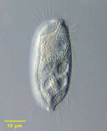
Dexiotricha tranquilla (Kahl,1926). DIC.
-
Dexiotricha tranquilla (Kahl,1926). Phase contrast.
-
Dexiotricha tranquilla (Kahl,1926).Brightfield.
-
Portrait of Balanonema dubium (Penard,1922) Kahl,1931, a philasterine scuticiciliate. The body is a slightly dorsoventrally flattened cylinder both ends of which are bluntly tapered into plug-like projections. There are subapical anterior and posterior grooves at the base of the terminal projections which spiral around half the cell circumference (both are well seen here). The cytostome is small (about 20% of the cell length), anterior and inconspicuous (not seen in this image). Kineties are longitudinal and cilia are sparse in the mid body and denser at the anterior and posterior ends. The subcentral macronucleus is spherical. The contractile vacuole is also subcentral. There is a long caudal cilium in some species. The cytoplasm of this species contains numerous small ring shaped granules similar to the glycogen granules of Dexiotricha granulosa, another philasterine scuticociliate. From sediment of slow-flowing organically enriched freshwater runoff stream near Boise, Idaho. Differential interference
-

Ventral infraciliature of Balanonema dubium (penard,1922) Kahl, 1931., a philasterine scuticiciliate. The body is a slightly dorsoventrally flattened cylinder both ends of which are bluntly tapered into plug-like projections. There are subapical anterior and posterior grooves at the base of the terminal projections which spiral around half the cell circumference. The cytostome is small (about 20% of the cell length), anterior and inconspicuous in vivo. Kineties are longitudinal and cilia are sparse in the mid body and denser at the anterior and posterior ends. The subcentral macronucleus is spherical. The contractile vacuole is also subcentral. There is a long caudal cilium in some species. The cytoplasm of this species contains numerous small ring shaped granules similar to the glycogen granules of Dexiotricha granulosa, another philasterine scuticociliate. From sediment of slow-flowing organically enriched freshwater runoff stream near Boise, Idaho. Stained by the silver carbonate technic (see Foissner, W.Europ. J. Protistol.27,313-330;1991) Brightfield.
-

Portrait of Balanonema dubium (penard,1922) Kahl,1931, a philasterine scuticiciliate. The body is a slightly dorsoventrally flattened cylinder both ends of which are bluntly tapered into plug-like projections. There are subapical anterior and posterior grooves at the base of the terminal projections which spiral around half the cell circumference (seen in accompanying images). The cytostome is small (about 20% of the cell length), anterior and inconspicuous (seen in accompanying image). Kineties are longitudinal and cilia are sparse in the mid body and denser at the anterior and posterior ends. The subcentral macronucleus is spherical. The contractile vacuole is also subcentral (both are seen well in this image). There is a long caudal cilium in some species. The cytoplasm of this species contains numerous small ring shaped granules similar to the glycogen granules of Dexiotricha granulosa, another philasterine scuticociliate. From sediment of slow-flowing organically enriched freshwater runoff stream near Boise, Idaho. Differential interference contrast.
-
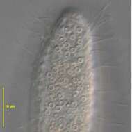
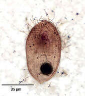
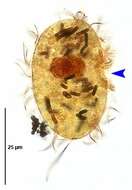
Ventral view of Dexiotrichides centralis (STOKES, 1886) , a sapropelic hymenostome ciliate. D. centralis is the only freshwater species of the genus. The cell is reniform and laterally compressed. The relatively large cytostome is located in the center of the body (seen well here). There are three adoral membranelles. There is an undulating membrane on the right margin of the cytostome. The somatic kineties are longitudinal. On the right surface there is an oblique row of longer cilia slanting posteriorly from the cytostome (seen here). There is a long caudal cilium. The contractile vacuole is terminal. The macronucleus is spherical . D. centralis is a facultative anaerobe. Easily confused with members of the similar genus, Dexiotricha. Collected from the bottom material in a freshwater aquaculture tub near Boise, Idaho in October 2003. DIC. DIC Song et al have described a marine species, Dexiotrichides pangi [Song et al . Dexiotrichides pangi n. sp. (Protozoa, Ciliophora, Scuticociliatia), a New Marine Ciliate from the North China Sea. J.Eukaryot. Microbiol., 50(2), pp. 114-122,2003.]
-

Left side of Dexiotrichides centralis (STOKES, 1886), a sapropelic scuticociliate. D. centralis is the only freshwater species of this genus. The cell is reniform and laterally compressed. The relatively large cytostome is located in the center of the body. There are three adoral membranelles. There is an undulating membrane on the right margin of the cytostome. The somatic kineties are longitudinal. On the right surface (not seen in this image) there is an oblique row of longer cilia slanting posteriorly from the cytostome. There is a long caudal cilium (seen here). The contractile vacuole is terminal. The spherical macronucleus is found in the anterior half. D. centralis is a facultative anaerobe. Easily confused with members of the similar genus, Dexiotricha. Collected from the bottom material in a freshwater aquaculture tub near Boise, Idaho in October 2003.DIC Song et al have described a marine species, Dexiotrichides pangi [Song et al . Dexiotrichides pangi n. sp. (Protozoa, Ciliophora, Scuticociliatia), a New Marine Ciliate from the North China Sea. J.Eukaryot. Microbiol., 50(2), pp. 114-122,2003.]
-
Dexiotrichides centralis (STOKES, 1886). Stained by the silver carbonate technique (Foissner,W. Europ. J. Protistol.27:313-330;1991).Brightfield.
-
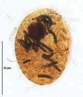
Lateral view (optical section) of Dexiotrichides centralis (STOKES, 1886). The blue arrowhead indicates the oral aperture. Densley stained endosymbiotic bacteria are seen in the cytoplasm. Stained by the silver carbonate technique (Foissner,W. Europ. J. Protistol.27:313-330;1991).Brightfield.
-
Right ventrolateral view (optical section) of the oral infraciliature of Dexiotrichides centralis (STOKES, 1886). The blue arrowhead indicates the oral aperture. Densley stained endosymbiotic bacteria are seen in the cytoplasm. Stained by the silver carbonate technique (Foissner,W. Europ. J. Protistol.27:313-330;1991).Brightfield.
-
Right ventrolateral view (optical section) of the oral infraciliature of Dexiotrichides centralis (STOKES, 1886). The blue, green and light blue arrowheads indicate the kinetosomes of the first (M1), second (M2) and third (M3) adoral membranelles respectively. The pink arrowhead indicates the undulating membrane on the right margin of the cytostome. The yellow arrowhead indicates the rhomboidal scutica, unciliated kinetosomes of the buccal anlage. Densley stained endosymbiotic bacteria are seen in the cytoplasm (white arrowhead). Stained by the silver carbonate technique (Foissner,W. Europ. J. Protistol.27:313-330;1991).Brightfield.