Hong–Liang Wang, Guo–Quan Wang, Wei–Hai Li
Zookeys
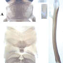
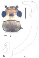
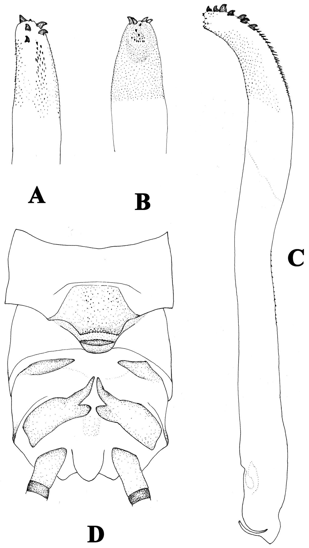
Figure 4.Neoperla mesostyla Li & Wang, sp. n. (male). A Head and pronotum, dorsal view B Terminalia, dorsal view C Hindleg (part of tarsi in this leg missing), lateral view D Aedeagus before eversion, lateral view E Aedeagus, lateral view.
Xue-Feng Qin, Dávid Murányi, Guo-Quan Wang, Wei-Hai Li
Zookeys
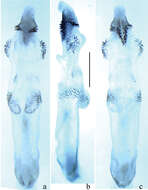
Figure 8.Neoperla xuansongae. Male a Head and pronotum, dorsal view b Aedeagus, lateral view with details in dorsal and ventral views. Scale bars: 0.5 mm.
Figure 1.Neoperla latispina Wang & Li, sp. n. Male. a Head and pronotum, dorsal view b Terminalia, dorsal and lateral views c Aedeagus before eversion, lateral view d Aedeagus before eversion, dorsal view e foreleg femur.
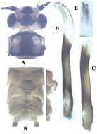
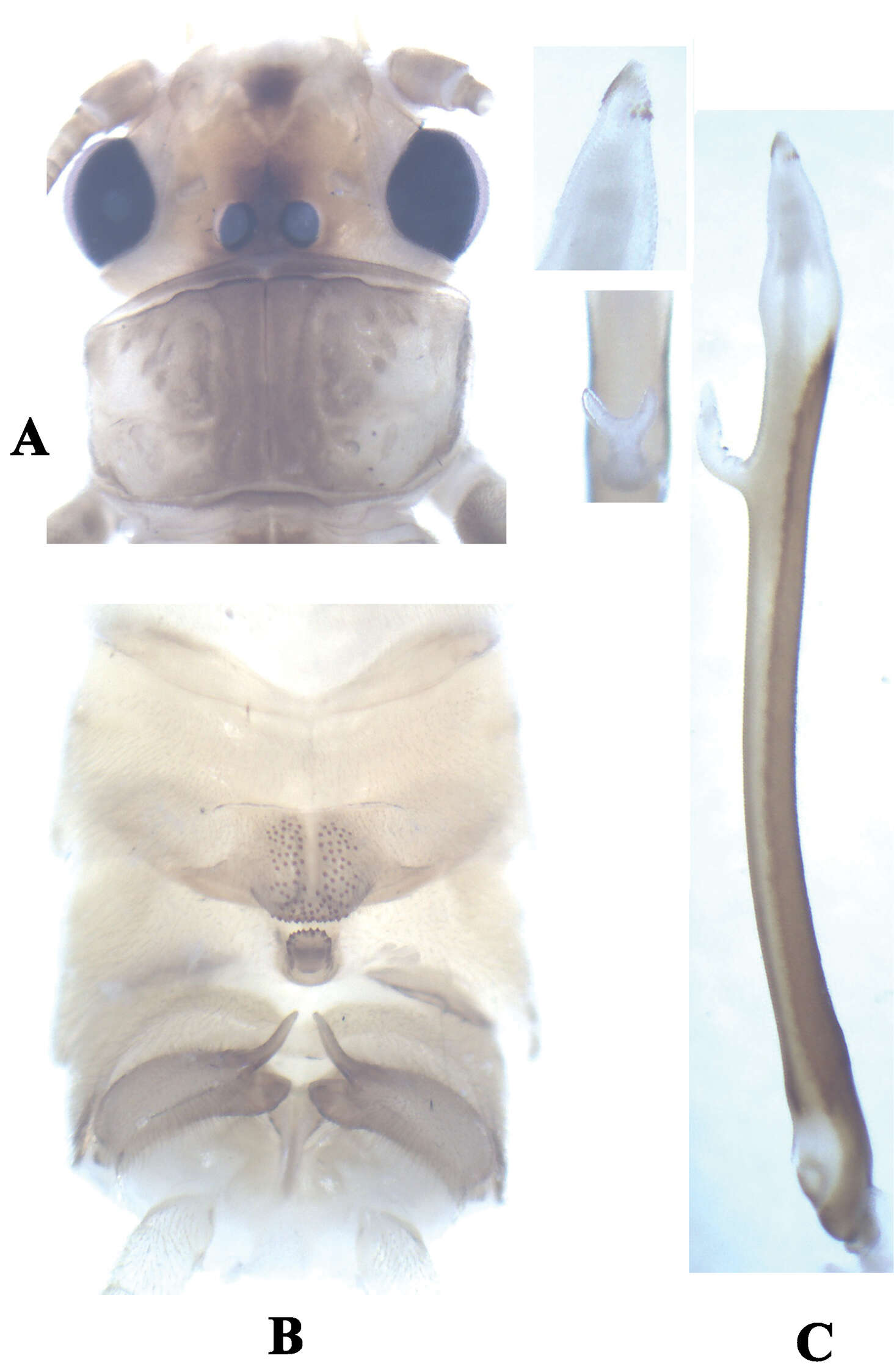
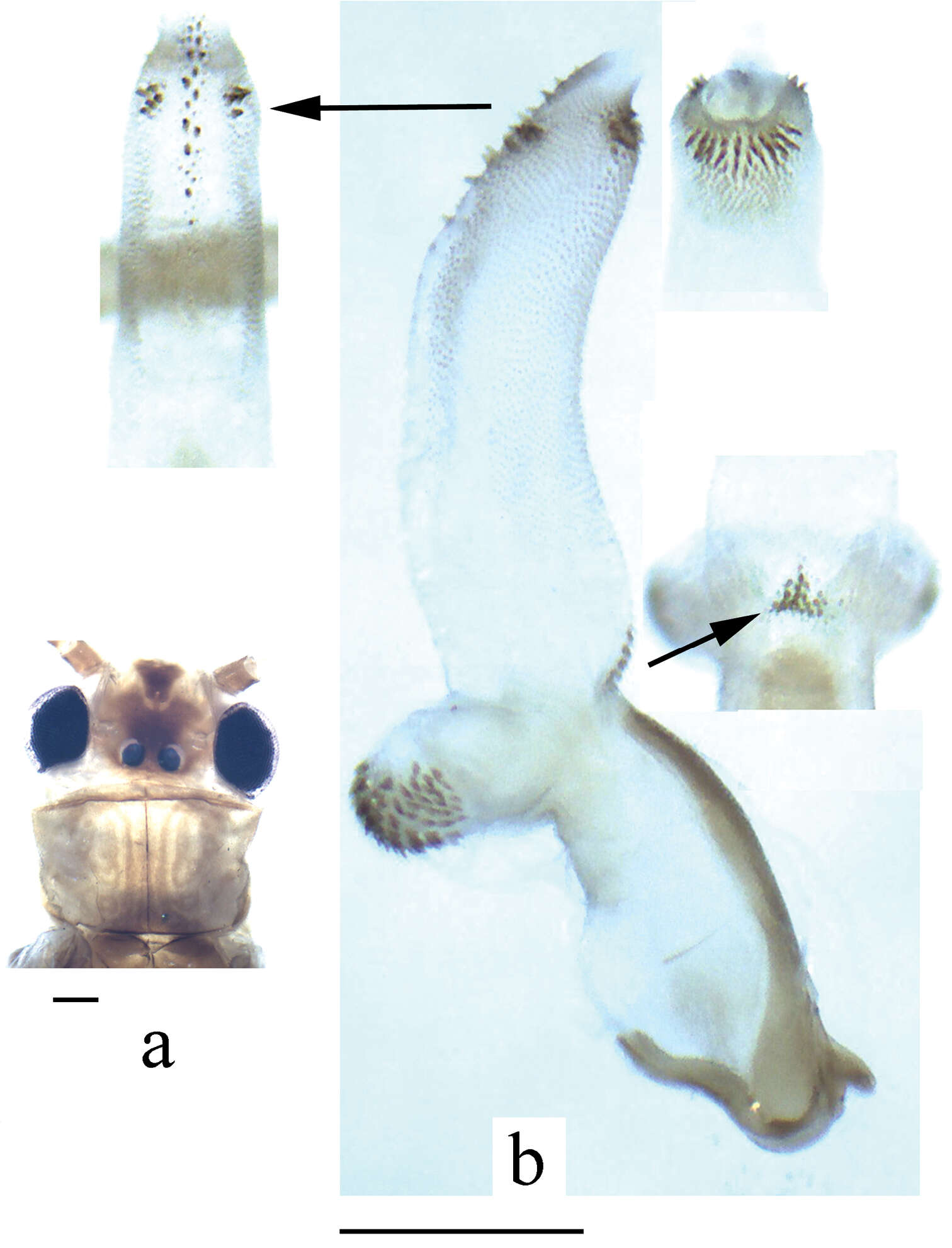
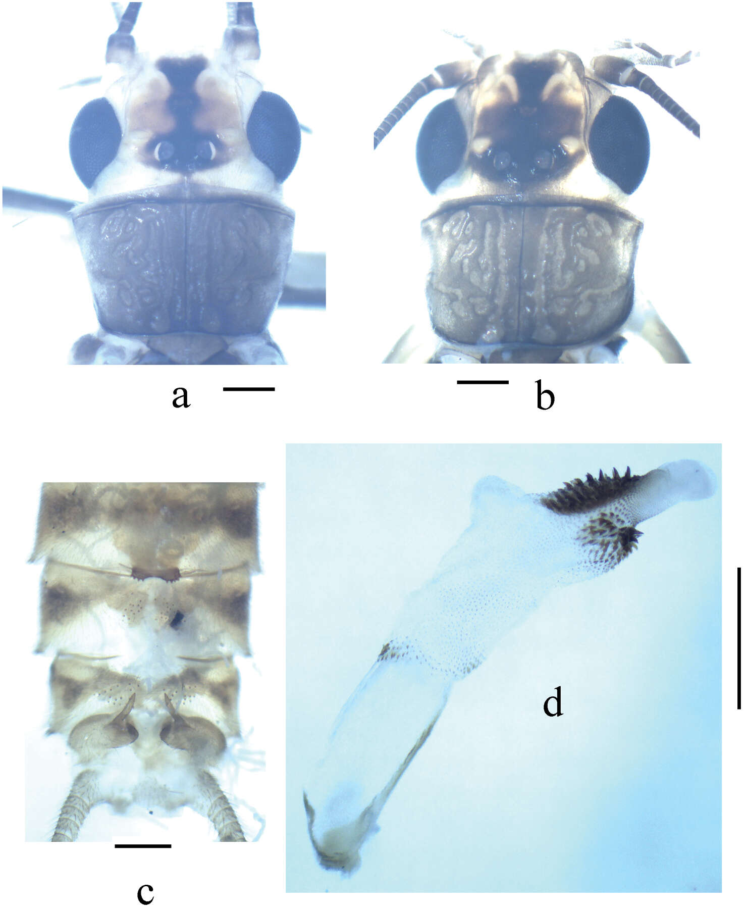
Figure 1.Neoperla nigromarginata Li & Zhang, sp. n. Male (a–e) a Head and pronotum, dorsal view b Terminalia, dorsal view c Terminalia, lateral view d Aedeagus before eversion, lateral view e Hindleg f Female subgenital fig, ventral view.
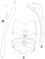
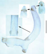
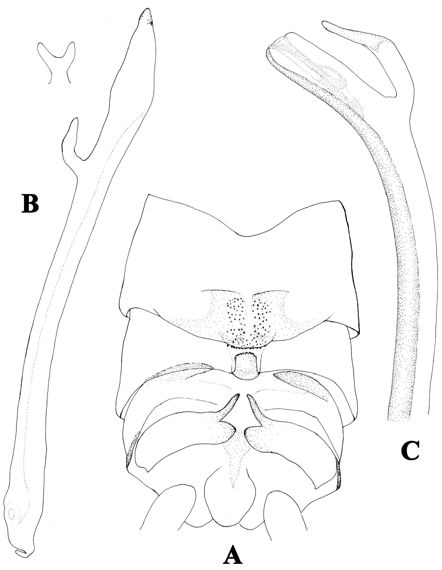
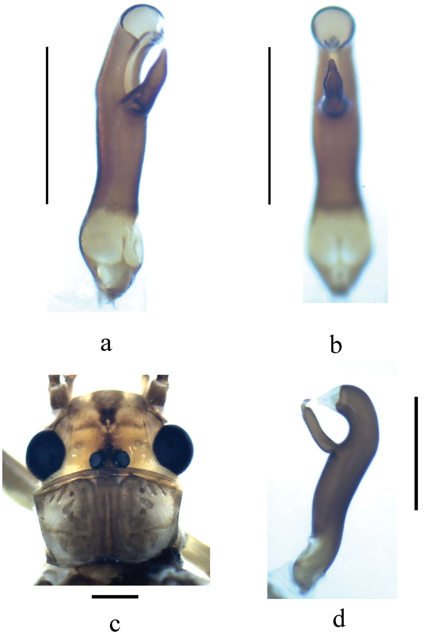
Figure 2.A–C Neoperla furcostyla Li and Qin,sp. n. (male). A Terminalia, dorsal view B Aedeagus, lateral view C Aedeagus of Neoperla forcipata Yang and Yang, lateral view.
Xue-Feng Qin, Dávid Murányi, Guo-Quan Wang, Wei-Hai Li
Zookeys
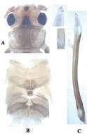
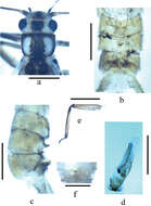
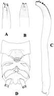
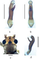
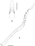
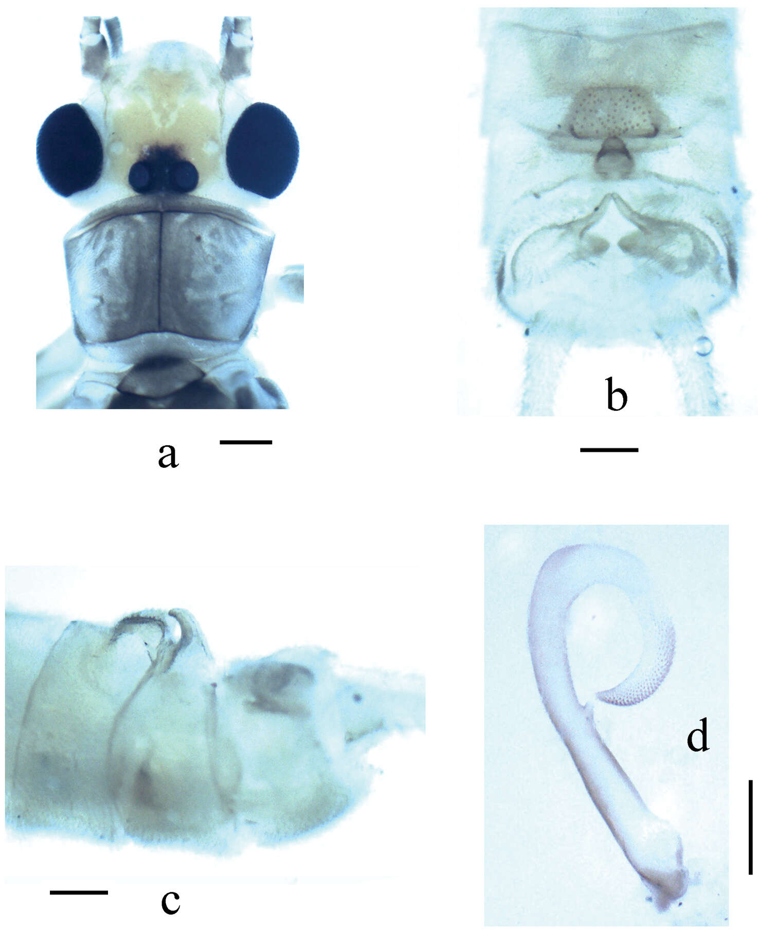
Figure 1.Neoperla brevistyla Li & Murányi, sp. n. Male a Head and pronotum, dorsal view b Hemitergal process, dorsal view c Aedeagus, lateral view. Scale bars: 0.5 mm.
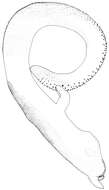
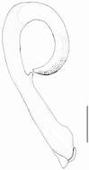
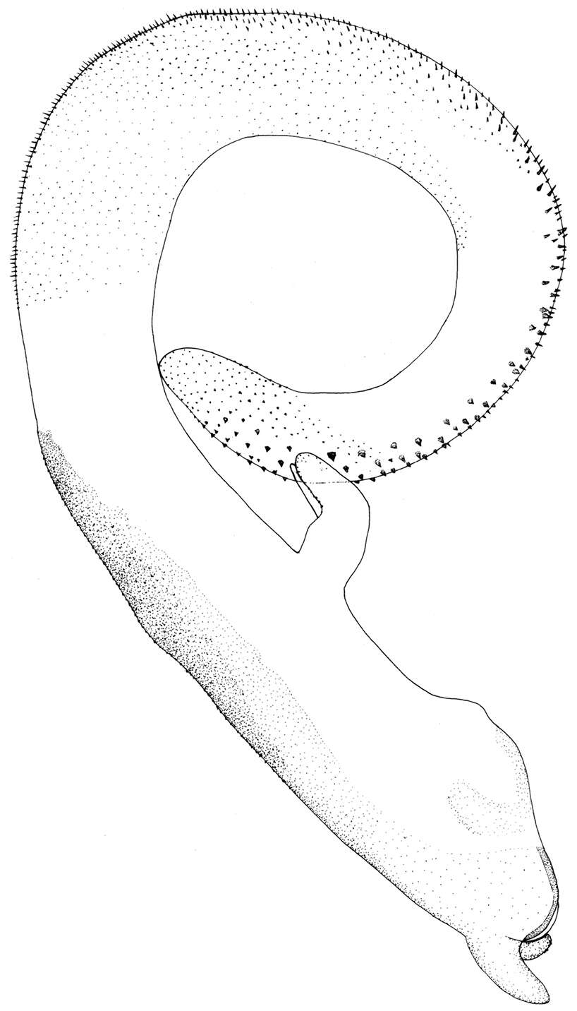
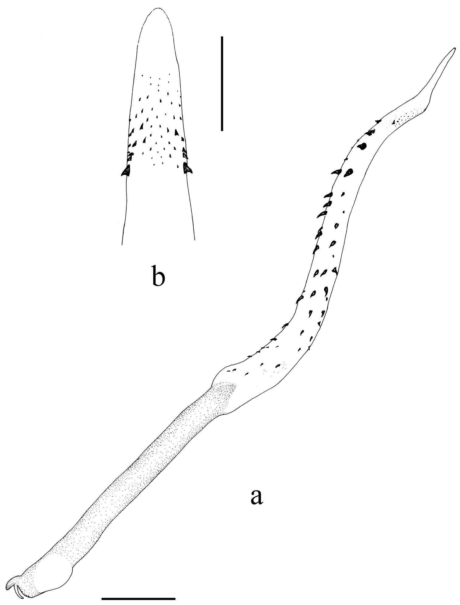
Figure 2.Neoperla nigromarginata Li & Zhang, sp. n. Male. a Dorsal aspect of aedeagal sac, top view b Aedeagus, lateral view. Note that the spines in b appear lightly pigmented and unclear, actually they are located on the lower surface of the sac, and are seen from beneath through the cuticle.
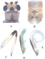
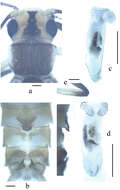
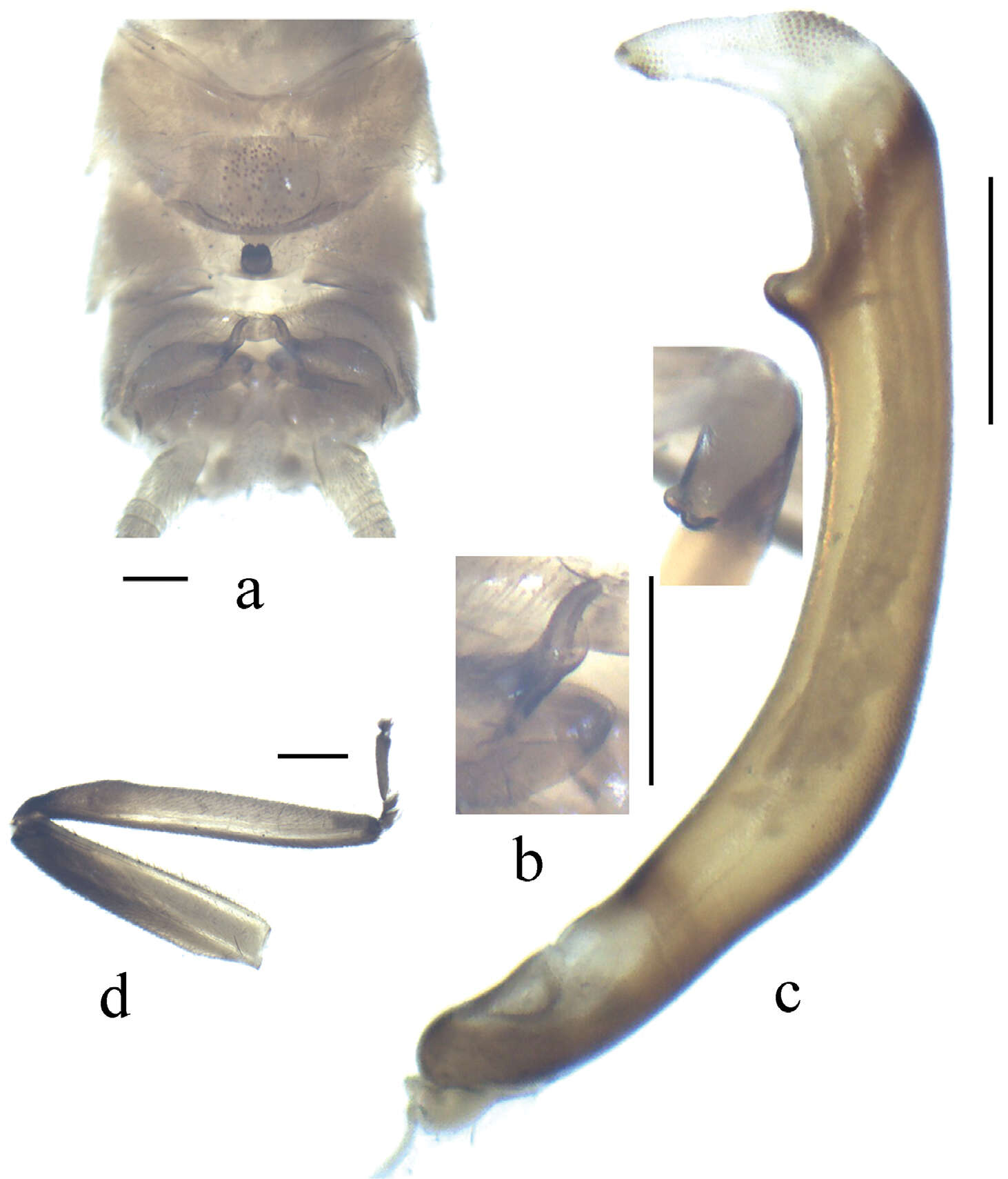
Figure 3.Neoperla similidella Li and Wang, sp. n. (male). A Head and pronotum, dorsal view B Terminalia, dorsal view C Aedeagus before eversion, lateral view D Aedeagus, lateral view E Aedeagal sac, dorsal view.
Xue-Feng Qin, Dávid Murányi, Guo-Quan Wang, Wei-Hai Li
Zookeys
Figure 2.Neoperla brevistyla Li & Murányi,sp. n. Male a Terminalia, dorsal view b Hemitergal process, dorsal view c Aedeagus, lateral view d Foreleg, lateral view. Scale bars: 0.5 mm.
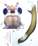
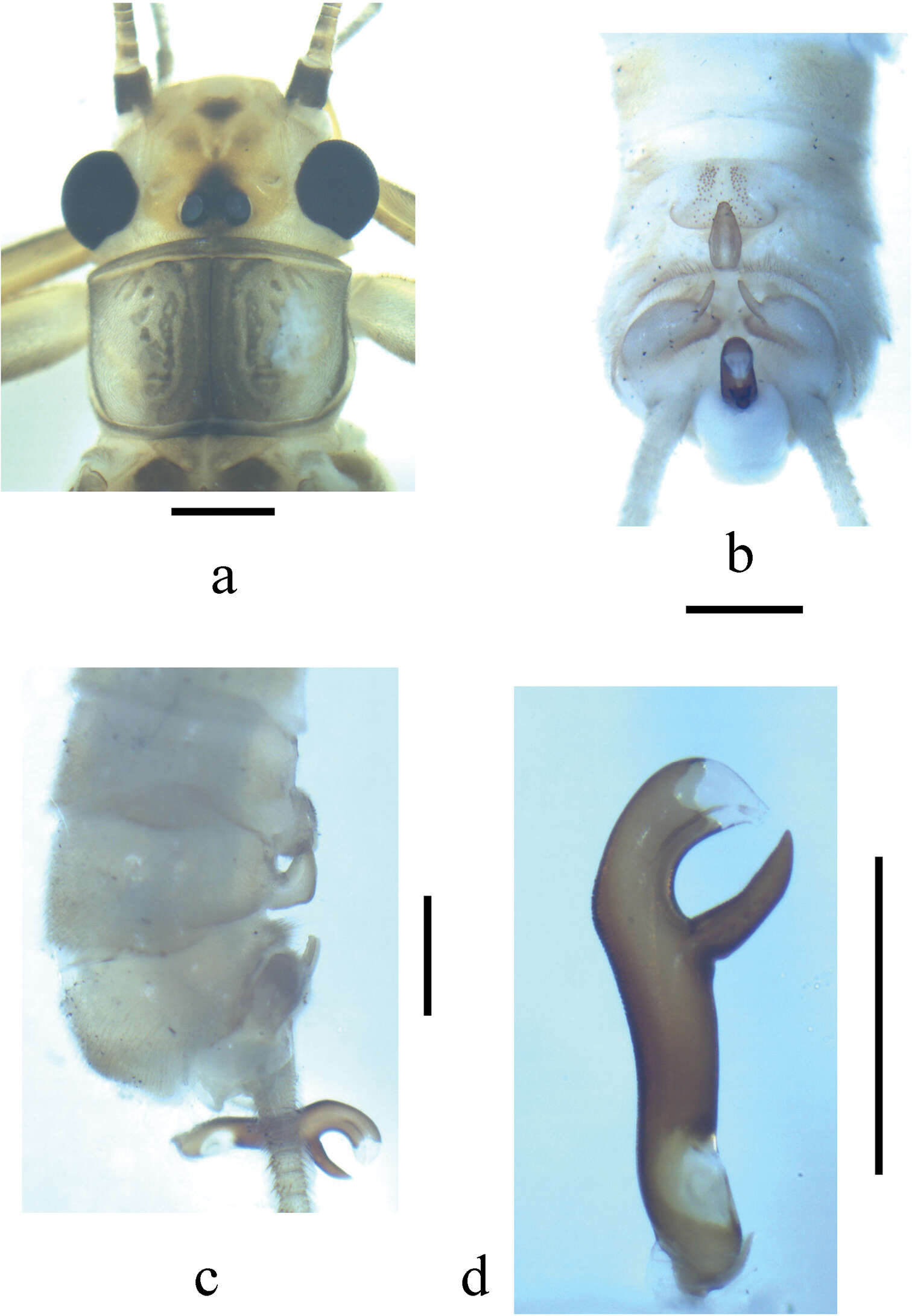
Figure 3.Neoperla mesospina Li and Wang, sp. n. Male. a Head and pronotum, dorsal view b Terminalia, dorsal view c Terminalia, lateral view d Aedeagus, lateral view.
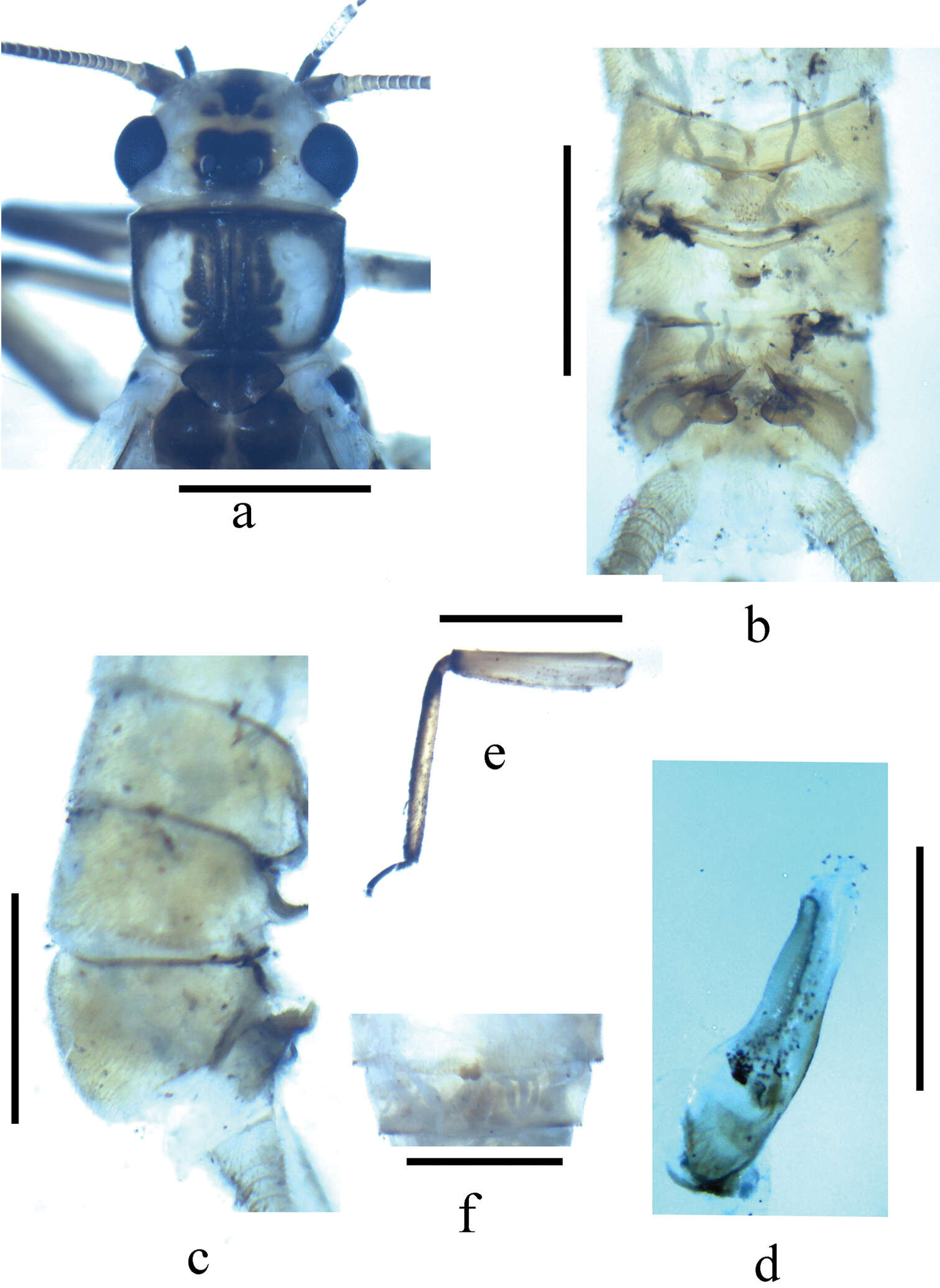
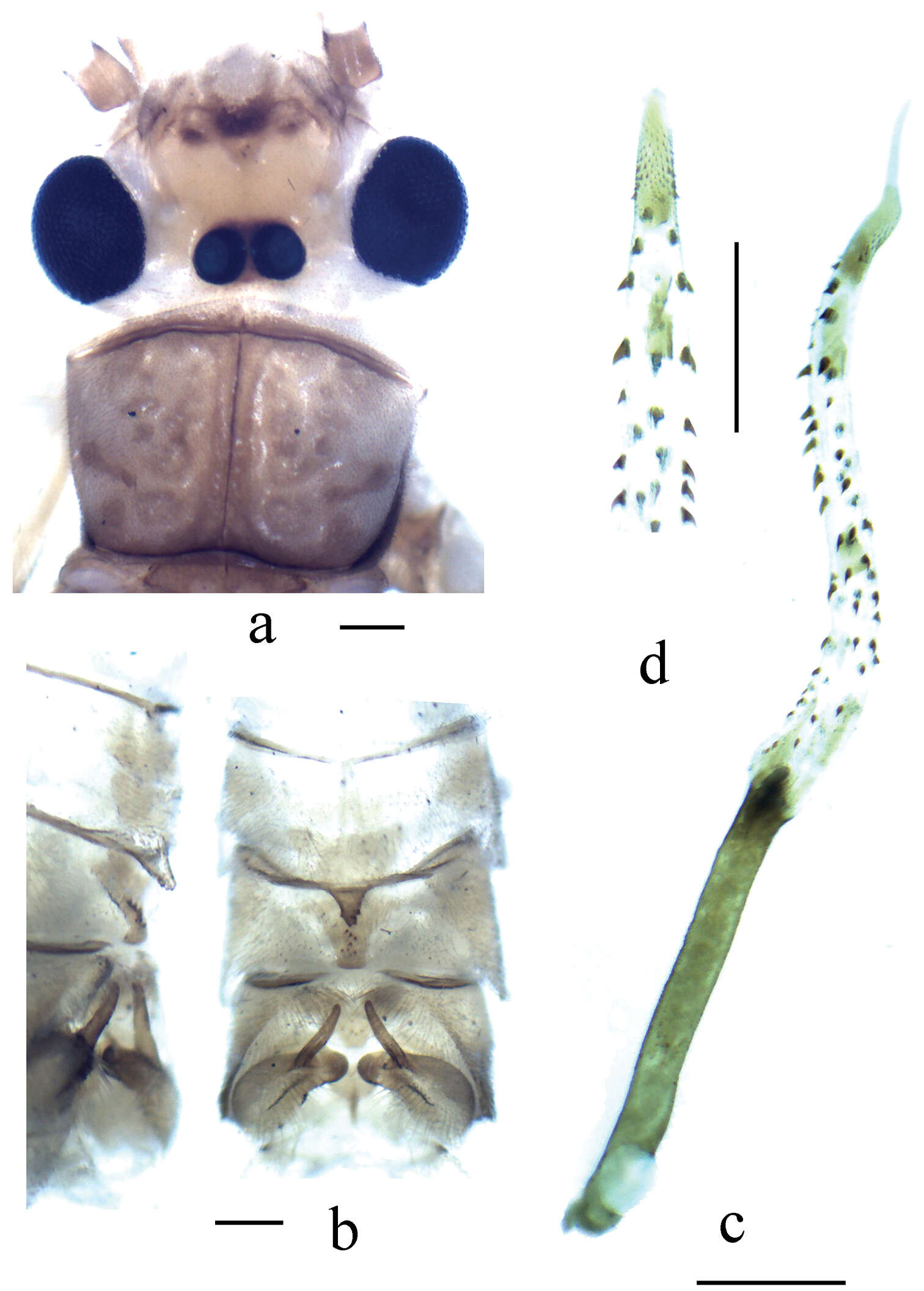
Figure 3.Neoperla similiflavescens Li & Zhang, sp. n. Male. a Head and pronotum, dorsal view b Terminalia, dorsal view c Terminalia, lateral view d Aedeagus, lateral view.
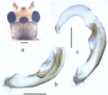
Figure 4.Neoperla similidella Li and Wang, sp. n. (male). A Aedeagal sac, dorsal view B Aedeagal sac, ventral view C Aedeagus, lateral view D Terminalia, dorsal view.
Figure 4.a–b Neoperla similiflavescens Li & Zhang, sp. n. Male. c–d Neoperla flavescens Chu. Male. a Aedeagus, oblique ventral view b Aedeagus, ventral view c Head and pronotum, dorsal view d Aedeagus, lateral view. Neoperla flavescens Chu for comparison. 1 male from Henan Province, Luoyang City, Song County, Cecun town, Muzhaling, 2012.VIII.19, Weihai Li.
Figure 5.Neoperla transversprojecta Du & Sivec, 2004. Male. a Head and pronotum (teneral specimen), dorsal view b Head and pronotum (older specimen), dorsal view c Terminalia, dorsal view d Aedeagus, lateral view.
Xue-Feng Qin, Dávid Murányi, Guo-Quan Wang, Wei-Hai Li
Zookeys
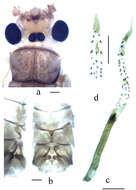
Figure 4.Neoperla lii. Male a Head and pronotum, dorsal view b Terminalia, dorsal view c Aedeagus, lateral view d Distal half of aedeagal sac, ventral view. Scale bars: 0.5 mm.
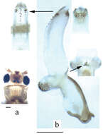
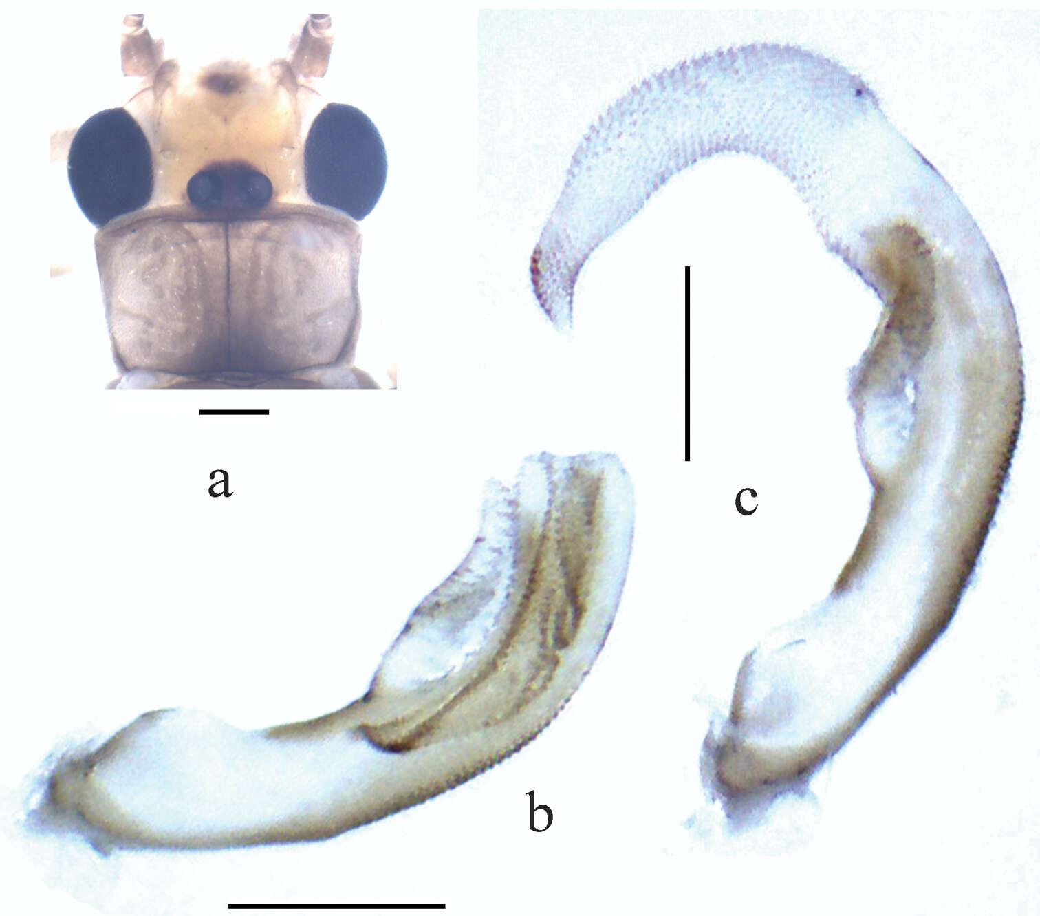
Figure 6.Neoperla yao Stark, 1987. Male. a Head and pronotum (teneral specimen), dorsal view b Aedeagus before eversion, lateral view c Aedeagus, lateral view.