“Psalidaster mordax sp.nov.
(Fig. K, 2-2g, 3-3 b; Plate XVIII, figs. 1, 2)
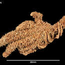
DIAGNOSIS. Rays 11; R 135 mm., r 27 mm., br 15-16 mm. Paratype R 110 mm., r 23 mm., br 12 mm. Rays flexible, slender, with evenly convex pliant abactinal surface uniformly beset with short soft papillae representing small spinelets, their associated crossed pedicellariae and smaller interspersed papulae; abactinal skin soft, not thick, not thrown into heavy welts, although finely wrinkled; marginal spines larger than abactinal, in two regular longiseries, carrying rosettes of pedicellariae; no actinal plates; adambulacral spines 2 (varying to 1 or 3), long, slender, terete, in a transverse series; actinostome small, mostly occluded by the long teeth (a pair to each mouth angle); furrows not wide; tube-feet slender, 4 ranked.
DESCRIPTION. The convex abactinal wall is beset with widely spaced slender acicular thorny spinelets (0.9-2 mm. long), the retracted sheath of which forms a soft collar at the base and carries 1 or 2 to 5 or 6 characteristic crossed pedicellariae. The integument is soft and until dried hides the skeleton. Between the spines are abundant papulae, singly or in two's and three's, and here and there a lanceolate straight pedicellaria. In alcoholic specimens the abactinal surface, under low magnification, has the appearance of being covered with low papillae—a confused mixture of papulae, skin-covered pedicellariae and spinelets. There is considerable difference between the two specimens in the number of pedicellariae. In the smaller, most of the abactinal spinelets of ray carry 1 or 2, and of the disk 2 or 3; whereas in the type the number is 5 or 6 for ray and 5–10 for disk. Madreporic body small (3 mm. diameter), situated at mid-r.
On the side of the ray are two longiseries of acicular spines conspicuously larger than the abactinal and surrounded by thick basal collars of crossed pedicellariae like the abactinal. The upper or superomarginal are 2-2.25 mm. long by 0.35 mm. just above base and carry 6-8 crossed pedicellariae, while the inferomarginal spines (sometimes 2 spines to a plate) are 2.9 mm. long by 0.5 mm. thick at base, and the fleshy sheath carries upward of 25 crossed pedicellariae. The spines correspond to about every fourth adambulacral plate and as the superomarginal plates are oriented obliquely to the inferomarginals the superomarginal spine is usually above the interval between 2 inferomarginal spines. There is a series of single inconspicuous intermarginal papulae which may represent encroachments of the abactinal system.
Between the inferomarginal and outer adambulacral spine is a narrow zone of bare skin devoid of papulae. The adambulacral spines are usually two in a transverse series on the inner half of plate; often there are 3, or only 1. They are long, slender (proximally 3.5 by 0.4 mm.), blunt, of nearly uniform diameter, and the proximal are a little longer than the inferomarginals. The furrow face of the adambulacral plates carries numerous small pedunculate straight pedicellariae of the common lanceolate form, 0.6–0.7 mm. in length, and 0.35 mm. broad at base.
The mouth-plates are small with a narrow actinostomial border, there being at each mouth angle two long "teeth" extending over the peristome; at outer end of each mouth pair are two spines intermediate in size between the teeth and the first adambulacrals. The narrow adoral carina, just back of mouth-plates, is composed of 5 or 6 pairs of juxtaposed adambulacrals which generally carry only one spine each; but sometimes a second smaller one is present on the fourth to sixth. The mouth-plates carry an abundance of small straight pedicellariae, exactly like the adambulacral.
Phylogenetically the abactinal skeleton is degenerating. It is composed of very numerous small plates forming a fantastically irregular reticulate structure, the irregularly spaced nodes of which are formed by 3- or 4-lobed primary plates, bearing single spinelets. The secondary plates are slender, overlapping, and the meshes are small, very unequal in size and irregular in shape. A meandering series of slightly larger carinal plates can be readily distinguished in a prepared specimen. Their spinelets are closer together than elsewhere because the carinal plates imbricate directly, or through one spineless plate between; whereas on the dorsolateral region 3 or 4 elongate ossicles may separate two primary plates. The form and imbrication of the marginals can best be appreciated from the figure (Fig. K, 3b). There is no trace of actinal plates. The above obtains on the proximal half of ray. Distally the mesh-work gradually becomes disconnected through the absence of intermediate ossicles until on the outer fourth of ray there are only spaced primary spiniferous plates. What really happens, of course, is that on the distal younger part of the ray the primary plates appear first, and only later, during further growth, the connecting ossicles develop. On the distal part of the ray the inferomarginals imbricate in a complete series. To the upper lobes of each is imbricated an oblique superomarginal. These are separated one from another by a conspicuous interval of membrane, encroached upon sometimes by a small disconnected abactinal plate. Even on the proximal half of ray the superomarginal plates are sometimes disconnected; or they are joined directly by overlapping lobes or by intermediate plates belonging to the abactinal system.
The crossed pedicellariae are diagnostic and recall those characteristic of Notasterias. The most frequent size varies between 0.7 and 0.93 mm. in length, but almost as numerous are various smaller sizes down to 0.6 mm., and a few as small as 0.5 mm. Each jaw is wide at the base and tapers to a narrow tip having 2 prominent teeth between which are usually several smaller ones. On the vertical inner edge of each jaw the denticles are numerous but very variable in details. At the attached end, the pedicellariae are wide and each jaw has a relatively large cavity for the accommodation of powerful "biting" muscles, as in the case of Notasterias.
The gonoducts open ventrally by prominent pores in the very constricted actinal interradial area. The pores are not symmetrically placed side by side but one is nearer margin than the other. Those of the female (smaller specimen) are larger than in the male (type). The small ovaries are attached lower on the side of the interbrachial septum (coalesced sides of rays) than the voluminous testes. The interbrachial septum contains calcareous plates. The tube-feet are crowded into 4 ranks and their ampullae are single, pyriform, voluminous.
TYPE LOCALITY. St. WS 821. Falkland Islands, 52° 55¾' S, 60° 55' W, 351-367 m., 2 specimens.”
(Fisher, 1940: 229-231)