-
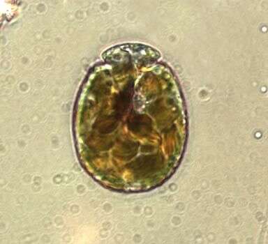
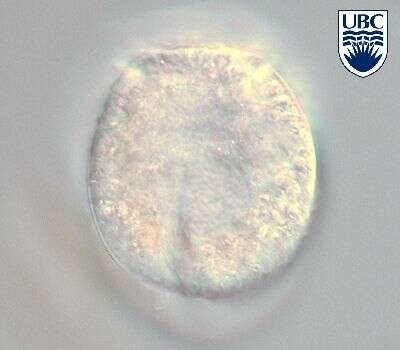
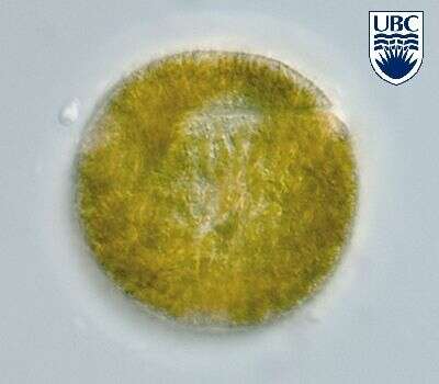
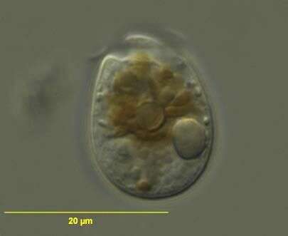
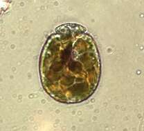
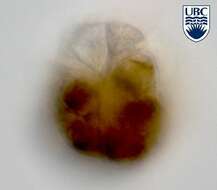
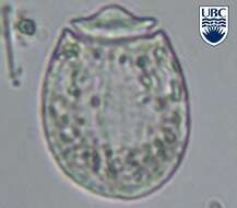
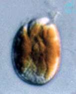
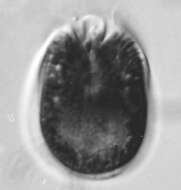
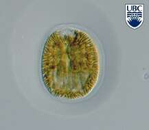
Amphidinium (am-fee-din-ee-um). The image shows a so far undescribed species, with many yellow-brown chloroplasts and a nucleus in the lower cell half. The cell is in an immobile stage, with an hyaline covering.
-
Amphidinium observed in marine muds and sandy sediments in the vicinity of Broome, Western Australia in September 2003. This work was supported by the Australian Biological Resources Study.
-
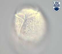
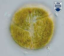
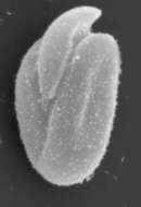
Amphidinium spec. 1.
-
Amphidinium spec. 1
-
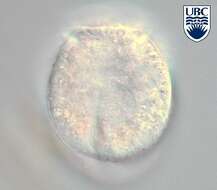
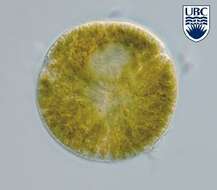
Amphidinium spec. 3.
-
Amphidinium spec. 3.
-
Amphidinium spec. 3.
-
Amphidinium spec. 3.
-
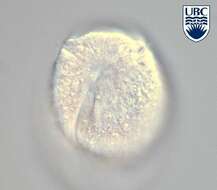
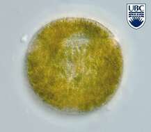
Amphidinium spec. 5.
-
Amphidinium spec. 5
-
Amphidinium spec. 5
-
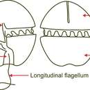
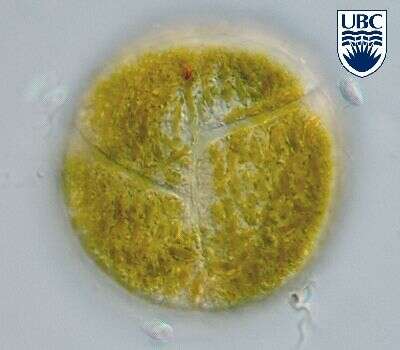
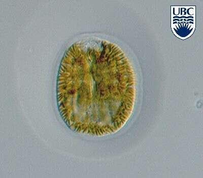
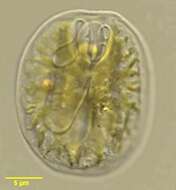
The genus Amphidinium (am-fee-din-ee-um) is a commonly encountered and speciose member of the dinoflagellates. There is a circumferential groove on the cell surface that runs around the anterior half of the cell, and a second one running along the length of the body. There is a flagellum in each groove. The asymmetrical location of the horizontal groove gives this genus its name (cf. Gymnodinium). Swimming species, with plastid and red inclusion. Differential interference contrast.
-
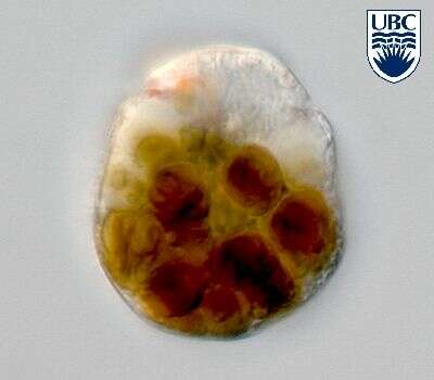
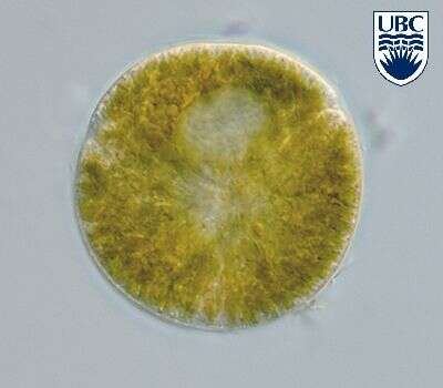
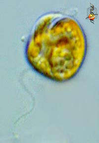
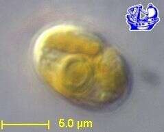
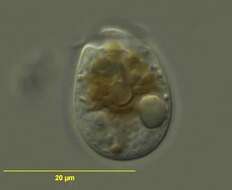
Amphidinium (am-fee-din-ee-um) carterae Hulburt 1957. The image shows a cell in ventral view with one yellow-brown chloroplast with multiple lobes. The epicone is small and turned to the left.
-
Cells oval from the ventral side, dorso-ventrally flattened. Length 11 - 17 microns, width 9 - 13 microns, breadth (lateral) approximately 6 microns, length to width ratio 1.2 - 1.6. Epicone crescent-shaped from the ventral side, clearly deflected towards the left. Cingulum beginning 0.3 - 0.4 of the cell length from the apex, midway across the ventral face, rising initially, then descending on the ventral side, distal end 0.5 of the cell length from the apex, 1 - 2 microns from the right margin of the ventral face. Sulcus beginning 1 - 2 microns below the proximal end of the cingulum, continuing to posterior. Nucleus in the posterior part of the hypocone, rounded. Chloroplast greenish-yellow, probably single with multiple lobes. Division is by binary fission in the motile cell. Central pyrenoid, approximately 3 microns diameter. A clonal culture has been made of this species.
-
Amphidinium carterae observed in marine muds and sandy sediments in the vicinity of Broome, Western Australia in September 2003. This image was taken using differential interference contrast optics. This work was supported by the Australian Biological Resources Study.
-
Amphidinium carterae observed in marine muds and sandy sediments in the vicinity of Broome, Western Australia in September 2003. This image was taken using differential interference contrast optics. This work was supported by the Australian Biological Resources Study.
-
Roscoff Culture Collection (strain number: 88 -strain name: CCMP1314).
-
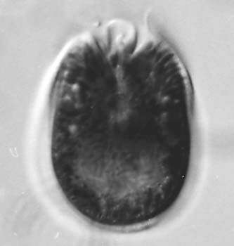
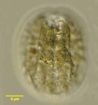
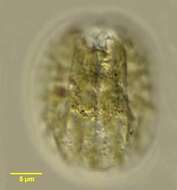
Cells oval from the ventral side, dorso-ventrally flattened. Length 26 - 34 microns, width 21 - 30 microns, length to width ratio 1.1 - 1.4. Cells ventrally flat, dorsally ridged into a series of approximately 7 - 8 'ribs' . Epicone small, club shaped, with a groove that begins just above the start of the cingulum and appears to widen into a depression near the apex. Hypocone forms a 'collar' around the epicone on the dorsal side. Cingulum incompletely encircling the epicone, distal end approximately 4 microns higher than proximal. Sulcus shallow and barely visible. Large (2 - 3 microns) pusule present 0.4 - 0.5 of the cell length from the apex, around the mid-line. Longitudinal flagellum originating just below the pusule. Indented around the inside of the periphery of the ventral side, 1 - 2 microns from the rim. Nucleus in the posterior part of the hypocone, oval to crescent shaped, 14 - 11 x 10 - 7 microns. Chloroplasts narrow, yellow-brown, 2 - 4 x 1 - 2 microns, radiating from the centre. Non-motile cells more rounded, with the hypocone encircling the epicone.
-
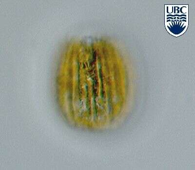
Amphidinium corrugatum observed in marine muds and sandy sediments in the vicinity of Broome, Western Australia in September 2003. This image was taken using differential interference contrast optics. This work was supported by the Australian Biological Resources Study.
-
Amphidinium corrugatum, from the dorsal side and showing the corrugations, observed in marine muds and sandy sediments in the vicinity of Broome, Western Australia in September 2003. This image was taken using differential interference contrast optics. This work was supported by the Australian Biological Resources Study.
-
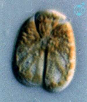
Amphidinium corrugatum Larsen et Patterson 1990.
-
Amphidinium corrugatum Larsen et Patterson 1990.
-
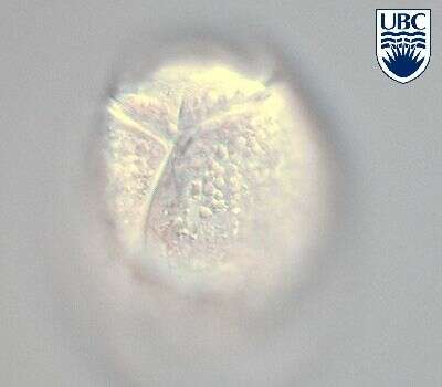
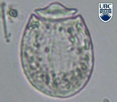
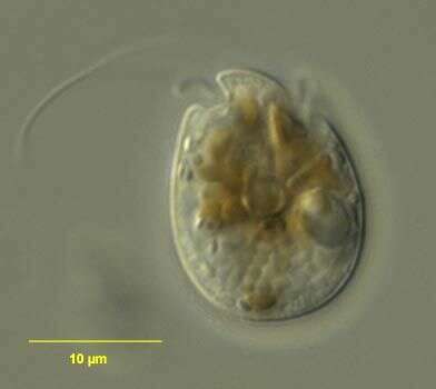
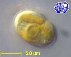
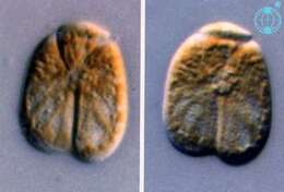
Amphidinium (am-fee-din-ee-um) herdmanii Kofoid & Swezy 1921. The image on the left shows a cell in ventral view. The epicone is small and turned to the left. The plastids are yellow-brown and radiating from the centre. The image on the right shows a mid-focus plane through a cell, showing the circular pyrenoid near the middle of the cell.
-
Amphidinium (am-fee-din-ee-um) herdmanii Kofoid & Swezy 1921. The image shows a cell in ventral view. The epicone is small and turned to the left. The plastids are yellow-brown and radiating from the centre. The hyaline area in the posterior of the cell is the nucleus.