-
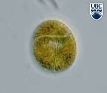
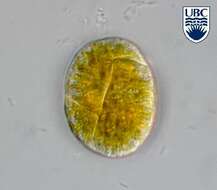
This is Amphidinium cf. glabrum in that it looks like but is not fully identical with the usual concept of this species.
-
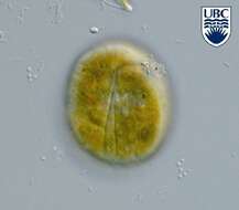
This is Amphidinium cf. glabrum in that it looks like but is not fully identical with the usual concept of this species.
-
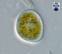
This is Amphidinium cf. glabrum in that it looks like but is not fully identical with the usual concept of this species.
-
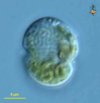
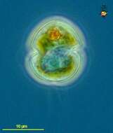
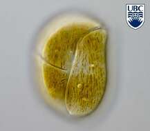
Karlodinium (car-low-din-ee-um) micrum (previously Gyrodinium galatheanum) has a equatorial flagellum lying in a groove (girdle or cingulum) near the centre of the cell and a second flagellum trailing behind the cell and arising in a longitudinal groove or sulcus. The large gray area posterior to the girdle is the nucleus. The orange element is probably a residue from ingested food (Rhodomonas). Phase contrast microscopy.
-
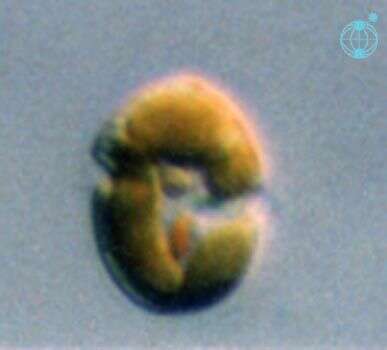
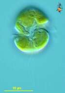
Karlodinium (car-low-din-ee-um) micrum (previously Gyrodinium galatheanum) has a equatorial flagellum lying in a groove (girdle or cingulum) near the centre of the cell and a second flagellum trailing behind the cell and arising in a longitudinal groove or sulcus. Differential interference microscopy. The grooves and plastids are emphasized in this image. Differential interference microscopy.
-
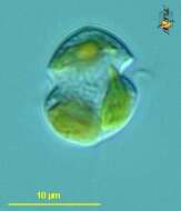
Karlodinium (car-low-din-ee-um) micrum (previously Gyrodinium galatheanum) has a equatorial flagellum lying in a groove (girdle or cingulum) near the centre of the cell and a second flagellum trailing behind the cell and arising in a longitudinal groove or sulcus. The granular region in the centre of the cell is the nucleus. Differential interference microscopy.
-
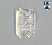
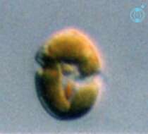
The epitheca of K. glaucum is both longer and wider than the hypotheca. The cingulum is displaced by about 4-5 times.
-
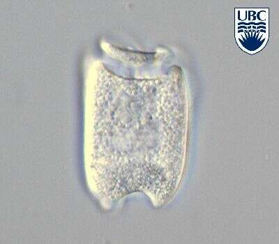
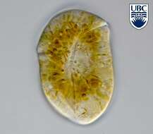
Togula britannica (Herdman) Flo Jorgensen, Murray et Daugbjerg 2004
-
Togula britannica (Herdman) Flo Jorgensen, Murray et Daugbjerg 2004
-
Togula britannica (Herdman) Flo Jorgensen, Murray et Daugbjerg 2004
-
Togula britannica (Herdman) Flo Jorgensen, Murray et Daugbjerg 2004
-
-
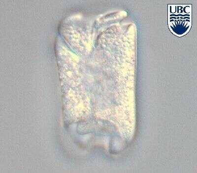
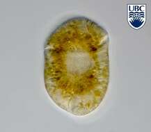
Togula jolla Flo Jorgensen, Murray et Daugbjerg 2004
-
Togula jolla Flo Jorgensen, Murray et Daugbjerg 2004
-
Togula jolla Flo Jorgensen, Murray et Daugbjerg 2004
-
Togula jolla Flo Jorgensen, Murray et Daugbjerg 2004
-
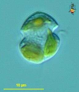
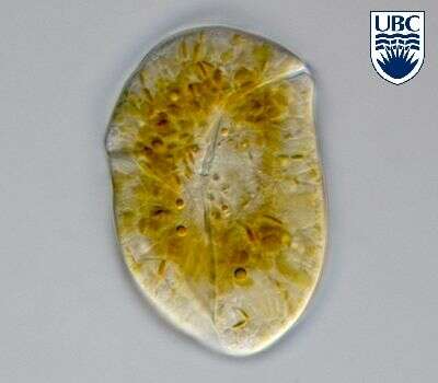
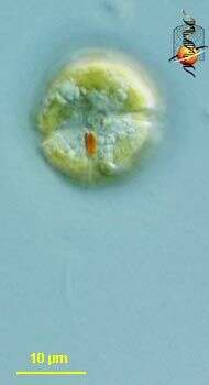
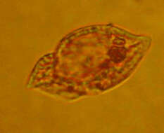
Gymnodinium (jim-no-din-ee-um), a so-called typical dinoflagellate. Most dinoflagellates have two flagella and they lie in grooves in the cell surface. The flagella are not evident here, but the grooves are. There is an circumferential groove (the girdle or cingulum) which wraps around the cell, and a longitudinal groove which extends from the point of flagellar insertion towards the back of the cell. This is an autotrophic dinoflagellate with numerous plastids with chlorophylls a and c. Also with an eyespot. Differential interference contrast.
-
Gymnodinium (jim-no-din-ee-um), a so-called typical dinoflagellate. Most dinoflagellates have two flagella and they lie in grooves in the cell surface. The flagella are not evident here, but the grooves are. There is an circumferential groove (the girdle or cingulum) which wraps around the cell, and a longitudinal groove which extends from the point of flagellar insertion towards the back of the cell. This is an autotrophic dinoflagellate with numerous plastids with chlorophylls a and c. The nucleus is the granular structure in the lower (hypocone) part of the cell. Differential interference contrast.
-
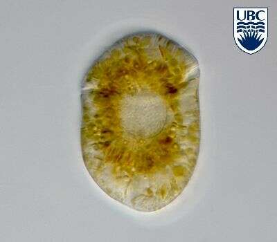
Gymnodinium (jim-no-din-ee-um), a so-called typical dinoflagellate. Most dinoflagellates have two flagella and they lie in grooves in the cell surface. The flagella are not evident here, but one groove -the circumferential groove - is. This is an autotrophic dinoflagellate with numerous plastids with chlorophylls a and c. The nucleus is the granular structure in the upper (epicone) part of the cell. Differential interference contrast.
-
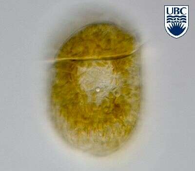
Gymnodinium (jim-no-din-ee-um), a so-called typical dinoflagellate. Most dinoflagellates have two flagella and they lie in grooves in the cell surface. The flagella are not evident here, but one groove -the circumferential groove - is., and the second (trailing) flagellum can be seen extending out the back of the cell. This is an autotrophic dinoflagellate with numerous plastids with chlorophylls a and c. Phase contrast.
-
-
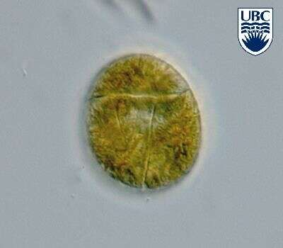
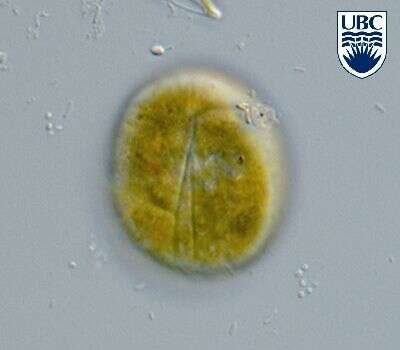
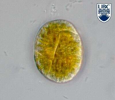
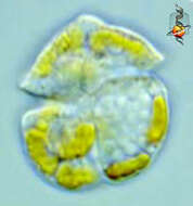
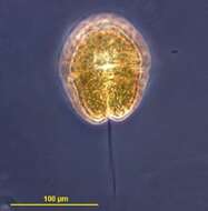
Gymnodinium (jim-no-din-ee-um) danicans Campbell 1973. The image shows a cell in ventral view. The red stigma is visible in the sulcal area. The plastids are yellow-brown and multiple.
-
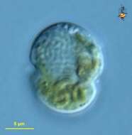
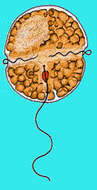
Image showing the off green colour of the chloroplasts of this dinoflagellate, the equatorial groove and the longitudinal (trailing) flagellum.
-
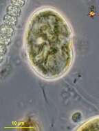
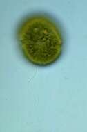
Dinoflagelate with chloroplasts. There are two flagella, one in the groove that runs around the middle of the body and the second lies in the longitudinal groove and extends behind the swimming cell.