-
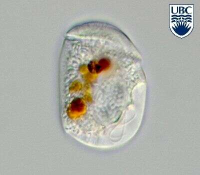
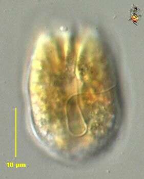
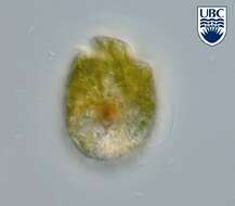
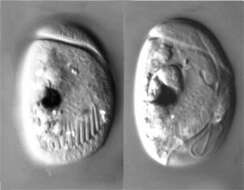
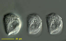
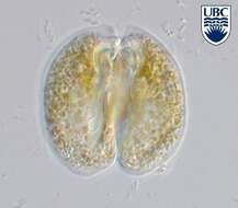
Amphidinium operculatum, showing its elongated plastids, observed in marine muds and sandy sediments in the vicinity of Broome, Western Australia in September 2003. This image was taken using differential interference contrast optics. This work was supported by the Australian Biological Resources Study.
-
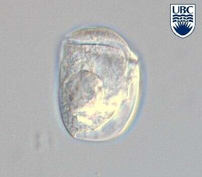
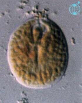
Amphidinium operculatum, observed in marine muds and sandy sediments in the vicinity of Broome, Western Australia in September 2003. This image was taken using differential interference contrast optics. This work was supported by the Australian Biological Resources Study.
-
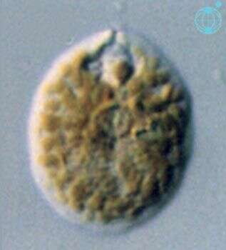
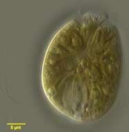
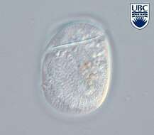
This is Amphidinium cf. operculatum in that it looks like but is not fully identical with the usual concept of this species.
-
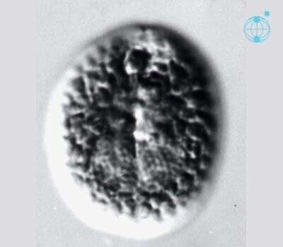
This is Amphidinium cf. operculatum in that it looks like but is not fully identical with the usual concept of this species.
-
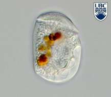
Amphidinium operculatum Claparede et Lachmann 1858.
-
Amphidinium operculatum Claparede et Lachmann 1858.
-
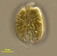
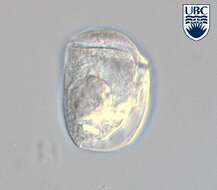
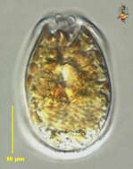
Amphidinium salinum observed in marine muds and sandy sediments in the vicinity of Broome, Western Australia in September 2003. This image was taken using phase contrast optics. This work was supported by the Australian Biological Resources Study.
-
Amphidinium salinum, from the dorsal side and showing its multiple extrusomes, observed in marine muds and sandy sediments in the vicinity of Broome, Western Australia in September 2003. This image was taken using differential interference contrast optics. This work was supported by the Australian Biological Resources Study.
-
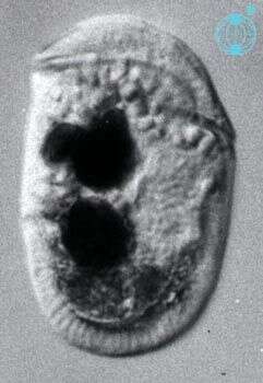
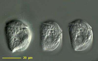
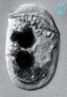
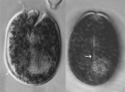
Amphidinium (am-fee-din-ee-um) semilunatum Herdman 1923. The images show cells in left lateral view. The cells contain no plastids. The cingulum is near the anterior end of the cell. The cell on the left is in a mid-focal plane, with food particles visible.
-
Amphidinium (am-fee-din-ee-um) semilunatum Herdman 1923. The image shows a cell in a mid-focal plane, with food particles visible. The cells contain no plastids. The cingulum is near the anterior end of the cell.
-
Amphidinium (am-fee-din-ee-um) semilunatum Herdman 1923. The image shows a cell in right lateral view. The nucleus is near the ventral side of the cell (right image side). The cell contains no plastids, but some food particles are visible.
-

Cells elliptical to oblong from the ventral side, dorso-ventrally flattened. Length 38 - 55 microns, width 16 - 24 microns, breadth (lateral) approximately 12 microns, length to width ratio 2.1 - 2.9. Epicone semi-circular, sloping towards the right. Cingulum relatively deep, initially rising on the ventral side, then descending with the right end tapering 2 - 3 microns from the sulcus. Proximal and distal ends of the cingulum displaced 10 - 12 microns. Transverse flagellum arising in a curved pocket approximately 10 microns long, leading from the proximal end of the cingulum. Sulcus a wedge-shaped indentation, approximately 0.75 of the cell length from the apex. Long narrow groove extending length of the cell from the proximal end of the cingulum to the sulcus. Apical groove originating at the proximal end of the cingulum, continuing in an anticlockwise spiral around the apex. Longitudinal flagellum arising in a pocket 0.3 - 0.4 of the cell length from the apex . Longitudinal flagellar pocket narrow, approximately 1 microns, following a slightly curved path to the sulcal indentation. Nucleus oval, 10 - 12 x 20 - 25 microns, in the central-right side of the hypocone. Chloroplasts absent. Small oil droplets and food particles often present. Fine longitudinal surface striations present, approximately 16 - 18 across the ventral side.
-
Amphidinium semilunatum Herdman 1923.
-
Amphidinium semilunatum Herdman 1923.
-
Amphidinium semilunatum Herdman 1923.
-
These are different focal planes of the gymnodinioid dinoflagellate, using DIC microscopy. Isolated by M. Virginia Sanchez Puerta from Little Sippewisset Pond, Woods Hole, MA, USA.
-
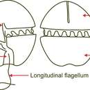
The genus Amphidinium (am-fee-din-ee-um) testudo is a commonly encountered and speciose member of the dinoflagellates. There is a circumferential groove on the cell surface that runs around the anterior half of the cell, and a second one running along the length of the body. There is a flagellum in each groove. The asymmetrical location of the horizontal groove gives this genus its name (cf. Gymnodinium). This is an atypical member of the genus, encountered adhering tightly to the substrate. The recurrent flagellum usually located under the cell. There is a second image of this cell showing the recurrent flagellum. The anterior flagellum is located around the very reduced epicone, or anterior part of the body. Differential interference contrast.
-
Amphidinium (am-fee-din-ee-um) testudo is a commonly encountered and speciose member of the dinoflagellates. There is a circumferential groove on the cell surface that runs around the anterior half of the cell, and a second one running along the length of the body. There is a flagellum in each groove. This photograph is of the ventral side and shows the recurrent flagellum. Differential interference contrast.
-
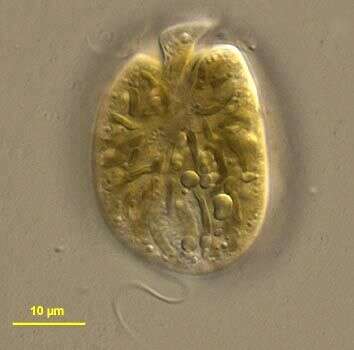
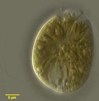
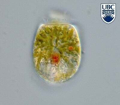
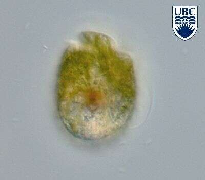
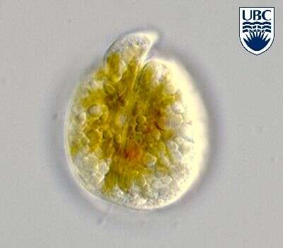
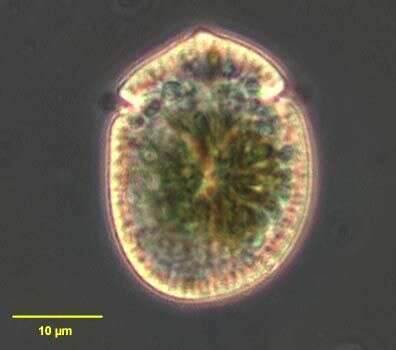
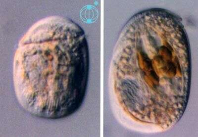
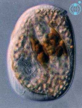
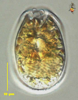
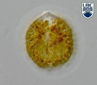
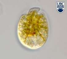
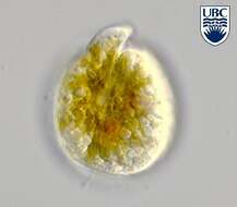
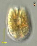
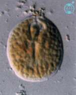
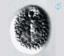
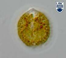
Amphidinium (am-fee-din-ee-um) testudo Herdman 1924. The image shows a cell in ventral view. The epicone is small, and does not protrude above the hypocone. The plastids are yellow-brown. This is a non-motile stage.
-
Amphidinium (am-fee-din-ee-um) testudo Herdman 1924. The image shows a cell in ventral view. The epicone is small, and does not protrude above the hypocone. The plastids are yellow-brown. The round pyrenoid is visible in the centre of the cell.
-
Amphidinium (am-fee-din-ee-um) testudo Herdman 1924. The image shows a cell in ventral view. The epicone is small, and does not protrude above the hypocone. The plastids are yellow-brown. This is a non-motile stage.
-
Cells rounded-oblong to pear shaped, dorso-ventrally flattened. Length 24 - 36 microns, width 19 - 26 microns, breadth (lateral) approximately 12 microns, length to width ratio 1.1 - 1.7. Epicone 2 - 4 microns wide, club-shaped, with two lobes, often slightly deflected to the left. Cingulum beginning 0.1 - 0.3 of the cell length from the apex, relatively wide (1 - 3 microns), incompletely encircling the epicone, distal end approximately 2 - 3 microns higher than proximal. Longitudinal groove (0.5 - 2 microns wide) present on the dorsal side, beginning at the apex and descending through the middle of the cell to 0.6 - 0.9 of the cell length from the apex. Hypocone forms a 'collar' around the epicone in dorsal view, this has a division in the middle due to the dorsal longitudinal groove. Sulcus indistinct. Longitudinal flagellum originating 2 - 3 microns below the proximal end of the cingulum. Nucleus in the posterior part of the hypocone, rounded to crescent shaped, may be to the left of the cell, 9 - 11 x 5 - 10 microns. Chloroplasts yellow-brown, 1 - 2 x 1 - 4 microns, often more concentrated in the centre of the cell. A central, pyrenoid-like structure sometimes observed. Indented around the inside of the periphery of the ventral side, 1 - 2 microns from the cell rim. Non-motile cells more circular, with the hypocone encircling the epicone, surrounded by a hyaline layer. Non-motile cells more commonly observed than motile cells.
-
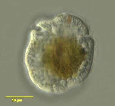
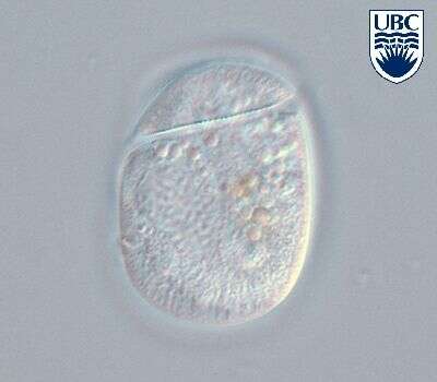
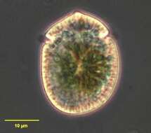
Amphidinium testudo Herdman 1924. Just dividing cell.
-
Amphidinium testudo Herdman 1924.