-
This image was taken using differential interference contrast optics. This work was supported by the Australian Biological Resources Study.
-
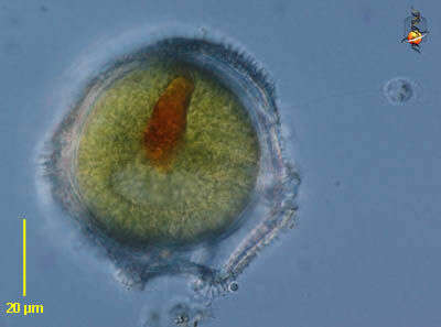
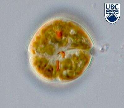
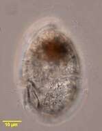
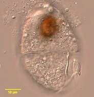
Durinskia baltica, showing its stigma, observed in marine muds and sandy sediments in the vicinity of Broome, Western Australia in September 2003. This image was taken using differential interference contrast optics. This work was supported by the Australian Biological Resources Study.
-
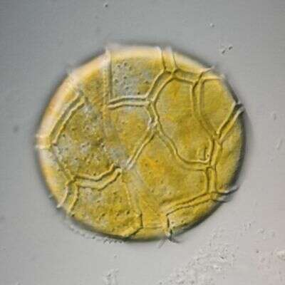
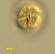
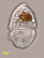
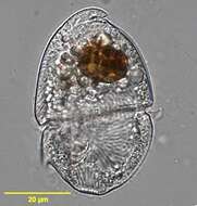
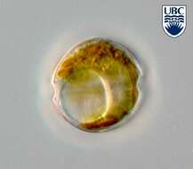
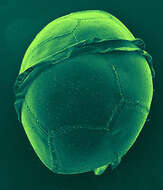
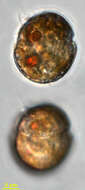
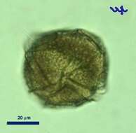
Peridinium (perry-din-ee-um) a dinoflagellate. This is one of the armoured dinoflagellate in which there are substantial skeletal elements in the cortical region of the cell. The groove which contains the circumferential flagellum has strongly developed margins. With chloroplasts. Encysted form - red inclusion resembles an eyespot but in this case we are advised this is more likely residues of food. Differential interference contrast.
-
This image was taken using differential interference contrast optics. This work was supported by the Australian Biological Resources Study.
-
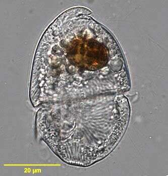
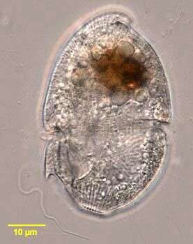
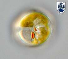
Durinskia baltica (Levander 1892) Carty et Cox 1986. This phototrophic species is also known from the plankton. Cell in ventral view, note the bright red stigma in the sulcal area.
-
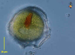
Peridinium (perry-din-ee-um) a dinoflagellate. This is one of the armoured dinoflagellate in which there are substantial skeletal elements in the cortical region of the cell. The groove which contains the circumferential flagellum has strongly developed margins. With chloroplasts. Differential interference contrast.
-
This image was taken using differential interference contrast optics with a closed condenser iris. This work was supported by the Australian Biological Resources Study.
-
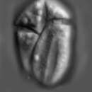
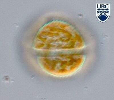
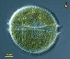
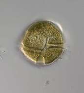
Durinskia baltica (Levander 1892) Carty et Cox 1986. Dorsal view showing the cingulum (transverse furrow) and golden-brown chloroplasts.
-
-
This image was taken using differential interference contrast optics. This work was supported by the Australian Biological Resources Study.
-
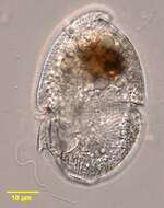
Durinskia baltica (Levander 1892) Carty et Cox 1986. Mid cell focus showing the large pusule.
-
Ventral view of a dinoflagellate of the genus Peridinium (Ehrenberg,1832). Collected from a freshwater pond near Boise,Idaho. DIC
-
This image was taken using differential interference contrast optics. This work was supported by the Australian Biological Resources Study.
-
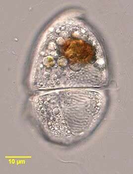
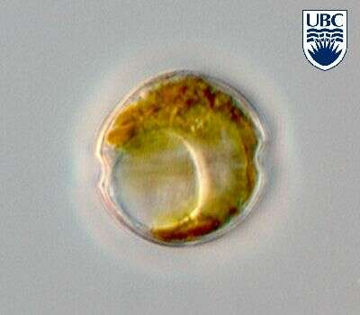
Durinskia baltica (Levander 1892) Carty et Cox 1986. Cell in ventral view. Visible are the bright red stigma (consisting of two parts) in the sulcal area, a red food body in the upper cell half, and golden-brown chloroplasts.
-
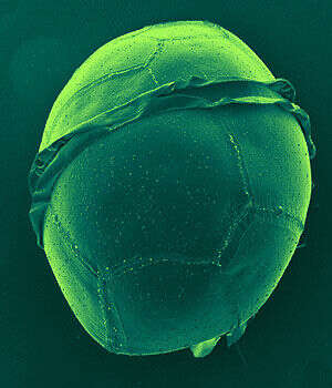
Dorsal view of the cell. The lists are the ridges on wirther side of the girdle.
-
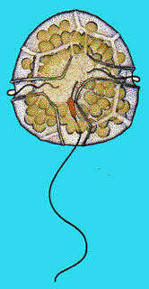
Durinskia_baltica. Collected by ATOL team at Oyster Pond near to Woods Hole, Massachusetts, during the Protistology Workshop at MBL. October-November 2005. Isolation and art by Adrian Reyes-Prieto.
-
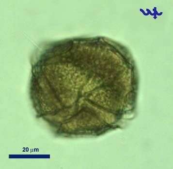
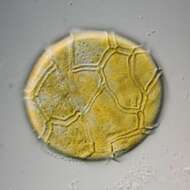
Peridinium penardii has skeletal elements (cellulose plates) in the cortical region of the cell. Species has chloroplasts. Collection from littoral region (stand of Phragmites) of a rain storage reservoir in Kiel (Schleswig-Holstein, Germany). This image was taken using Zeiss Universal with Olympus C7070 CCD camera.
-
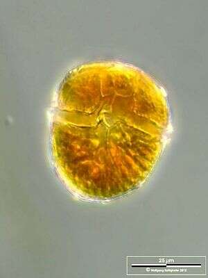
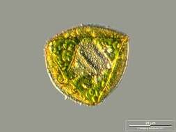
Amphidiniopsis korewalensis cells round to oval from the ventral side, dorso-ventrally flattened, 24 - 35 microns long, 20-30 microns wide, 15 - 23 microns broad. Thecal plates present: an apical pore, 4 apical plates, 3 anterior intercalary plates, 6 precingular plates, an x plate, 4 cingular plates, possibly 4 sulcal plates, 6 postcingular plates, 2 posterior plates. Epicone small and relatively flat, hypocone large and rounded. Average epicone/hypocone ratio 0.2-0.25. A small apical hook points to the left lateral cell side, over the round apical pore. Plates quite thin and boundaries not visible with the light microscope, covered in scattered small pores, approximately 0.2 microns diameter. Small protrusions often present on the posterior sulcal plate. Possesses no chloroplast or eyespot. Nucleus large (20-25 x 12-15 microns), elliptical and positioned centrally. Numerous colourless globules present
-
Scale bar indicates 25 µm. Sample from a wetland at the Pillersee (Tyrol, Austria). The image was built up using several photomicrographic frames with manual stacking technique. Images were taken using Zeiss Universal with Olympus C7070 CCD camera.Image under Creative Commons License V 3.0 (CC BY-NC-SA).
-
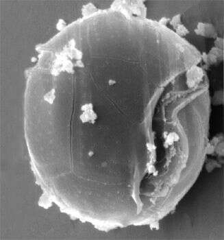
Cyst. Scale bar indicates 25 µm. Sample from a wetland at the Pillersee (Tyrol, Austria). The image was built up using several photomicrographic frames with manual stacking technique. Images were taken using Zeiss Universal with Olympus C7070 CCD camera.Image under Creative Commons License V 3.0 (CC BY-NC-SA).
-
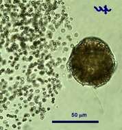
Peridinium gatunense is a large (ca. 50 um diameter) dinoflagellate, the most common bloom-forming species in Lake Kinneret, and the most studied species from this lake. Its winter-spring blooms give the water coffee-brown color. The blooms are patchy, the brown color patches can be observed from the distance. These blooms, reported to occur each spring since the 1950s, were characteristic of Lake Kinneret till the mid 1990s. In recent years Peridinium gatunense failed to bloom in some of the years, whereas in others its bloom was even more intense than recorded previously.
-
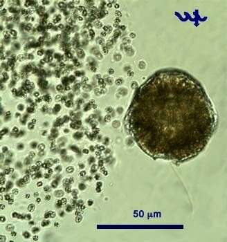
Peridinium gatunense, a large dinoflagellate (ca. 50 um diameter) is the main bloom-forming species in the plankton of Lake Kinneret. At the height of the bloom it forms patches in which cell densities are particularly high and the water gets coffee color. Peridinium has 2 unequal flagella, the longitudinal "whiplash" flagellum is seen in this picture, the second, transverse flagellum is hidden in the transverse groove or cingulum. A Microcystis colony is seen next to the Peridinium, note the large difference in cell size. This specimen was sampled in the littoral in June 2006.
-
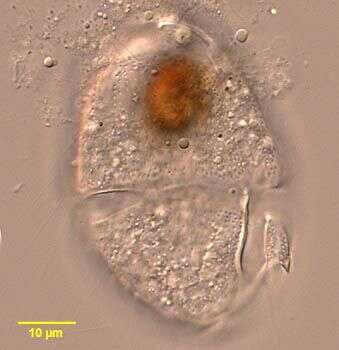
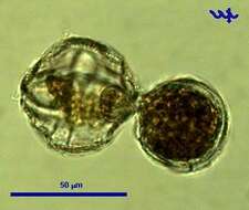
A protoplast of Peridinium is leaving its thecae. Usually this happens during adverse conditions, e.g. lack of nutrients or when being subjected to very strong light - or as part of cell division. In this case, there is no cell division.
-
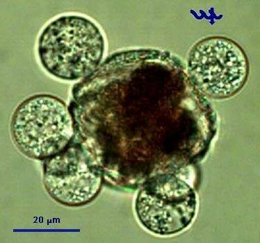
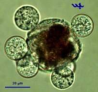
This cell of Peridinium gatunense is infected by several individuals of Phlyctochytrium sp, a chytrid fungus. Note that the Peridinium protoplasm is separated from the thecae and condensed, an indication that the cell is dead or dying. The fungal sporangia are at a developed stage with zoospores about to emerge out of in order to infect new Peridinium cells.