-
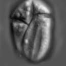
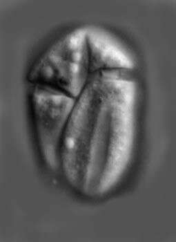
Amphidinium (am-fee-din-ee-um) pellucidum Herdman 1922. The image shows a cell in ventral view. The cell is colourless (contains no plastids). The cingulum is near the anterior end of the cell and is slightly descending. There is an apical groove present.
-
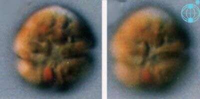
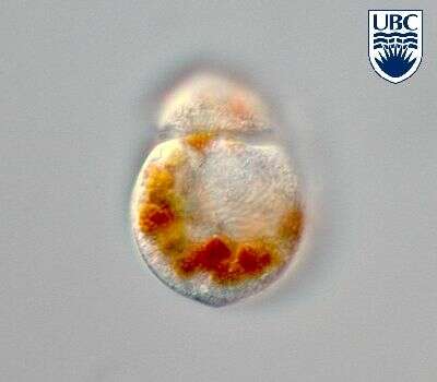
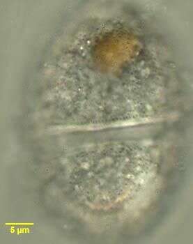
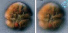
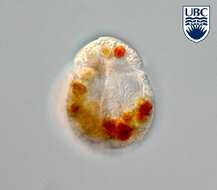
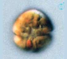
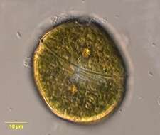
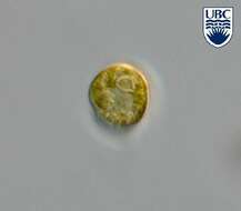
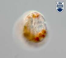
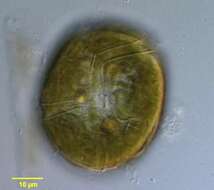
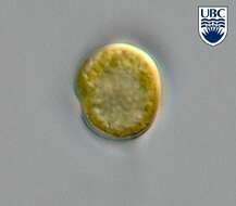
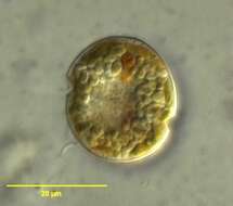
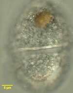
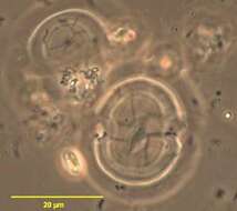
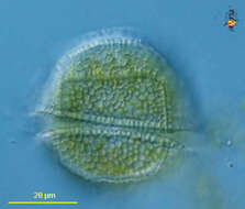
Durinskia (dew-rink-see-a) baltica (Levander 1894) Carty and Cox 1986. The images show cells in ventral view. The red stigma is visible in the sulcal area. The plastids are yellow-brown and multiple. The cingulum is in the middle of the cell.
-
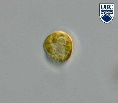
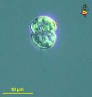
Pfiesteria (fist-ear-ee-a) small colourless dinoflagellate equatorial girdle in which one flagellum lies, another trailing. Probably misleading reports in the literature suggest that this organism has very polymorphic life cycles - held to be misleading because the case is based on non-pure cultures and supporting evidence includes images of taxa which clearly are related to other kinds of organisms. In certain areas within the US, this organism is held to be a major environmental threat because it is said to cause fish kills, but the less hysterical perspective is that the organism, with limited variation in form, may attack damaged tissue and eat it - hence the association with unhappy fish. Because of the issues of putative toxicity, correct identification needs expert input, which can be obtained from other sites such as
IOC Harmful Algal Bloom Program. Differential interference microscopy.
data on this strain.
-
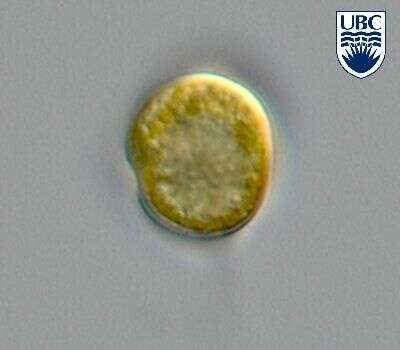
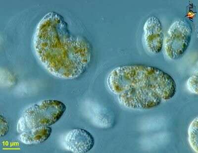
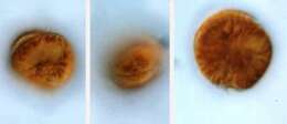
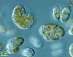
Coolia (coo-lee-a) monotis Meunier 1919. The images show swimming cells. The cells are flattened in the anterior-posterior plane, slightly asymmetrically. The cell on the left is in posterior-lateral view. The nucleus is visible. The cell in the middle is in ventral-lateral view. The cingulum is visible in the middle of the cell. The cell on the right is in posterior view. The cells contain yellow-brown plastids.
-
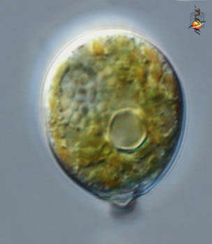
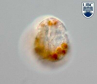
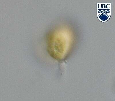
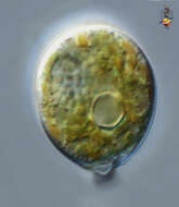
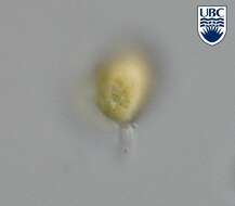
Stylodinium (style-owe-din-ee-um) is a rather unusual dinoflagellate, in which one phase of the life history is as a stalked and unflagellated cell - as here. Other forms include cysts, flagellates and heliozoan like organisms. The dinoflagellate nucleus with its condensed chromosomes is seen upper right. The circular structure is /are starch deposits around a pyrenoid. Probably related to Pfiesteria, a genus gaining (undue?) significance for its potential as a source of fish killer. Differential interference contrast.
-
Amphidinium pellucidum
-
Durinskia (dew-rink-see-a) baltica (Levander 1894) Carty and Cox 1986. The image shows a cell in ventral view. The stigma is visible in the sulcal area. The plastids are yellow-brown and multiple. The cingulum is in the middle of the cell.
-
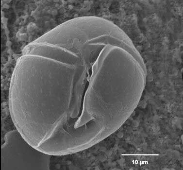
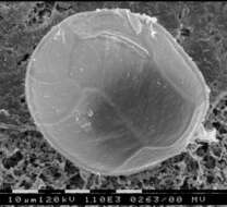
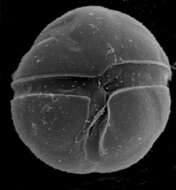
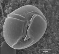
Ostreopsis lenticularis, scanning electron microscope image. This image was taken by Shauna Murray of a sample from Raine Island, northern Great Barrier Reef, Australia. This work was supported by the Australian Biological Resources Study.
-
Stylodinium littorale motile cells are oval from the dorsal side. Length 12 - 20 microns, width 9 15 microns. Non-motile cells are a similar shape, surrounded by a hyaline layer, attached to the substrate by a colourless stalk, approximately 15 microns long and 3 microns wide, from the apex. On the motile cell, cingulum begins approximately 0.5 of the cell length from the apex, in the centre of the cell, descending across the dorsal side, not continuing onto the ventral side. Cytoplasm full of small yellow-green plastids. Colourless globules also present. Pyrenoid (2 3 microns) present in the centre of the cell. Nucleus in the hypocone, 3 4 microns wide. Motile cell very fast swimming.
-
Amphidinium pellucidum
-
Durinskia (dew-rink-see-a) baltica (Levander 1894) Carty and Cox 1986. The image shows a cell in ventral view. The red stigma is visible in the sulcal area. The plastids are yellow-brown and multiple. The cingulum is in the middle of the cell.
-
Bysmatrum, from the dorsal side, observed in marine muds and sandy sediments in the vicinity of Broome, Western Australia in September 2003. This image was taken using differential interference contrast optics. This work was supported by the Australian Biological Resources Study.
-
Stylodinium littorale Horiguchi et Chihara 1983
-
Amphidinium pellucidum
-
Durinskia baltica cells are round from the ventral side, slightly dorso-ventrally flattened, 16 - 28 microns long, 15 - 27 microns wide, and approximately 19 microns broad. Thecal plates present: an apical pore, a canal plate, 4 apical plates, 2 anterior intercalary plates, 7 precinguluar plates, 5 cinguluar plates, 4 sulcal plates, 5 postcingular plates, 2 posterior plates. Second apical intercalary plate much larger than the first and extending across the dorsal side. Cingulum 2-3 microns wide, consists of 5 plates, including a transitional plate. The large anterior sulcal plate forming a list over the left sulcal plate. Thecal plates smooth, with scattered pores (about 0.2 microns). Pores form a row along the edge of the cingulum. Nucleus round, in the centre or in the left side of the cell, approximately 7 microns. Reddish stigma, approximately 4 microns long, present in the sulcus. Yellow- brown plastids, 2 - 3 microns diameter, scattered throughout the cell.
-
Bysmatrum, from the ventral side, observed in marine muds and sandy sediments in the vicinity of Broome, Western Australia in September 2003. This image was taken using differential interference contrast optics. This work was supported by the Australian Biological Resources Study.
-
Stylodinium littorale Horiguchi et Chihara 1983
-
DIfferential interference microscopy.
-
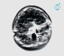
Durinskia baltica, observed in marine muds and sandy sediments in the vicinity of Broome, Western Australia in September 2003. This image was taken using differential interference contrast optics. This work was supported by the Australian Biological Resources Study.
-
Bysmatrum observed in marine muds and sandy sediments in the vicinity of Broome, Western Australia in September 2003. This image was taken using scanning electron microscopy. This work was supported by the Australian Biological Resources Study.
-
Stylodinium littorale Horiguchi et Chihara 1983
-
This image was taken using differential interference contrast optics. This work was supported by the Australian Biological Resources Study.
-
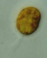
Durinskia baltica theca observed in marine muds and sandy sediments in the vicinity of Broome, Western Australia in September 2003. This image was taken using phase contrast optics. This work was supported by the Australian Biological Resources Study.
-
Peridinium (perry-din-ee-um) a dinoflagellate. This is one of the armoured dinoflagellate in which there are substantial skeletal elements in the cortical region of the cell. The groove which contains the circumferential flagellum has strongly developed margins. With chloroplasts. Differential interference contrast.