-
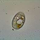
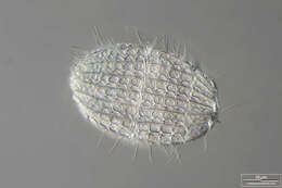
Coleps hirtus This ciliat belongs to prostomatid group. Coleps is one of nature´s little scavengers. It can eat residual tissue attached to cast off exoskeletons of arthropods, or will attack wounded organisms. Scale bar indicates 10 µm. Collection from a tropical freshwater aquarium. This image was taken using Zeiss Universal with Olympus C7070 CCD camera.Image under Creative Commons License V 3.0 (CC BY-NC-SA). Place name: Tropical freshwater aquarium Latitude: 54.3018013 Longitude: 10.07120132 Dieser Ciliat gehört zur Gruppe der Prostomatida. Coleps ist einer der kleinsten Aasfresser in der Natur. Er vertilgt z. B. Restgewebe toter Gliederfüßler, sie greifen auch verwundete Organismen an. Multiebenen-Abbildung, manuell gestapelt. Der Messbalken markiert eine Länge von 10 µm. Aus einem Süßwasseraquarium für tropische Fische in Kiel. Mikrotechnik: Zeiss Universal, Kamera: Olympus C7070. Creative Commons License V 3.0 (CC BY-NC-SA). For permission to use of (high-resolution) images please contact postmaster@protisten.de.
-
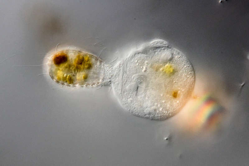
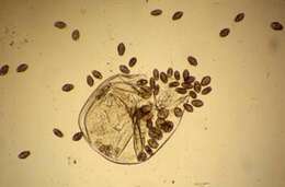
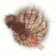
Coleps hirtus Coleps feeding on a living peritrich. The specimen was gathered in the pond Birkensee near Rödelsee (Lower Franconia, Germany). Sampling date 7/2018.Copyright Dr. Rainer Meisch, Würzburg, Germany.Images were taken using Zeiss Axioplan with Canon DSLR Image under Creative Commons License V 3.0 (CC BY-NC-SA). Place name: Pond Birkensee near Rödelsee (Lower Franconia, Germany) Latitude: 49.71819841 Longitude: 10.27807474 Coleps beim Angriff auf ein lebendes Glockentierchen. Probe aus dem Birkensee bei Rödelsee (Unterfranken). Datum der Aufsammlung: 7/2018. Copyright Dr. Rainer Meisch, Würzburg. Mikrotechnik: Zeiss Axioplan, Kamera: Canon DSLR. Creative Commons License V 3.0 (CC BY-NC-SA). For permission to use of (high-resolution) images please contact postmaster@protisten.de.
-
Coleps hirtus This ciliat belongs to prostomatid group. Coleps is one of nature´s little scavengers. It can eat residual tissue attached to cast off exoskeletons of arthropods, or will attack wounded organisms. Scale bar indicates 10 µm. Collection from a tropical freshwater aquarium. This image was taken using Zeiss Universal with Olympus C7070 CCD camera.Image under Creative Commons License V 3.0 (CC BY-NC-SA). Place name: Tropical freshwater aquarium Latitude: 54.3018013 Longitude: 10.07120132 Dieser Ciliat gehört zur Gruppe der Prostomatida. Coleps ist einer der kleinsten Aasfresser in der Natur. Er vertilgt z. B. Restgewebe toter Gliederfüßler, sie greifen auch verwundete Organismen an. Multiebenen-Abbildung, manuell gestapelt. Der Messbalken markiert eine Länge von 10 µm. Aus einem Süßwasseraquarium für tropische Fische in Kiel. Mikrotechnik: Zeiss Universal, Kamera: Olympus C7070. Creative Commons License V 3.0 (CC BY-NC-SA). For permission to use of (high-resolution) images please contact postmaster@protisten.de.
-
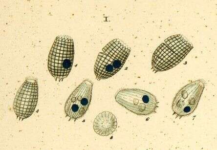
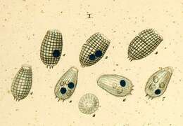
Body with regurarly arranged ectoplasmic plates. Cytostome at anterior end, surrounded by slightly longer cilia . Often spinous projection at or near posterior end.
-
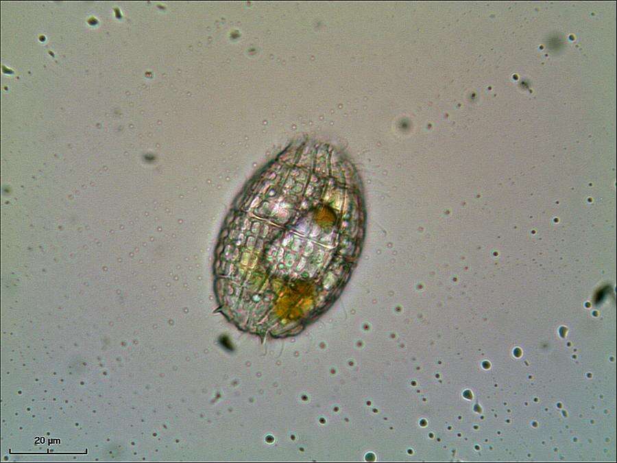
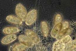
Coleps is one of nature's little scavengers. It will attach dead and decaying metazoa, eating residual pieces of tissue on cast off exoskeletons or attaching organisms that are wounded. It will also eat detrital material. This species retains chloroplasts from algal food - a phenomenon referred to as kleptoplasty. Chloroplasts are derived from blue-green algae that have survived symbiotically within other cells, and kleptoplasty exploits aspects of the autonomy of these organelles. Phase contrast microscopy.
-
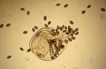
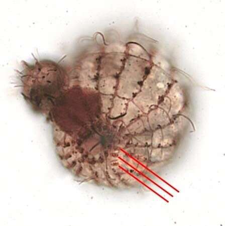
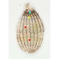
Coleps is a ciliate and one of nature's little scavengers. In nature it can eat remanent tissue attached to cast off exoskeletons of arthropods, or will attack wounded organisms. Here about 50 cells crowd round the remains of a Daphnia.
-
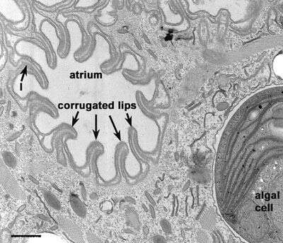
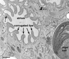
Opening to oral region at the anterior end of Coleps hirtus. Corrugated lips containing lamellae (l) surround the atrium. The oral region is bordered by cilia. Rods extend inward (see Figs. 12 and 13) from these cilia. Toxicysts and mucocysts lie in the cytosol. An algal cell, enclosed in a smooth membrane is also present. EM taken on 3/24/69 by R. Allen with Philips 300 TEM. Neg. 14,800X. Bar = 0.5µm.
This image is available in Richard Allen's collection.
-
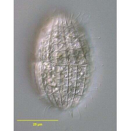
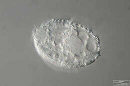
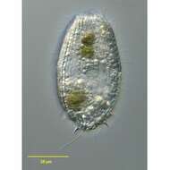
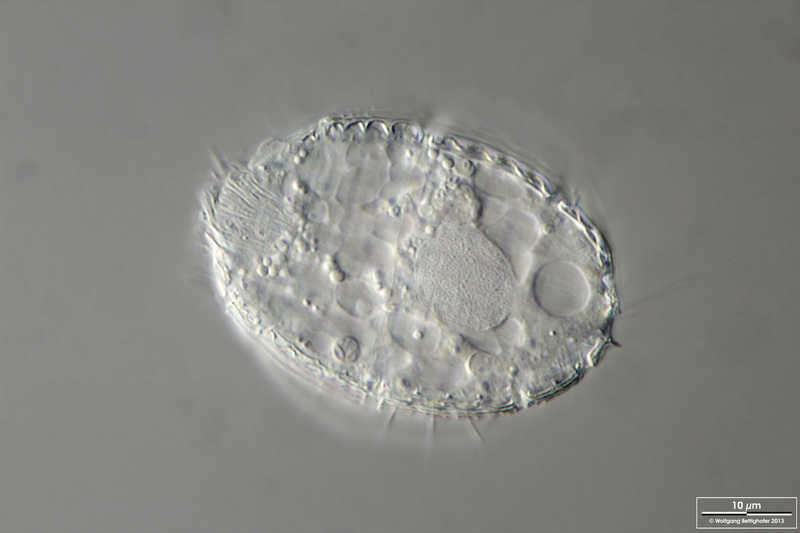
In vivo portrait of Coleps hirtus (MÃLLER,1786) NITZCH,1827.Collected from a freshwater pond near Boise, Idaho.DIC.
-
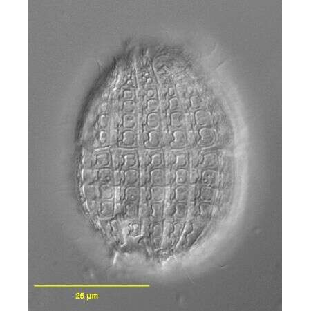
In vivo portrait of Coleps hirtus (MÃLLER,1786) NITZCH,1827.Collected from a freshwater pond near Boise, idaho.DIC.
-
Squashed specimen of Coleps hirtus (MÃLLER,1786) NITZCH,1827.Collected from a freshwater pond near Boise, Idaho.DIC.
-
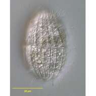
Infraciliature of Coleps hirtus (MÃLLER,1786) NITZCH,1827.Collected from a freshwater pond near Boise, Idaho.Stained by the silver carbonate technique (see Foissner, W. Europ. J. Protistol., 27:313-330;1991).Brightfield.
-
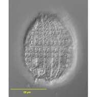
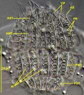
Infraciliature of Coleps hirtus (MÃLLER,1786) NITZCH,1827.The anterior apical cytostome is ringed by an undulating membrane composed of dikinetids (light blue arrowhead).Just posterior to this is a ring of pectinelles composed of two closely spaced dikinetids (yellow arrowhead).The monokinetids of the somatic kineties are located in notches in the cortical plates.Each somatic kinetid (red arrowheads) is associated with a smaller posterior parasomal sac.These parasomal sacs can be easily confused with kinetids in siolver stained specimens. Collected from a freshwater pond near Boise, Idaho.Stained by the silver carbonate technique (see Foissner, W. Europ. J. Protistol., 27:313-330;1991).Brightfield.
-
-
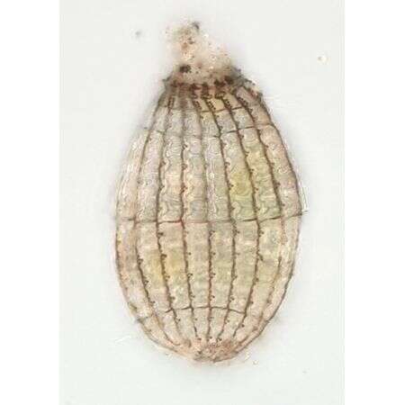
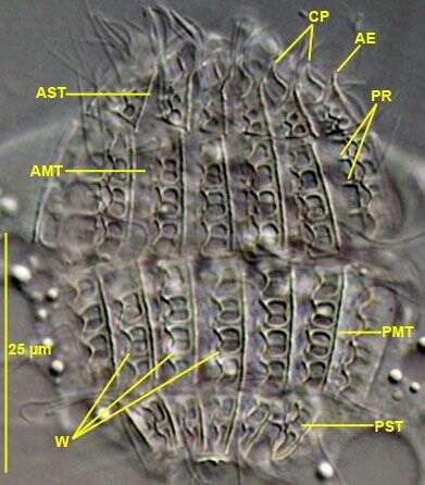
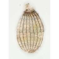
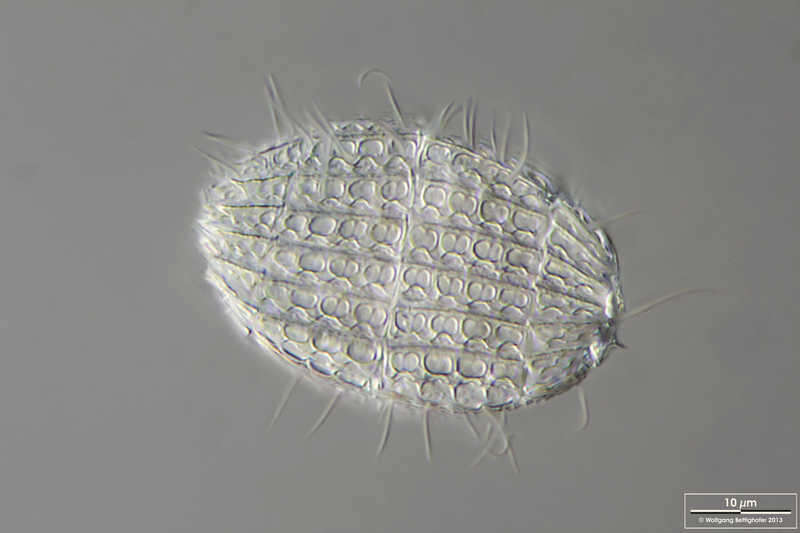
Armor plates of squashed Coleps hirtus (MÃLLER,1786) NITZCH,1827.CP=circumoral tier of plates.AE= acute end of anterior secondary tier (AST) plates.AMT=anterior main tier of plates.PMT=posterior main tier of plates.PST=posterior secondary tier of plates. The caudal tier of plates is not visible here.PR= plate process. W= plate windows (pretzel-shaped in the \""hirtus type\"" of armor plates).Collected from a freshwater pond near Boise, Idaho.DIC.
-