-
Rozas de Puerto Real, Comunidad de Madrid, Espaa
-
Valganon, La Rioja, Spain
-
, Andaluca, Espaa
-
Lumbreras, La Rioja, Spain
-
Melgar de Tera, Castille and Leon, Spain
-
Lumbreras, La Rioja, Spain
-
Lumbreras, La Rioja, Spain
-
Vilobi Del Penedes, Catalonia, Spain
-
Lumbreras, La Rioja, Spain
-
Arboli, Catalonia, Spain
-
Ribadelago de Franco, Castille and Leon, Spain
-
Ribadelago, Castille and Leon, Spain
-
Ribadelago de Franco, Castilla y Len, Espaa
-
Ribadelago, Castille and Leon, Spain
-
Ribadelago, Castille and Leon, Spain
-
Ribadelago de Franco, Castille and Leon, Spain
-
-
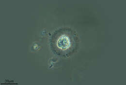
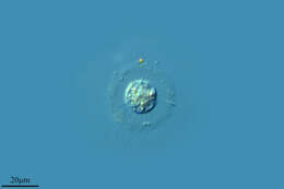
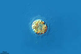
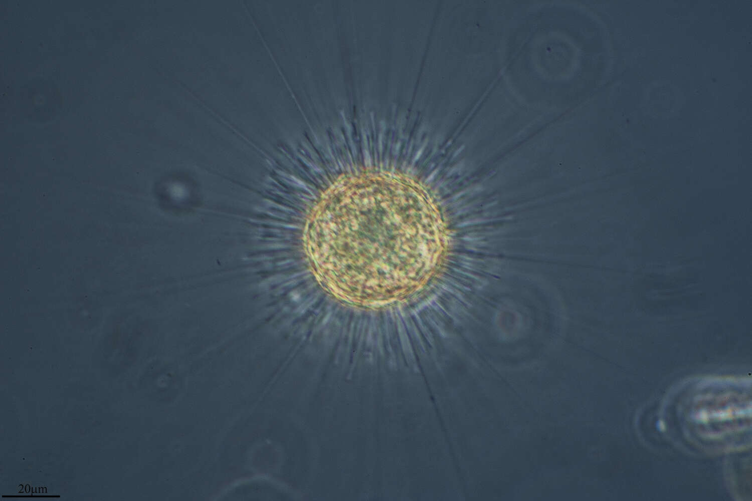
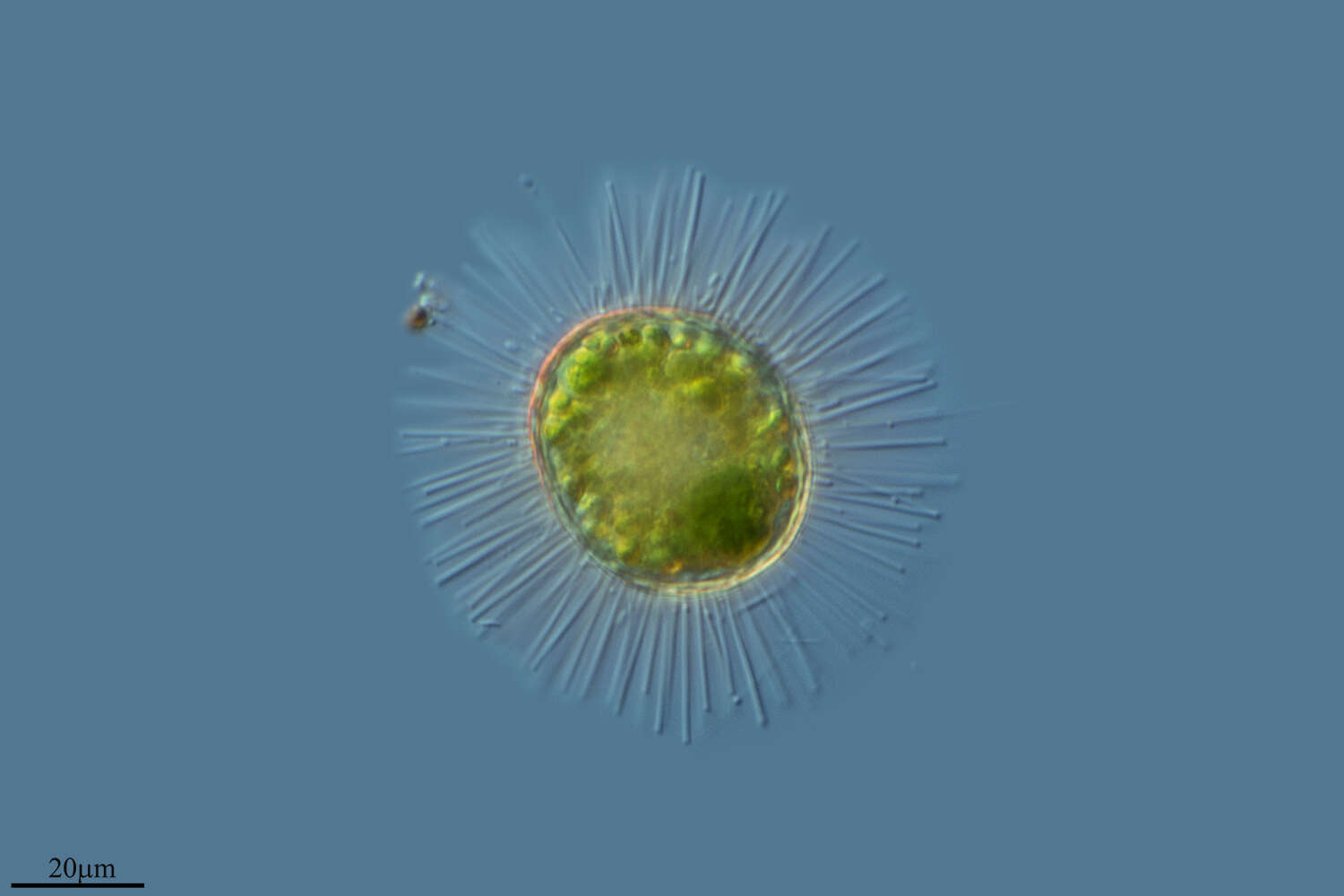
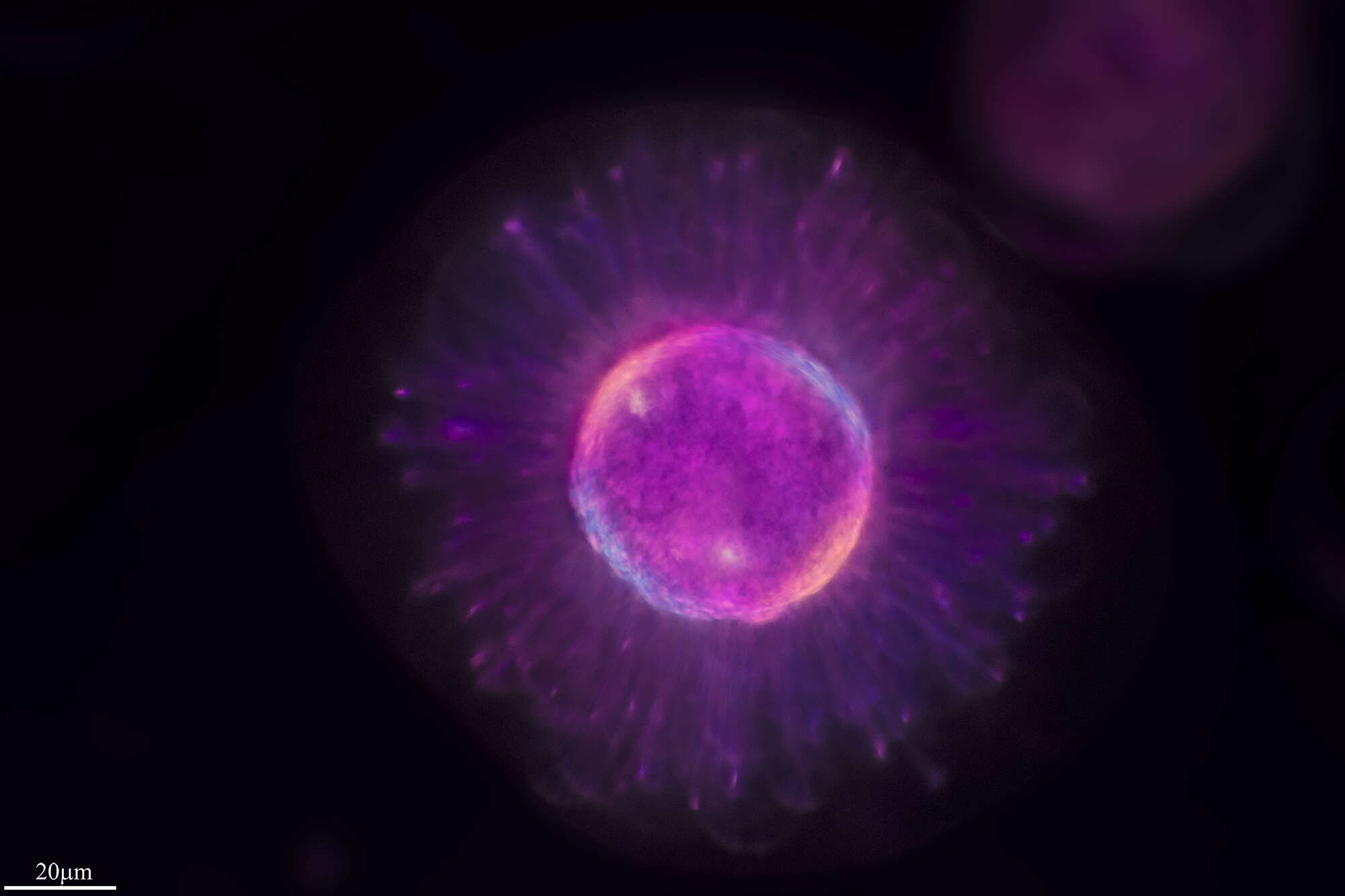
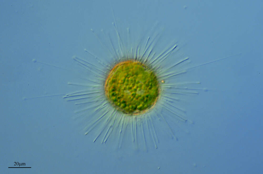
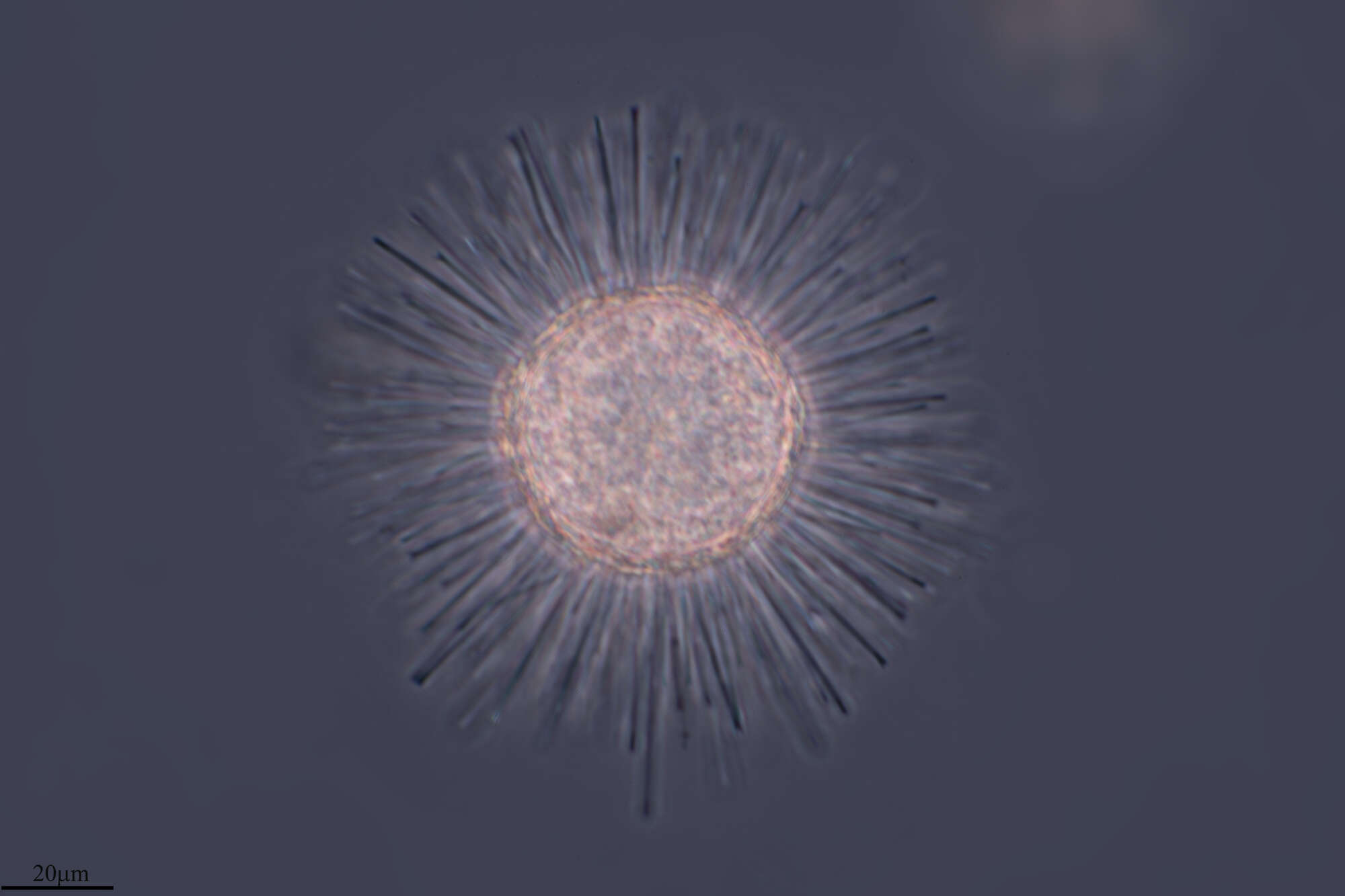
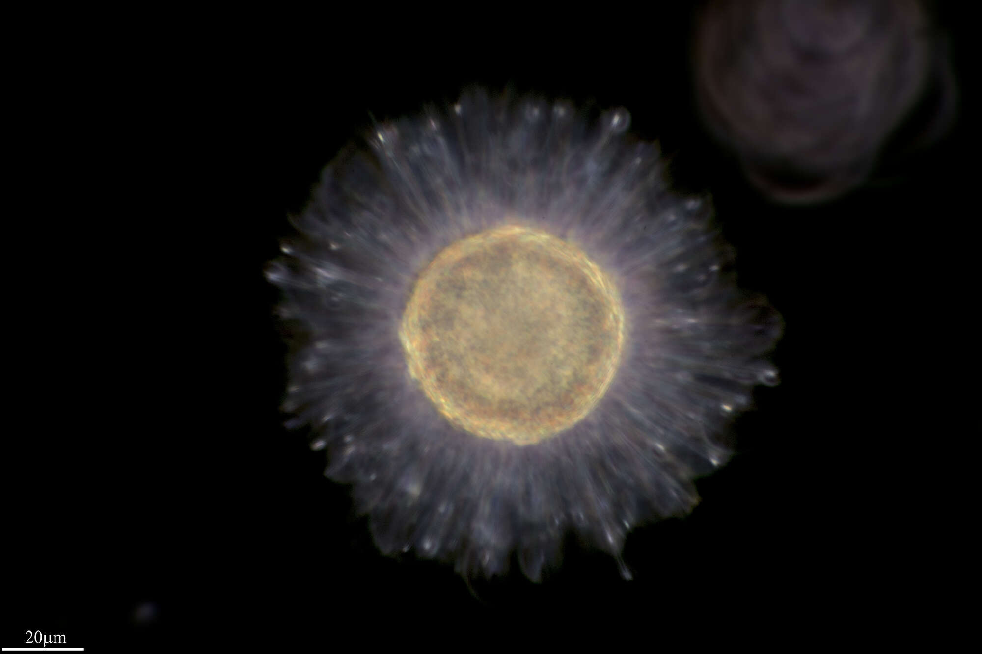
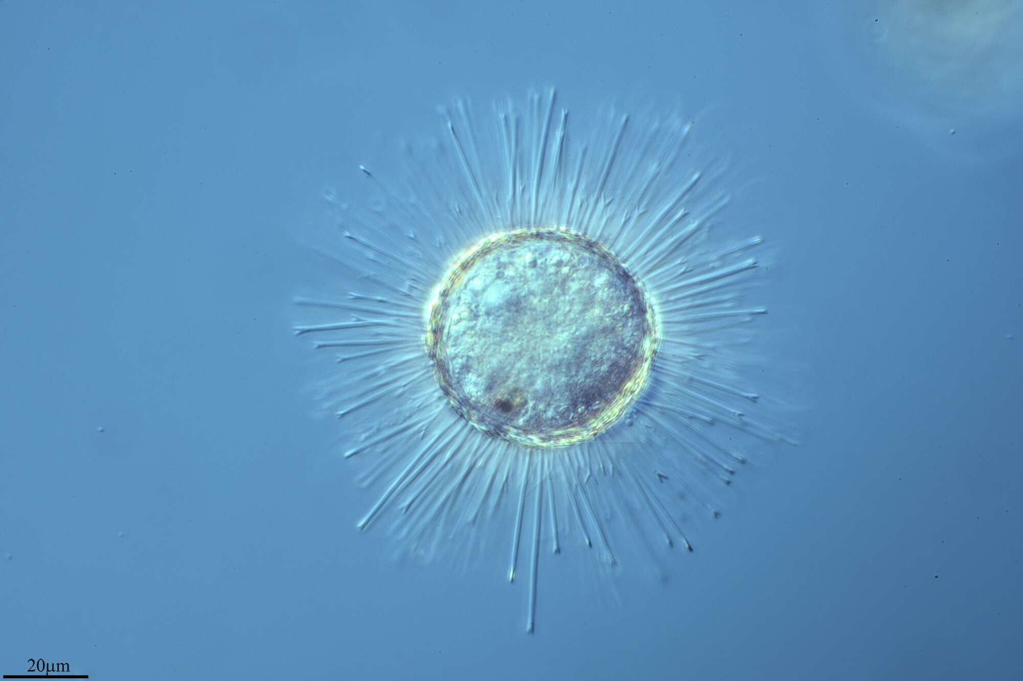
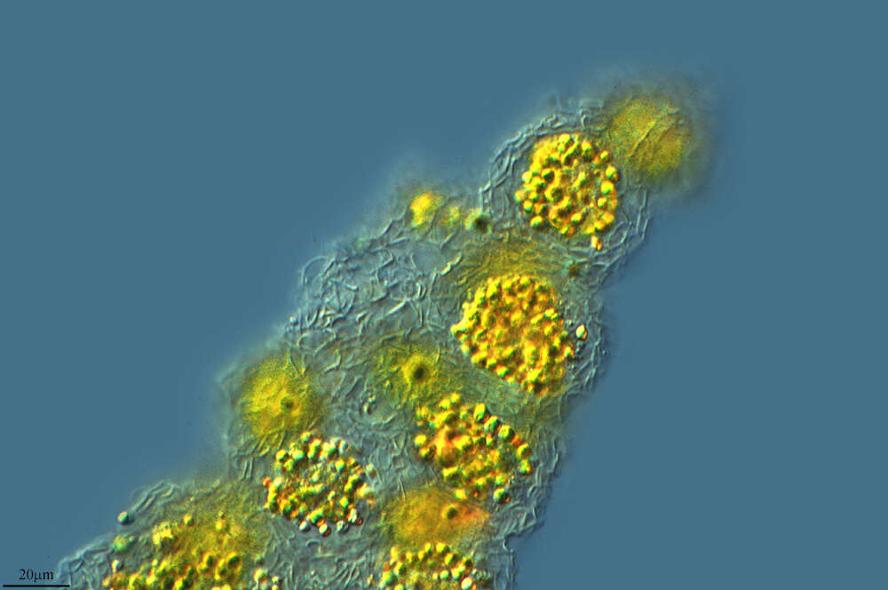
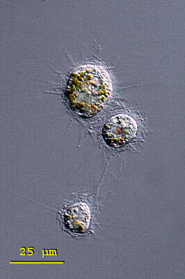
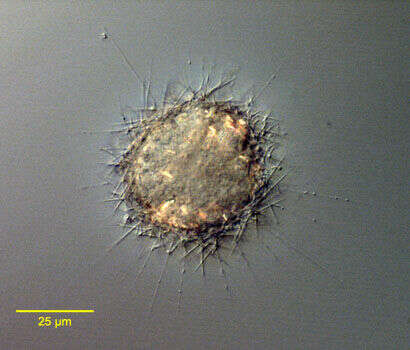
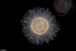
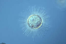
Raphidiophrys (raff-fid-ee-off-riss) ambigua is a solitary species in this genus of centrohelid heliozoa. It forms three kinds of spindle shaped spicules that accumulate around the pseudopodia. The first form are spindle shaped and pointed at both ends, the second form are medium sized, broadly spindle shaped and rounded at the ends, and the final form are very small (5 - 6 mm long) and elliptically shaped. The spicules are embedded in a gelatinous envelope of the cell. Cytoplasm without symbiotic algae but often coloured greenish, brownish or yellowish by food vacuoles. Three closely arranged specimen of Raphidiophrys ambigua, possibly shortly after cell division. Note the yellow brownish food vacuoles that are characteristic of this species. From a pond near Konstanz, Germany.Differential interference contrast.
-
Single whole plate scale viewed by transmission electron microscopy. The siliceous scales are formed within the cell and then form a loose layer or periplast around the outside of the cell.
-
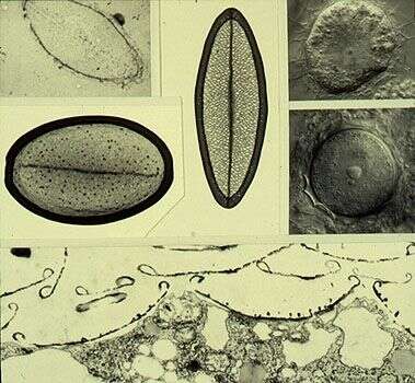
Composite image showing single scales (upper left). Upper left corner treated with hydrofluoric acid to show siliceous nature, vertical scale from trophic cell (Nomarski image upper right corner), larger scale from the cyst (middle right image). The lower image is a transmission electron micrograph of a thin section showing the scales adhering to the outer surface of the cell.
-
Various teratological (malformed) scales. Transmission electron micrographs of whole scales.
-
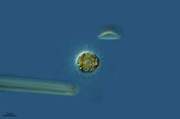
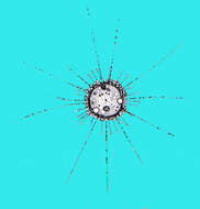
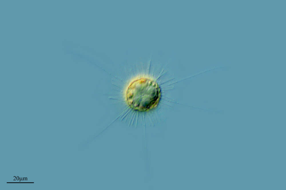
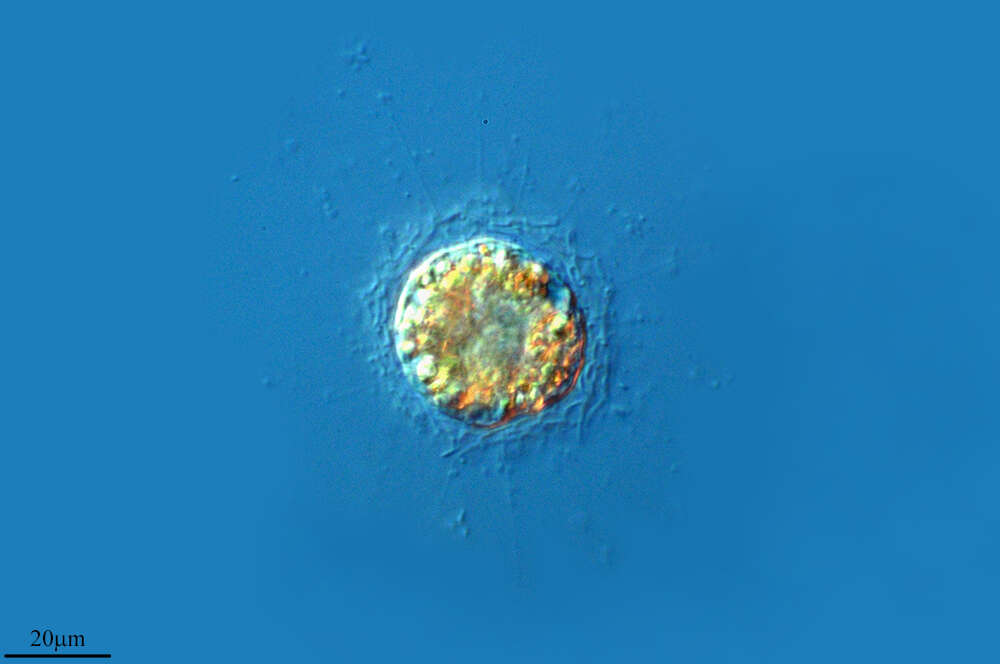
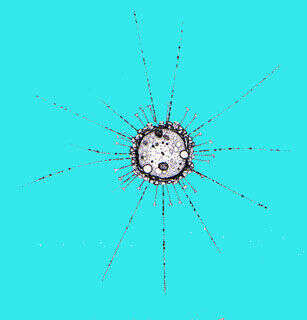
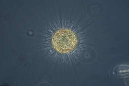
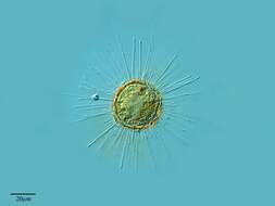
Portarit of the centroheliozoan Parasphaerastrum marina (Ostenfeld,1904)Mikrjukov,1996.The periplast is composed of tangential solid slightly curved siliceous rod with pointed ends. Radial elements are absent.Extrusomes are visible along the axopodia. The cytoplasm contains refractile crystalline inclusions. Cells may form pseudocolonies (aggregates of cells without cytoplasmic connections).Collected from a commercial marine aquarium in Boise, Idaho.October, 2005.DIC.
-
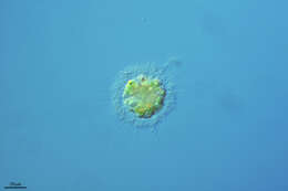
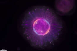
Tangential periplast elements of the centroheliozoan Parasphaerastrum marina (Ostenfeld,1904)Mikrjukov,1996.The periplast is composed of tangential solid slightly curved siliceous rod with pointed ends. Radial elements are absent.Extrusomes are visible along the axopodia. The cytoplasm contains refractile crystalline inclusions. Cells may form pseudocolonies (aggregates of cells without cytoplasmic connections).Collected from a commercial marine aquarium in Boise, Idaho.October, 2005.DIC.
-
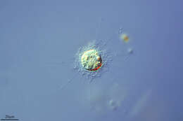
Portarit of the centroheliozoan Parasphaerastrum marina (Ostenfeld,1904)Mikrjukov,1996.The periplast is composed of tangential solid slightly curved siliceous rod with pointed ends. Radial elements are absent.Extrusomes are visible along the axopodia. The cytoplasm contains refractile crystalline inclusions. Cells may form pseudocolonies (aggregates of cells without cytoplasmic connections).Collected from a commercial marine aquarium in Boise, Idaho.October, 2005.DIC.