-
-
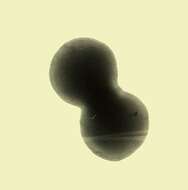
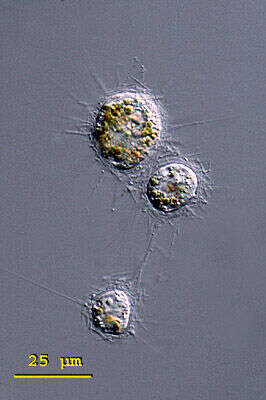
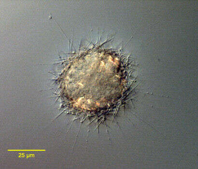
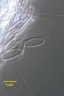
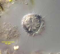
Raphidiophrys (raff-fid-ee-off-riss) ambigua is a solitary species in this genus of centrohelid heliozoa. It forms three kinds of spindle shaped spicules that accumulate around the pseudopodia. The first form are spindle shaped and pointed at both ends, the second form are medium sized, broadly spindle shaped and rounded at the ends, and the final form are very small (5 - 6 mm long) and elliptically shaped. The spicules are embedded in a gelatinous envelope of the cell. Cytoplasm without symbiotic algae but often coloured greenish, brownish or yellowish by food vacuoles. Three closely arranged specimen of Raphidiophrys ambigua, possibly shortly after cell division. Note the yellow brownish food vacuoles that are characteristic of this species. From a pond near Konstanz, Germany.Differential interference contrast.
-
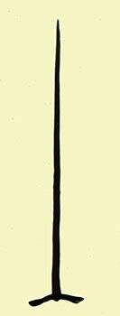
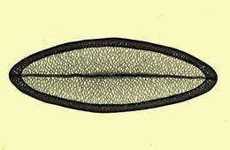
Single whole plate scale viewed by transmission electron microscopy. The siliceous scales are formed within the cell and then form a loose layer or periplast around the outside of the cell.
-
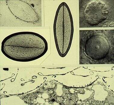
Composite image showing single scales (upper left). Upper left corner treated with hydrofluoric acid to show siliceous nature, vertical scale from trophic cell (Nomarski image upper right corner), larger scale from the cyst (middle right image). The lower image is a transmission electron micrograph of a thin section showing the scales adhering to the outer surface of the cell.
-
Various teratological (malformed) scales. Transmission electron micrographs of whole scales.
-
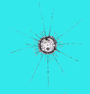
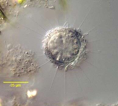
Portarit of the centroheliozoan Parasphaerastrum marina (Ostenfeld,1904)Mikrjukov,1996.The periplast is composed of tangential solid slightly curved siliceous rod with pointed ends. Radial elements are absent.Extrusomes are visible along the axopodia. The cytoplasm contains refractile crystalline inclusions. Cells may form pseudocolonies (aggregates of cells without cytoplasmic connections).Collected from a commercial marine aquarium in Boise, Idaho.October, 2005.DIC.
-
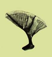
Tangential periplast elements of the centroheliozoan Parasphaerastrum marina (Ostenfeld,1904)Mikrjukov,1996.The periplast is composed of tangential solid slightly curved siliceous rod with pointed ends. Radial elements are absent.Extrusomes are visible along the axopodia. The cytoplasm contains refractile crystalline inclusions. Cells may form pseudocolonies (aggregates of cells without cytoplasmic connections).Collected from a commercial marine aquarium in Boise, Idaho.October, 2005.DIC.
-
Portarit of the centroheliozoan Parasphaerastrum marina (Ostenfeld,1904)Mikrjukov,1996.The periplast is composed of tangential solid slightly curved siliceous rod with pointed ends. Radial elements are absent.Extrusomes are visible along the axopodia. The cytoplasm contains refractile crystalline inclusions. Cells may form pseudocolonies (aggregates of cells without cytoplasmic connections).Collected from a commercial marine aquarium in Boise, Idaho.October, 2005.DIC.
-
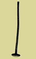
Detail of the siliceous scales of the centroheliozoan, Polyplacocystis symmetrica (Penard, 1904) Mikrjukov, 1996. An axopodium with a bead-like extrusome is visible below the scale to the viewer's left. Collected from a freshwater aquaculture pond near Boise, Idaho. November 2003. DIC.
-
Portrait of the centroheliozoan, Polyplacocystis symmetrica (Penard, 1904) Mikrjukov, 1996. Collected from a freshwater aquaculture pond near Boise, Idaho. November 2003. DIC.
-
Portrait of the centroheliozoan, Pseudoraphidocystis glutinosa (Penard,1904) Mikrjukov, 1997. The periplast consists of two types of siliceous elements; ellipsoid tangential plate scales which lack a hollow marginal rim and short radial funnel-shaped structure all of approximately equal length.Bead-like extrusomes are seen along the axopodia. Collected from a freshwater pond near Boise, Idaho. DIC.
-
Portrait of the centroheliozoan, Pseudoraphidocystis glutinosa (Penard,1904) Mikrjukov, 1997. The periplast consists of two types of siliceous elements; ellipsoid tangential plate scales which lack a hollow marginal rim and short radial funnel-shaped structure all of approximately equal length. Collected from a freshwater pond near Boise, Idaho. DIC.
-
Portrait of the centroheliozoan, Pseudoraphidocystis glutinosa (Penard,1904) Mikrjukov, 1997. The periplast consists of two types of siliceous elements; ellipsoid tangential plate scales which lack a hollow marginal rim and short radial funnel-shaped structure all of approximately equal length.Bead-like extrusomes are seen along the axopodia. Collected from a freshwater pond near Boise, Idaho. DIC.
-
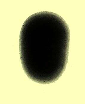
Transmission electron micrograph of a whole plate scale from the surface of a cell.
-
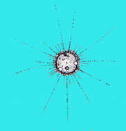
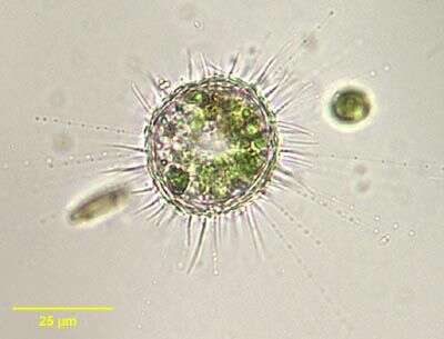
Portrait of Acanthocystis aculeata HERTWIG & LESSER, 1874, a heliozoan with long thin radiating axopodia containing bead-like extrusomes and layered surface plates with shorter curved sharp projecting spines. This individual contains zoochlorellae. From freshwater pond near Boise, Idaho. Brightfield.
-
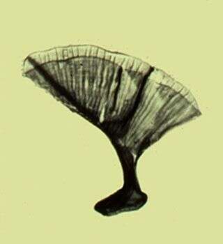
Whole mount preparation of the spine scale of this species. Transmission electron micrograph.
-
Transmission electron micrograph of a plate scale that coats the cell body.
-
Whole leaf scale, transmission electron micrograph.
-
Spine scale, whole mount, transmission electron micrograph.
-
Acanthocystis penardi (WAILES,1925).
-
Acanthocystis penardi (WAILES,1925).DIC.
-
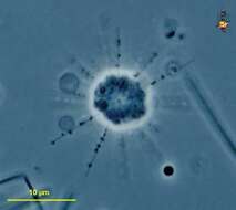
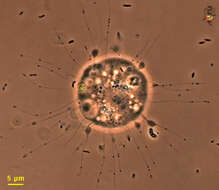
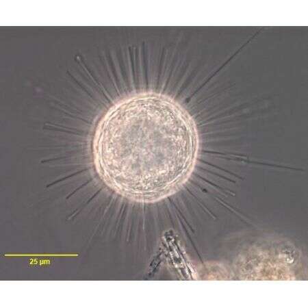
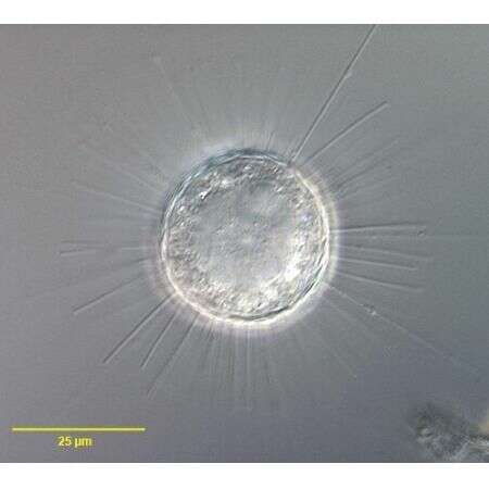
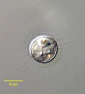
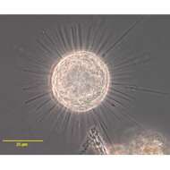
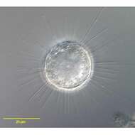
Oxnerella (ox-ner-ell-a) is a centrohelid heliozoon - the most speciose group of heliozoa. As with other heliozoa, it has radiating arms which intercept swimming prey which are captured by the extrusomes (the lumps on the arms) and then ingested. This genus includes spicules with fine spicules. Those spicules may be so delicate as not to be readily visible (if at all) by light microscopy. Heterophrys is similar but has delicate organic spicules. These may be very hard to see by light microscopy, and this can easily lead to misidentification. Phase contrast.
-
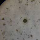
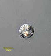
Oxnerella (ox-nerr-ell-a) is a centrohelid heliozoon, distinguished from the other centrohelids because there are no spicules or other materials around the outside of the cell. As with other centroheliozoa, the axopods are thin, parallel sides, and the extrusomes seem relatively large. Phase contrast micrograph.
-
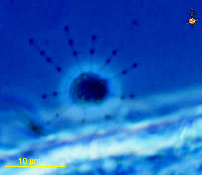
Small centrohelid heliozoon, thin axopodia carry kinetocysts - a type of extrusome - that look like granules on the axopodia. The cell body seems to lack any surrounding layer of mucus, spines or scales. Phase contrast microscopy.