Diagnostic Description
provided by Fishbase
Hisonotus oliveirai is diagnosed from all species ofHisonotus, except H. insperatus, H. luteofrenatus and H. paresi, by having odontodes forming longitudinally aligned rows (one odontode after the other, but not necessarily forming parallel series) on head and trunk (vs. odontodes not forming longitudinally aligned rows). It differs also from all congeners except H. insperatus, H. luteofrenatus, H. paresi, and H. piracanjuba, by the possession of a pair of rostral plates at the tip of the snout (vs. a single rostral plate). In addition, it further differs from all congeners except H. bockmanni, H. chromodontus, H. insperatus, H. luteofrenatus, and H. paresi by having a functional v-shaped spinelet (vs. spinelet non-functional, square-shaped, or absent). It can be separated from H. bockmanni and H. paresi by lacking contrasting dark geometric spots on the anterodorsal region of the body (vs. presence); from H. insperatus by having small, inconspicuous odontodes forming rows on the head and trunk (vs. large, conspicuous odontodes forming rows on the head and the trunk), head depth 51.6?59.2% HL (vs. 44.3?48.7% HL) and suborbital depth 20.9?25.5% HL (vs. 16.6?20.1% HL); from H. luteofrenatus by having caudal peduncle depth 10.8?12.5% SL (vs. 8.9?10.2% SL) and snout length 46.9?52.2% HL (vs. 67.0?75.3% HL); from H. paresi by a having head depth 51.6?59.2% HL (vs. 42.4?47.7% HL), 11-18 premaxillary teeth (vs. 6?10), and 11-15 dentary teeth (vs. 4?7); from H. piracanjuba by having caudal peduncle depth 10.8?12.5% SL (vs. 8.3?9.5% SL), and snout length 46.9?52.2% HL (vs. 67.7?72.7% HL) (Ref. 95507).
Morphology
provided by Fishbase
Dorsal soft rays (total): 9; Analsoft rays: 6; Vertebrae: 27
Curculionichthys oliveirai: Brief Summary
provided by wikipedia EN
Curculionichthys oliveirai is a species of catfish in the family Loricariidae. It is a freshwater species native to South America, where it occurs in tributaries of the Ivaí River in Brazil. The species reaches 3 cm (1.2 inches) SL and was named for Claudio Oliveira, a professor from São Paulo State University, Botucatu, São Paulo, after his contributions to the studies of Neotropical freshwater fishes.
- license
- cc-by-sa-3.0
- copyright
- Wikipedia authors and editors
Description
provided by Zookeys
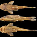
Morphometric data presented in Table 1. Maximum body length 28.4 mm SL. Dorsal profile of head slightly convex to straight from upper part of rostrum to posterior margin of nares, convex from eyes to posterior margin of parieto-supraoccipital, and straight to dorsal-fin origin. Dorsal profile of trunk slightly concave and descending from dorsal-fin origin to end of dorsal-fin base, straight to caudal peduncle. Ventral profile strongly concave from snout tip to opercular region; convex from opercular region to anal-fin origin; concave to caudal-fin insertion. Greatest body depth at dorsal-fin origin (18.6−23.9% SL). Greatest body width at opercular region, gradually decreasing towards snout and caudal fin. Cross-section of caudal peduncle almost ellipsoid; rounded laterally and almost flat dorsally and ventrally.
Head rounded in dorsal view, snout round to slightly pointed. Dorsal and ventral series of odontodes along anterior margin of snout completely covering its tip; odontodes larger than remaining ones on head. Odontodes on head and trunk hypertrophied and arranged in longitudinal rows (most prominent on head). Eyes moderately small (13.9−17.6% in HL), dorsolaterally positioned. Lips roundish with papillae uniformly distributed on base of dentary and premaxilla and slightly decreasing distally. Lower lip larger than upper lip; its border fringed. Maxillary barbel present; joined to lower lip by membrane for half its length. Teeth slender and bicuspid; mesial cusp larger than lateral cusp. Premaxillary teeth 11−18. Dentary teeth 11−15.
Dorsal-fin ii,7; dorsal-fin spinelet short and V-shaped; dorsal-fin lock functional; dorsal-fin origin slightly posterior to pelvic-fin origin. Tip of adpressed dorsal fin almost reaching end of anal-fin base. Pectoral-fin i,6; its tip almost reaching middle of pelvic-fin unbranched ray length when depressed. Pectoral axillary slit present between pectoral-fin insertion and lateral process of cleithrum. Pectoral spine supporting odontodes on ventral, anterior and dorsal surfaces. Pelvic-fin i,5; tip of pelvic-fin longest ray almost reaching anal-fin origin when depressed in females and reaching anal-fin origin in males. Pelvic-fin unbranched ray with dermal flap along its dorsal surface in males. Anal-fin i,5; its tip reaching 7th or 8th plate from its origin. Caudal-fin i,14,i; distal margin forked. Adipose-fin absent. Total vertebrae 27.
Body covered with bony plates except above lower lip, around pectoral and pelvic-fin origins and on dorsal-fin base. Cleithrum and coracoid totally exposed. Arrector fossae partially to completely enclosed by ventral lamina of coracoids. Abdomen entirely covered by plates (Fig. 3A); abdomen covered by large, elongate lateral plate series, formed by two lateral rows, approximately of same size; median plates formed by two patterns of plate distributions; first, median plate series not reaching anal shield plates with lateral plate series beginning to contact each other at middle of abdomen; second, median plate series reaching anal shield and lateral plate series remaining separate; anal plates series covered by large square or triangular plates. Body entirely covered laterally by plates (Fig. 3B); mid-dorsal plates poorly developed and reaching middle of dorsal-fin base; median plates series continuous in median portion of body; mid-ventral plates reaching vertical through end of dorsal-fin base.
Parts of dorsal head bone plates presented in Fig. 3C. Snout tip formed by one pair of square rostral plates (r). Nasal (n) rectangular, forming anterior medial nostril margin, posterior nasal margin contacting frontals (f), anterior and lateral margins contacting pre-nasals (pn). Pre-nasals (pn) positioned posterior to rostral plates (r); formed by two large square-shaped plates, one small and triangular and one elongated and rectangular between nares. Posterodorsal head plates consist of compound pterotic (cpt), parieto-supraoccipital (soc) and frontal (f; largest bones of head), prefrontal (pf) and sphenotic (sp). Compound pterotic (cpt) covered with few and small, unclustered fenestra. Lateral surface of head illustrated in Fig. 3D. Posterior rostrum plates pr1-pr2 smallest, rectangular shaped; pr4-pr3 largest, first rectangular and second square. Complete infraorbital plate series (io1-io5), present just above posterior rostrum series, all covered by laterosensory canal system; io2 largest and io5 smallest; io3, io4 and io5 forming inferior orbital margin of eyes. Preopercle (pop) elongate and rectangular, covered by laterosensory canal; preopercle present under pr4, io4 and io5, and upper cp1, cp2 and op. Subocular cheek plates (cp1-cp2) and opercle (op) form posterior lateral margin of head.
- license
- cc-by-3.0
- copyright
- Fábio F. Roxo, Cláudio H. Zawadzki, Waldo P. Troy
- bibliographic citation
- Roxo F, Zawadzki C, Troy W (2014) Description of two new species of Hisonotus Eigenmann & Eigenmann, 1889 (Ostariophysi, Loricariidae) from the rio Paraná-Paraguay basin, Brazil ZooKeys 395: 57–78
- author
- Fábio F. Roxo
- author
- Cláudio H. Zawadzki
- author
- Waldo P. Troy
Distribution
provided by Zookeys
Hisonotus oliveirai is only known from four small to medium-sized streams, the ribeirão Salto Grande, ribeirão Keller, rio Mourão, and the ribeirão Cambira, all tributaries of the rio Ivaí in the upper rio Paraná basin (Fig. 5A).
- license
- cc-by-3.0
- copyright
- Fábio F. Roxo, Cláudio H. Zawadzki, Waldo P. Troy
- bibliographic citation
- Roxo F, Zawadzki C, Troy W (2014) Description of two new species of Hisonotus Eigenmann & Eigenmann, 1889 (Ostariophysi, Loricariidae) from the rio Paraná-Paraguay basin, Brazil ZooKeys 395: 57–78
- author
- Fábio F. Roxo
- author
- Cláudio H. Zawadzki
- author
- Waldo P. Troy