-
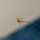
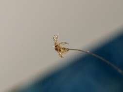
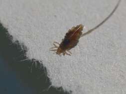
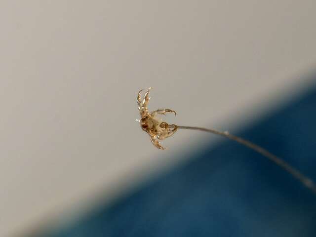
This week’s podcast is guaranteed to make your scalp crawl—but don’t worry, it’s most likely all in your head, and not on it. We’ll visit entomologist Richard Pollack to learn about an insect that’s the bane of parents and school principals everywhere—or is it? Ari Daniel Shapiro explains. Photo Credit: Gilles San Martin, CC BY-SA
Download a transcript of this podcast read moreDuration: 5:04Published: Wed, 08 Aug 2012 13:52:49 +0000
-
Hovedhår
-
Centers for Disease Control/Division of Parasitic Diseases and Malaria
EOL staff
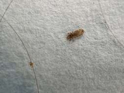
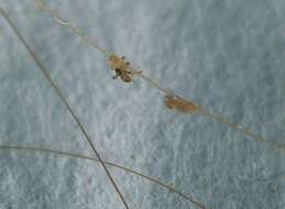
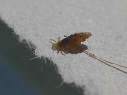
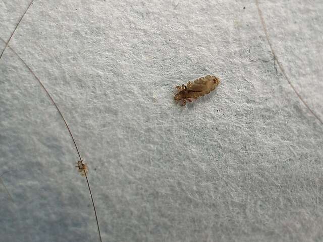
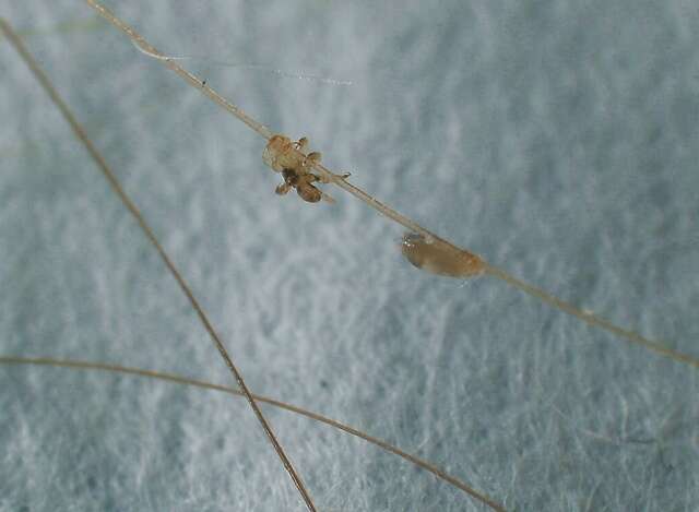
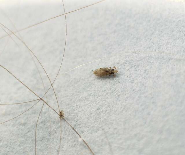
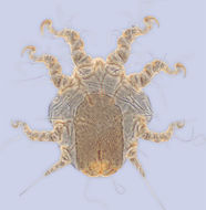
Life cycle of the Head Louse (Pediculus humanus capitis), an ectparasite of humansThe life cycle of the Head Louse (Pediculis humanus capitis) has three stages: egg, nymph, and adult. Eggs: Nits are Head Lice eggs. They are hard to see and are often mistaken for dandruff or hair spray droplets. Nits are laid by the adult female and are cemented at the base of the hair shaft nearest the scalp (1). They are 0.8 mm by 0.3 mm, oval and usually yellow to white. Nits take about 1 week to hatch (range 6 to 9 days). Viable eggs are usually located within 6 mm of the scalp. Nymphs: The egg hatches to release a nymph (2). The nit shell then becomes a more visible dull yellow and remains attached to the hair shaft. The nymph looks like an adult head louse, but is about the size of a pinhead. Nymphs mature after three molts (3, 4) and become adults about 7 days after hatching. Adults: The adult louse is about the size of a sesame seed, has 6 legs (each with claws), and is tan to grayish-white (5). In persons with dark hair, the adult louse will appear darker. Females are usually larger than males and can lay up to 8 nits per day. Adult lice can live up to 30 days on a person’s head. To survive , adult lice need to feed on blood several times daily. Without blood meals, the louse will die within 1 to 2 days off the host.From
Centers for Disease Control Parasites and Health website.
-
Hovedhår
-
Hovedhår
-
Hovedhår
-
Hovedhår
-
Hovedhår
-
Hovedhår
-
Hovedhår
-
-
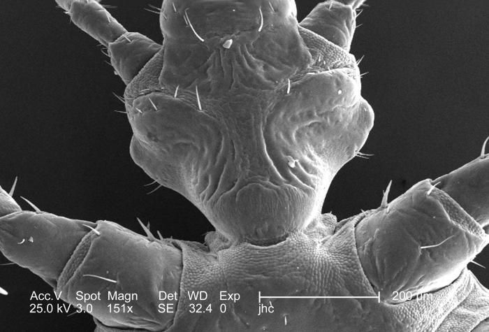
From a ventral perspective, and at a low magnification of 151x, this 2006 scanning electron micrograph (SEM) depicted an enlarged view of the chitinous, exoskeletal surface of a female louse, Pediculus humanus var. corporis, in the region where the organisms forelegs and hean attached to its thoracic region. In this particular view, the exoskeleton seems to be composed of interlocking plates, which is not far from the case, in order to provide flexibility to this patent joint, the chitinous components were arranged in a plate-like manner, attached to one another with thin, by strong layers of exoskeletal chitin. Chitin is a molecule made up of bound units of acetylglucosamine, which is joined in such a way as to allow for increased points at which hydrogen bonding can occur. In this way chitin provides increased strength, and durability as an exoskeletal foundation.Created: 2006
-
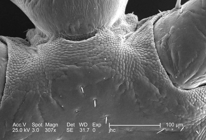
From a ventral perspective, and at a moderate magnification of 307x, this 2006 scanning electron micrograph (SEM) depicted an enlarged view of the chitinous, exoskeletal surface of a female louse, Pediculus humanus var. corporis, in the region where the organisms forelegs attached to its thoracic region. In this particular view, the exoskeleton seems to be composed of interlocking plates, which is not far from the case, in order to provide flexibility to this patent joint, the chitinous components were arranged in a plate-like manner, attached to one another with thin, by strong layers of exoskeletal chitin. Chitin is a molecule made up of bound units of acetylglucosamine, which is joined in such a way as to allow for increased points at which hydrogen bonding can occur. In this way chitin provides increased strength, and durability as an exoskeletal foundation.Created: 2006
-
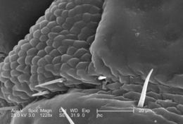
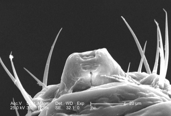
From a ventral perspective, and at a relatively high magnification of 1228x, this 2006 scanning electron micrograph (SEM) depicted an enlarged view of the chitinous, exoskeletal surface of a female louse, Pediculus humanus var. corporis, in the region where the right antennal scape attached to its cephalic region, or head. In this particular view, the exoskeleton seems to be composed of interlocking plates, which is not far from the case, in order to provide flexibility to this patent joint, the chitinous components were arranged in a plate-like manner, attached to one another with thin, by strong layers of exoskeletal chitin. Chitin is a molecule made up of bound units of acetylglucosamine, which is joined in such a way as to allow for increased points at which hydrogen bonding can occur. In this way chitin provides increased strength, and durability as an exoskeletal foundation.Created: 2006
-
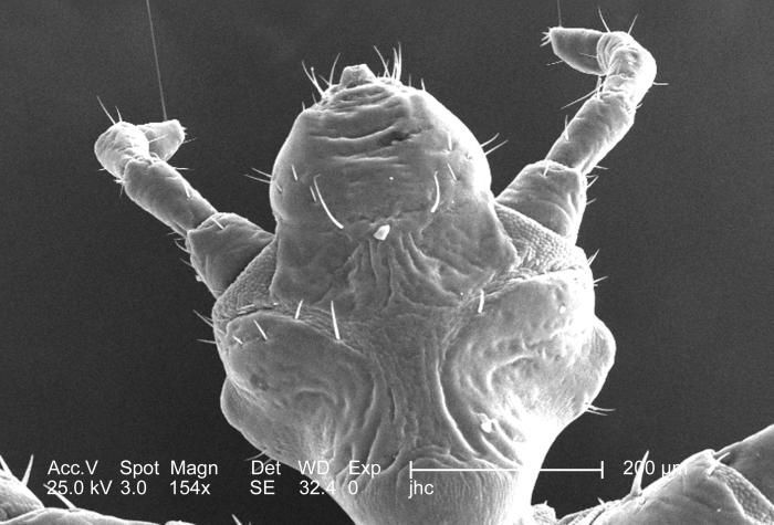
This was one of five scanning electron micrographic (SEM) images (PHIL# 9243 9247), successively magnified at higher and higher values, which focused on the head region of a female body louse, Pediculus humanus var. corporis from a ventral perspective. At a high magnification of 1228x, this SEM revealed some of the insects exoskeletal morphology exhibited by the cephalic region. Highlighted in this view is the insects cone-shaped mouth, which is surrounded by a number of setae, or sensorial hairs, which provide the organism with informational feedback about its environment such as chemistry and temperature.Created: 2006
-
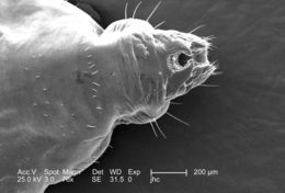
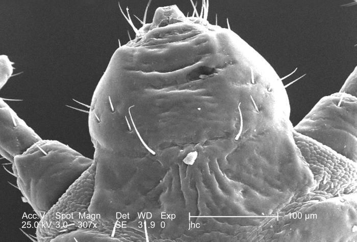
This was one of five scanning electron micrographic (SEM) images (PHIL# 9243 9247), successively magnified at higher and higher values, which focused on the head region of a female body louse, Pediculus humanus var. corporis from a ventral perspective. At a moderate magnification of 307x, this SEM revealed some of the insects exoskeletal morphology exhibited by the cephalic region. Note the two partially visible, bilaterally situated antennae composed of three main segments: visible here was the most proximal scape and the pedicle, and not in the field of view, the multi-segmented flagellum. The antennae, and the insects body sport sensorial hairs known as "setae, both of which provided the organism with a "picture of its environment, by taking readings in thermal, chemical, and mechanical changes encountered in its immediate surroundings. Its cone-shaped mouth is located at the very top of the image.Created: 2006
-
This was one of five scanning electron micrographic (SEM) images (PHIL# 9243 9247), successively magnified at higher and higher values, which focused on the head region of a female body louse, Pediculus humanus var. corporis from a ventral perspective. At a relatively low magnification of 154x, this SEM revealed some of the insects exoskeletal morphology exhibited by the cephalic region. Note the two bilaterally situated antennae composed of three main segments: the most proximal scape, a pedicle, and the multi-segmented flagellum. The antennae, and the insects body sport sensorial hairs known as "setae, both of which provided the organism with a "picture of its environment, by taking readings in thermal, chemical, and mechanical changes encountered in its immediate surroundings.Created: 2006
-
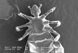
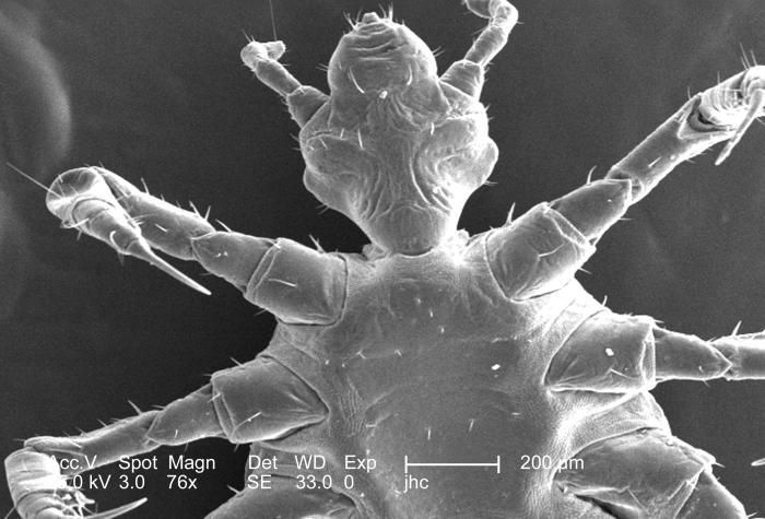
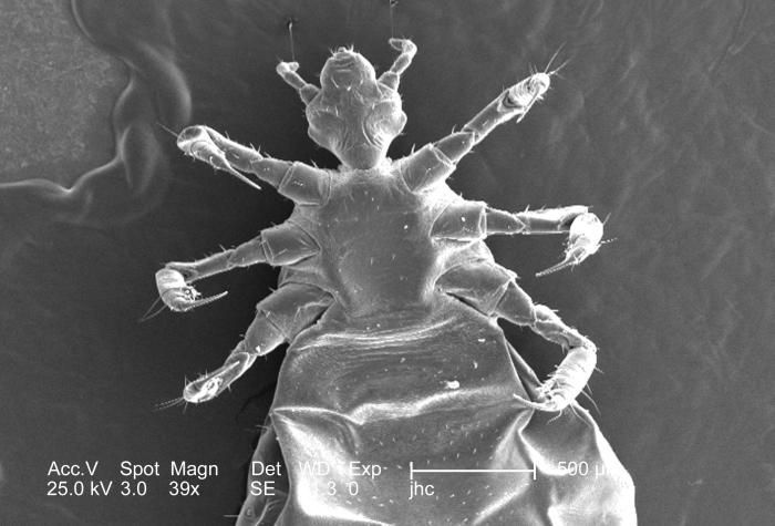
This was one of five scanning electron micrographic (SEM) images (PHIL# 9243 9247), successively magnified at higher and higher values, which focused on the head region of a female body louse, Pediculus humanus var. corporis from a ventral perspective. At a relatively low magnification, this SEM revealed some of the insects exoskeletal morphology exhibited by its cephalic, or head region, thoracic, and proximal abdominal regions. Of interest is the jointed configuration of its six extremities, from which it derived its classification in the phylum of Athropoda, i.e., Arthro from "joint, and poda from "leg"). Also note the sensorial hairs known as setae, which really arent hairs at all, but chitinous exoskeletal extensions, unlike mammalian hairs, which are made up of keratin, and make mammals unique in this regard.Created: 2006
-
This was one of five scanning electron micrographic (SEM) images (PHIL# 9243 9247), successively magnified at higher and higher values, which focused on the head region of a female body louse, Pediculus humanus var. corporis from a ventral perspective. At a relatively low magnification, this SEM revealed some of the insects exoskeletal morphology exhibited by its cephalic, or head region, thoracic, and proximal abdominal regions. Of interest is the jointed configuration of its six extremities, from which it derived its classification in the phylum of Athropoda, i.e., Arthro from "joint, and poda from "leg").Created: 2006
-
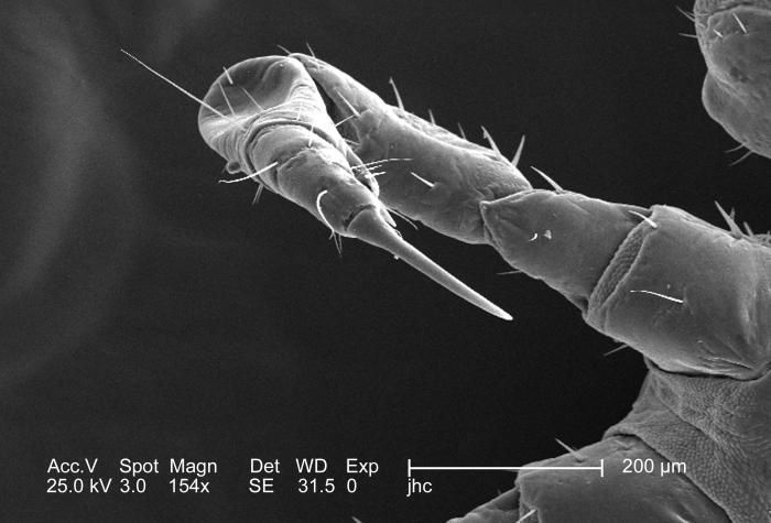
At half the magnification of PHIL# 9241, this 2006 scanning electron micrograph (SEM) depicted a dorsal view of the right flexed foreleg of a female body louse, Pediculus humanus var. corporis, in its entirty. What is visible includes the most distal segment, known as the pretarsus, followed by the more proximal tarsus, then the tibia, femur, and trochanter, and finally the most proximal segment, the coxa. In the case of the louse, the leg segments are very stout, and end in claws, which it used to firmly grasp clothing, or a hosts hair shafts. Note how the exoskeletal covering appears to possess an almost fabric-like appearance to the chitinous exoskeleton, which acts to add flexibility at the coxa-trochanteric joint.Created: 2006
-
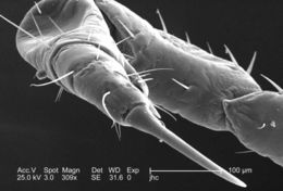
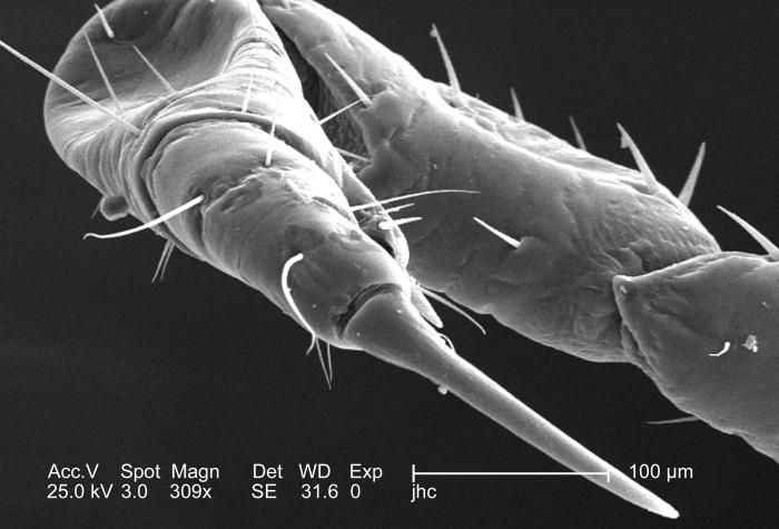
At a moderate magnification of 309x, this 2006 scanning electron micrograph (SEM) depicted an enlarged dorsal view of the right flexed foreleg of a female body louse, Pediculus humanus var. corporis. The entire leg is not quite visible, but what is visible includes the most distal segment, known as the pretarsus, followed by the more proximal tarsus, then the tibia, femur, and trochanter. The final segment, of the six segments from which each leg is composed, the coxa, is visible under reduced magnification, in PHIL# 9242. In the case of the louse, the leg segments are very stout, and end in claws, which it used to firmly grasp clothing, or a hosts hair shafts.Created: 2006
-
This 2006 scanning electron micrograph (SEM), under a low magnification of only 76x, revealed the distal tip of the abdominal region of a female body louse, Pediculus humanus var. corporis from a dorsal perspective. Some of the morphologic characteristics seen in this image include the two gonopodia, which are located dorsal to the larger two setae-bearing claspers. It is into this notch that the male would insert the aedeagus, or penis during the process of copulation. This notch, identifying the louse as a female is observable to the naked eye, whereas, in the male louse, the distal abdomen is rounded, and not concave.Created: 2006
-
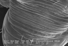
At a low magnification of 299x, this 2006 scanning electron micrograph (SEM) depicted an enlarged view of the chitinous, exoskeletal surface of a male louse, Pediculus humanus var. corporis. In this particular view, the exoskeleton seems to be configured in accordion-like convolutions, which is visible with the naked eye on the insects abdomen. Chitin is a molecule made up of bound units of acetylglucosamine, which is joined in such a way as to allow for increased points at which hydrogen bonding can occur. In this way chitin provides increased strength, and durability as an exoskeletal foundation. Note the sparse amount of setae, or sensorial hairs in this area of the abdominal surface.Created: 2006
-
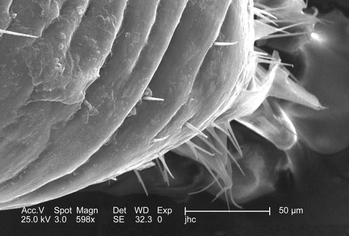
Magnified 598x, this 2006 scanning electron micrograph (SEM) depicted an enlarged view of the chitinous, exoskeletal surface of a male louse, Pediculus humanus var. corporis. In this particular view, the exoskeleton seems to be configured in accordion-like convolutions, which is visible with the naked eye on the insects abdomen. Chitin is a molecule made up of bound units of acetylglucosamine, which is joined in such a way as to allow for increased points at which hydrogen bonding can occur. In this way chitin provides increased strength, and durability as an exoskeletal foundation. Note the sparse amount of setae, or sensorial hairs in this area of the abdominal surface.Created: 2006