-
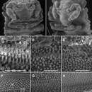
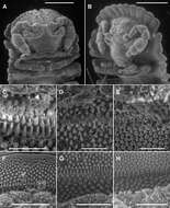
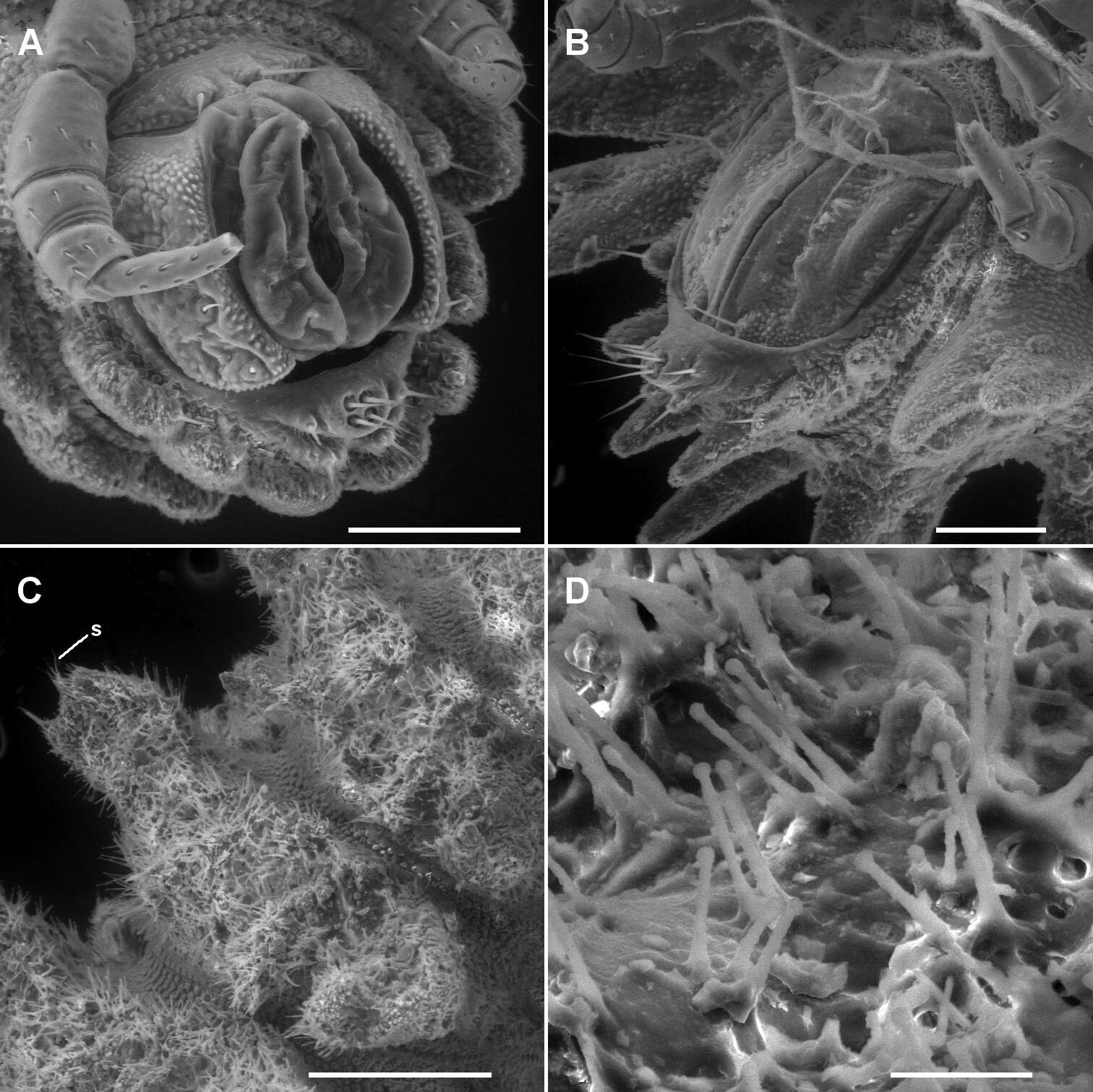
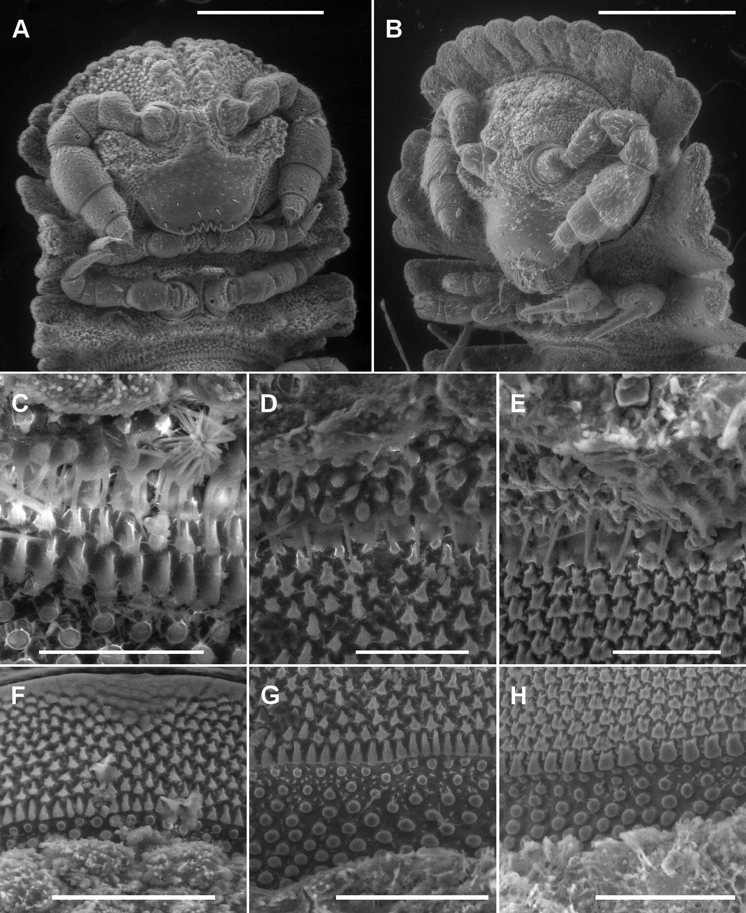
Figure 2.A, B Ventral views of head of Asticopyrgodesmus maiala sp. n., male paratype ex ANIC 64-000220 (A) and Notopyrgodesmus weiri sp. n., male paratype ANIC 64-000249 C, D, E Views of lobe-and-spike limbus on midbody rings (anterior at top) of Asticopyrgodesmus maiala sp. n., male paratype ex ANIC 64-000220 (C), Nephopyrgodesmus eungella sp. n., male paratype ex ANIC 64-000231 (D) and Notopyrgodesmus kulla sp. n., male paratype ex ANIC 64-000243 (E) F, G, H Views of prozonite sculpture on midbody rings (anterior at top) of Asticopyrgodesmus lamingtonensis sp. n., male paratype ex ANIC 64-000217 (F), Notopyrgodesmus eungella sp. n., male paratype ex ANIC 64-000231 (G) and Notopyrgodesmus kulla sp. n., male paratype ex ANIC 64-000243 (H). Scanning electron micrographs of uncoated specimens; scale bars: A = 0.5 mm, B = 0.2 mm, C, D, E, H = 0.05 mm, F, G = 0.1 mm.
-
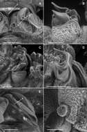
Figure 3.A–E Left lateral views of anterior end. A Asticopyrgodesmus maiala sp. n., holotype B Nephopyrgodesmus eungella sp. n., male paratype ex ANIC 64-000231 C Notopyrgodesmus kulla sp. n., male paratype ex ANIC 64-000242 D Nephopyrgodesmus lanosus sp. n., holotype E Nephopyrgodesmus weiri sp. n., holotype F Nephopyrgodesmus weiri sp. n., male paratype ANIC 64-000251, dorsal view of anterior end. Images not to same scale.
-
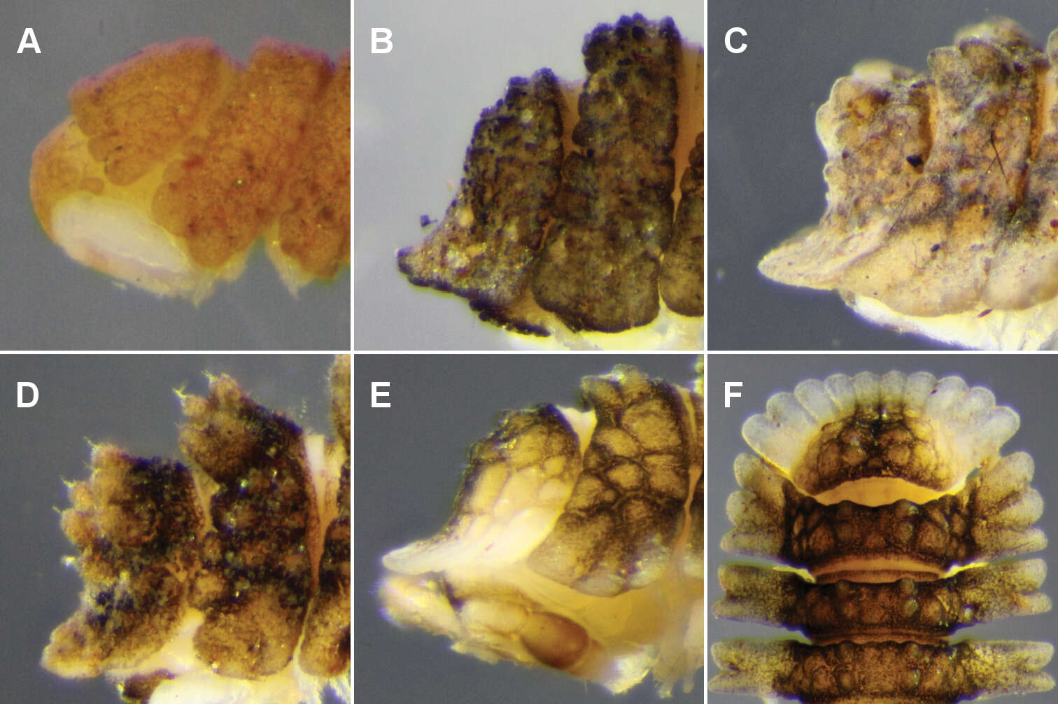
Figure 4.Left lateral views of posterior end of holotype. A Asticopyrgodesmus maiala sp. n. B Asticopyrgodesmus lamingtonensis sp. n. C Nephopyrgodesmus eungella sp. n. D Notopyrgodesmus kulla sp. n. E Notopyrgodesmus lanosus sp. n. F Notopyrgodesmus weiri sp. n. Images not to same scale.
-
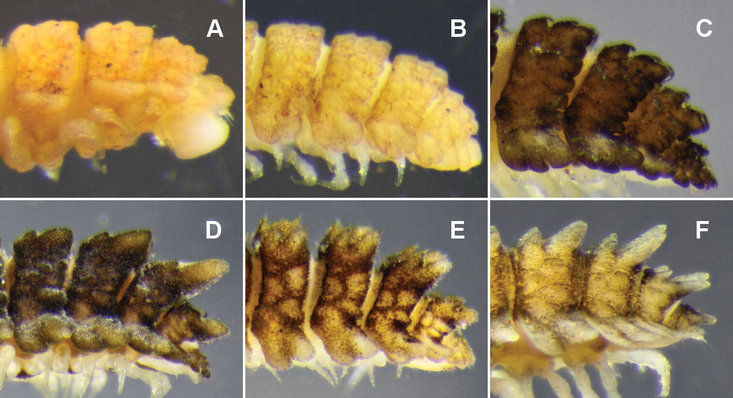
Figure 5.Right dorsolateral views of midbody, anterior to right. A Asticopyrgodesmus maiala sp. n., holotype B Asticopyrgodesmus lamingtonensis sp. n., holotype C Nephopyrgodesmus eungella sp. n., holotype D Notopyrgodesmus kulla sp. n., male paratype ex ANIC 64-000242 E Notopyrgodesmus lanosus sp. n., holotype F Notopyrgodesmus weiri sp. n., holotype. Images not to same scale. White circles: ozopore or porostele.
-
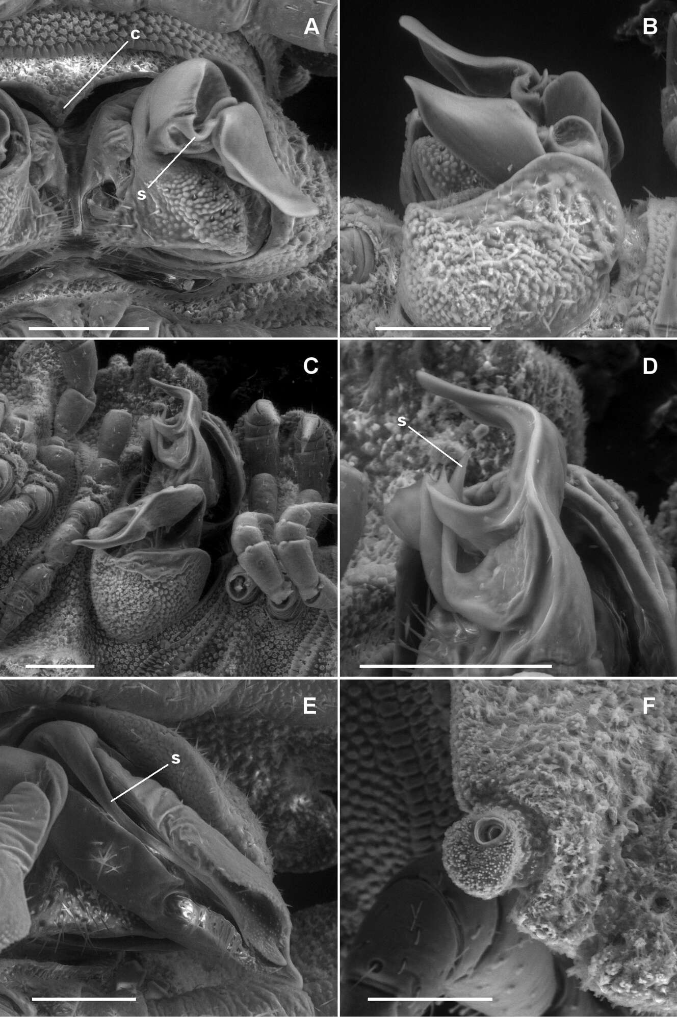
Figure 7.A, B Asticopyrgodesmus maiala sp. n., male paratypes ex ANIC 64-000220. A Ventral view (anterior at top) of left gonopod; c = aperture constriction, s = solenomere B Left lateral view of gonopods in situ C, D Asticopyrgodesmus lamingtonensis sp. n., male paratype ex ANIC 64-000217 C Left ventrolateral view of gonopods in situ D Close-up of view in C; s = solenomere. (See also Fig. 8A) E Nephopyrgodesmus eungella sp. n., male paratype ex ANIC 64-000231, ventral view of left gonopod telopodite; s = solenomere (see also Fig. 8B) F Notopyrgodesmus kulla sp. n., male paratype ex ANIC 64-000243, left lateral view of midbody porostele. Scanning electron micrographs of uncoated specimens; scale bars = 0.1 mm.
-
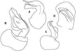
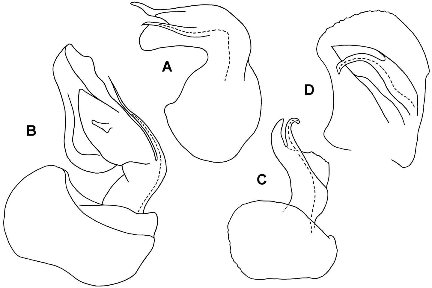
Figure 8.A Medial view of right gonopod telopodite of Asticopyrgodesmus lamingtonensis sp. n., paratype ex ANIC 64-000217 (see also Figs 7C, 7D) B Posterior view of right gonopod of Nephopyrgodesmus eungella sp. n., paratype ex ANIC 64-000231 (see also Fig. 7E) C Anterior view of left gonopod of Notopyrgodesmus kulla sp. n., QM S92795 D Medial view of right gonopod (retracted) of Notopyrgodesmus kulla sp. n., paratype ex ANIC 64-000239. Setation not shown; dashed line indicates course of prostatic groove. Drawings not to same scale.
-
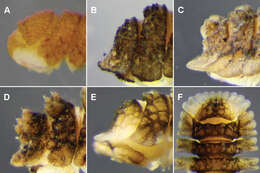
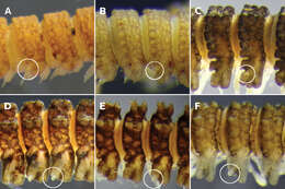
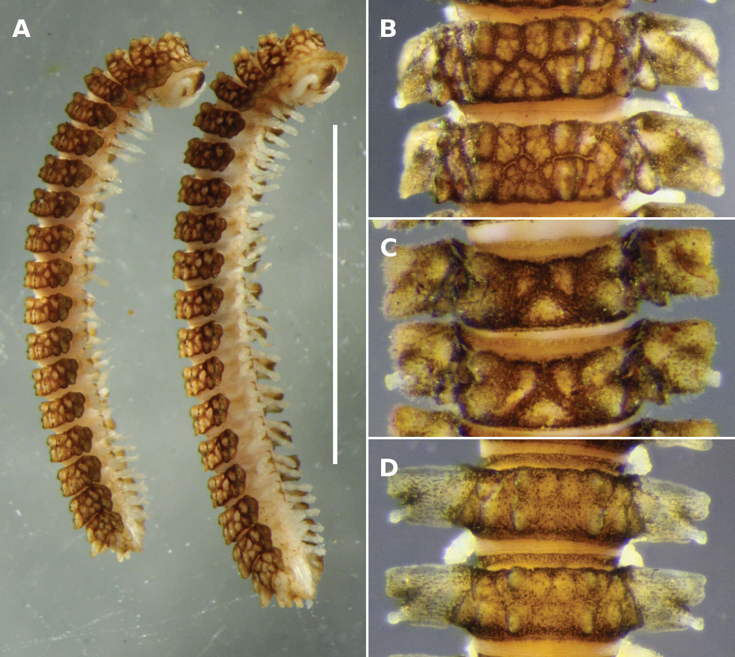
Figure 10.A Notopyrgodesmus kulla sp. n., male (left) and female (right) paratypes ex QM S92793. Scale bar = 5 mm B–D Dorsal views of midbody rings of males, not to same scale B Notopyrgodesmus kulla sp. n., paratype ex QM S92793 C Notopyrgodesmus lanosus sp. n., holotype D Notopyrgodesmus weiri sp. n., paratype ANIC 64-000251.
-
Figure 3.A–E Left lateral views of anterior end. A Asticopyrgodesmus maiala sp. n., holotype B Nephopyrgodesmus eungella sp. n., male paratype ex ANIC 64-000231 C Notopyrgodesmus kulla sp. n., male paratype ex ANIC 64-000242 D Nephopyrgodesmus lanosus sp. n., holotype E Nephopyrgodesmus weiri sp. n., holotype F Nephopyrgodesmus weiri sp. n., male paratype ANIC 64-000251, dorsal view of anterior end. Images not to same scale.
-
Figure 4.Left lateral views of posterior end of holotype. A Asticopyrgodesmus maiala sp. n. B Asticopyrgodesmus lamingtonensis sp. n. C Nephopyrgodesmus eungella sp. n. D Notopyrgodesmus kulla sp. n. E Notopyrgodesmus lanosus sp. n. F Notopyrgodesmus weiri sp. n. Images not to same scale.
-
Figure 5.Right dorsolateral views of midbody, anterior to right. A Asticopyrgodesmus maiala sp. n., holotype B Asticopyrgodesmus lamingtonensis sp. n., holotype C Nephopyrgodesmus eungella sp. n., holotype D Notopyrgodesmus kulla sp. n., male paratype ex ANIC 64-000242 E Notopyrgodesmus lanosus sp. n., holotype F Notopyrgodesmus weiri sp. n., holotype. Images not to same scale. White circles: ozopore or porostele.
-
Figure 6.A, B Ventrolateral views of telson of Asticopyrgodesmus maiala sp. n., male paratype ex ANIC 64-000220 (A) and Notopyrgodesmus weiri sp. n., male paratype ANIC 64-000249 (B) C Right lateral view of midbody ring of Notopyrgodesmus kulla sp. n., male paratype ex ANIC 64-000243, anterior to upper right; s = ‘spine’ on tip of paramedian tubercle D Close-up of hair-like cuticular outgrowths on specimen in C Scanning electron micrographs of uncoated specimens; scale bars: A, B = 0.1 mm, C = 0.25 mm, D = 0.025 mm.
-
Figure 10.A Notopyrgodesmus kulla sp. n., male (left) and female (right) paratypes ex QM S92793. Scale bar = 5 mm B–D Dorsal views of midbody rings of males, not to same scale B Notopyrgodesmus kulla sp. n., paratype ex QM S92793 C Notopyrgodesmus lanosus sp. n., holotype D Notopyrgodesmus weiri sp. n., paratype ANIC 64-000251.
-
Figure 2.A, B Ventral views of head of Asticopyrgodesmus maiala sp. n., male paratype ex ANIC 64-000220 (A) and Notopyrgodesmus weiri sp. n., male paratype ANIC 64-000249 C, D, E Views of lobe-and-spike limbus on midbody rings (anterior at top) of Asticopyrgodesmus maiala sp. n., male paratype ex ANIC 64-000220 (C), Nephopyrgodesmus eungella sp. n., male paratype ex ANIC 64-000231 (D) and Notopyrgodesmus kulla sp. n., male paratype ex ANIC 64-000243 (E) F, G, H Views of prozonite sculpture on midbody rings (anterior at top) of Asticopyrgodesmus lamingtonensis sp. n., male paratype ex ANIC 64-000217 (F), Notopyrgodesmus eungella sp. n., male paratype ex ANIC 64-000231 (G) and Notopyrgodesmus kulla sp. n., male paratype ex ANIC 64-000243 (H). Scanning electron micrographs of uncoated specimens; scale bars: A = 0.5 mm, B = 0.2 mm, C, D, E, H = 0.05 mm, F, G = 0.1 mm.
-
Figure 3.A–E Left lateral views of anterior end. A Asticopyrgodesmus maiala sp. n., holotype B Nephopyrgodesmus eungella sp. n., male paratype ex ANIC 64-000231 C Notopyrgodesmus kulla sp. n., male paratype ex ANIC 64-000242 D Nephopyrgodesmus lanosus sp. n., holotype E Nephopyrgodesmus weiri sp. n., holotype F Nephopyrgodesmus weiri sp. n., male paratype ANIC 64-000251, dorsal view of anterior end. Images not to same scale.
-
Figure 4.Left lateral views of posterior end of holotype. A Asticopyrgodesmus maiala sp. n. B Asticopyrgodesmus lamingtonensis sp. n. C Nephopyrgodesmus eungella sp. n. D Notopyrgodesmus kulla sp. n. E Notopyrgodesmus lanosus sp. n. F Notopyrgodesmus weiri sp. n. Images not to same scale.
-
Figure 5.Right dorsolateral views of midbody, anterior to right. A Asticopyrgodesmus maiala sp. n., holotype B Asticopyrgodesmus lamingtonensis sp. n., holotype C Nephopyrgodesmus eungella sp. n., holotype D Notopyrgodesmus kulla sp. n., male paratype ex ANIC 64-000242 E Notopyrgodesmus lanosus sp. n., holotype F Notopyrgodesmus weiri sp. n., holotype. Images not to same scale. White circles: ozopore or porostele.
-
Figure 6.A, B Ventrolateral views of telson of Asticopyrgodesmus maiala sp. n., male paratype ex ANIC 64-000220 (A) and Notopyrgodesmus weiri sp. n., male paratype ANIC 64-000249 (B) C Right lateral view of midbody ring of Notopyrgodesmus kulla sp. n., male paratype ex ANIC 64-000243, anterior to upper right; s = ‘spine’ on tip of paramedian tubercle D Close-up of hair-like cuticular outgrowths on specimen in C Scanning electron micrographs of uncoated specimens; scale bars: A, B = 0.1 mm, C = 0.25 mm, D = 0.025 mm.
-
Figure 7.A, B Asticopyrgodesmus maiala sp. n., male paratypes ex ANIC 64-000220. A Ventral view (anterior at top) of left gonopod; c = aperture constriction, s = solenomere B Left lateral view of gonopods in situ C, D Asticopyrgodesmus lamingtonensis sp. n., male paratype ex ANIC 64-000217 C Left ventrolateral view of gonopods in situ D Close-up of view in C; s = solenomere. (See also Fig. 8A) E Nephopyrgodesmus eungella sp. n., male paratype ex ANIC 64-000231, ventral view of left gonopod telopodite; s = solenomere (see also Fig. 8B) F Notopyrgodesmus kulla sp. n., male paratype ex ANIC 64-000243, left lateral view of midbody porostele. Scanning electron micrographs of uncoated specimens; scale bars = 0.1 mm.