Comprehensive Description
provided by Smithsonian Contributions to Zoology
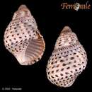
Planaxis sulcatus (Born, 1780)
Buccinum sulcatum Born, 1780:258, pl. 10: figs. 3, 6.
Buccinum pyramidale Gmelin, 1791:3488.
Planaxis sulcata Lamarck, 1822:51.—Quoy and Gaimard, 1833:486, pl. 33: figs. 25–39.
Planaxis undulata, Lamarck, 1822:51.
Planaxis sulcatus Lamarck.—Sowerby, 1822(12), pl. 1: fig. 1.
Planaxis buccinoides Deshayes, 1828:13.
Planaxis sulcatus (Born).—Sowerby, 1877, pl. 1: fig. 4.—Morlet, 1889:144.—Fischer, 1891:164.—Dautzenberg and Fischer, 1905:139–140.
This ubiquitous snail occurs in large populations throughout the Indo-Pacific among intertidal, rocky environments where it is frequently found on large rocks, stones, and beachrock in the low to midtidal zones. It grazes on microalgae covering rocky substrates in moderate to low energy habitats. When exposed to air during low tides, groups of snails frequently cluster together in crevices and depressions and are highly visible.
SHELL MORPHOLOGY (Figure 1).—Shell solid, wide, moderately elongate, reaching 35 mm in length, and comprising about 7 inflated whorls. Whorls are sculptured with incised spiral lines and grooves, the penultimate whorl having 6 grooves. Body whorl very large, wide, with an ovate aperture a little less than one-half the shell length. Outer lip smooth, slightly scalloped and denticulate within. Interior of aperture sculptured with deep grooves paralleling outer spiral grooves. Columella concave with slight callus and prominent parietal tooth at edge of anal canal. Base of body whorl moderately constricted with short, wide, anterior canal. Suture deeply incised. Larval shell has 3.5 whorls. Protoconch I with 1.5 smooth whorls. Protoconch II sculptured with fine subsutural axial plaits and two primary spiral cords, and a secondary presutural spiral of minute pustules (Figure 1E, F). Well-developed sinusigeral notch present in planktotrophic forms. Shell has white background color but usually of an overall brown-black aspect due to many spiral dark brown to black blotches. Interior of aperture purple, columella white. Periostracum thin, brown. Operculum large, lenticular, paucispiral with subterminal nucleus (Figure 1G).
EXTERNAL ANATOMY (Figure 2A).—The head-foot is black although the bases of the cephalic tentacles may be dusky white and the metapodium a yellowish dirty white. The sole of the foot is white. In females, there is a ciliated groove on the right side of the foot beginning at the distal end of the oviduct and ending in a swollen birth pore on the neck beneath the right cephalic tentacle (Figure 2A, bp). The mantle edge is slightly scalloped (Figure 2A, me).
MANTLE CAVITY ORGANS.—The osphradium is in the form of a very thin ridge flanked on each side by a thin, densely ciliated strip. The ctenidium is broad and composed of shallow, triangular filaments. There is a raised ridge at the basal side of the ctenidium adjacent to the osphradium. The hypobranchial gland is wide, thick, whitish in color, and divided into raised transverse ridges. A raised glandular pad of the hypobranchial gland lies adjacent to the distal end of the osphradium where it curves into the inhalant siphon. The intestine is wide and filled with many transversely stacked fecal pellets. The distal end of the pallial oviduct opens anterior to the rectum.
ALIMENTARY TRACT.—The buccal mass is very large, filling the snout (Figure 3D, bm). A small pair of chitinous jaws is situated at the edge of the inner lips. A long, robust radula curves under the buccal mass and dorsally around the nerve ring, terminating in the radular sac, which lies to the right of the nerve ring (Figure 3F). Buccal muscles are well developed.
Radula (Figure 3A, B). The radular ribbon is long, a little over one-half the shell length and has about 5 rows of teeth per millimeter. The rachidian tooth (Figure 3A) is squarish in shape, with a pentagonal basal plate on which there are a pair of basal lateral cusps, a rounded basal margin and a long thin lateral extension on each side. The cutting edge is convex and has a single broad spoon-like cusp. The rachidian formula is . The lateral tooth (Figure 3B) is rhomboidal and has a cutting edge with a broad, straight-edged central cusp flanked on each side by two small outer and inner denticles. There is a long lateral extension that extends downward and twists where it anchors the tooth to the basal membrane. The marginal teeth (Figure 3B) are long, narrow and expanded at the tips. The tip of the inner marginal tooth has five wide denticles while the tip of the outer marginal tooth has nine digitate denticles. Wide flange on outer margin of marginal tooth.
The paired salivary glands (Figure 3F, sg) are minute, coiled tubes with separate origins that begin behind the nerve ring but join into a single mass that is spread over the anterior dorsal surface of the esophageal gland. The salivary glands pass through the nerve ring as two uncoiled simple tubes, coiling again and uniting anterior to the nerve ring as a large mass that lies on the posterior dorsal surface of the buccal mass. The two anterior salivary ducts lie close together, but separate before emptying into the dorsolateral sides of the buccal mass.
The midesophagus widens immediately behind the nerve ring expanding laterally and thickening to form a wide esophageal gland. In section, the inner wall of the esophageal gland is folded into many deep longitudinal lamellae. The epithelial cells lining the esophageal gland stain deep purple with hemotoxylin (Figure 3E, eg). The esophageal gland extends posteriorly nearly the length of the mantle cavity before constricting to form the posterior esophagus.
The large stomach (Figure 2B) has a wide, posteriorly bilobed, raised pad that is T-shaped in profile and is highly muscular and mobile, particularly in its dorsal lips (Figure 2B, rp). This pad serves to separate the particulate matter entering the stomach from the esophagus on the right from the sorted particles being transported on the left toward the anterior typlosoles. The two typhlosoles (Figure 2B, t), located in the anterior portion of the stomach, most of the sorting area of the stomach (Figure 2B, sa), and the style sac (Figure 2B, ss) are weakly ciliated. The cuticularized gastric shield (Figure 2B, gs) is large. A short protostyle is present in freshly collected specimens. The single opening to the digestive gland (Figure 2B, ld) is close to the esophageal opening (Figure 2B, es) and lies adjacent to the left base of the bilobed pad.
REPRODUCTIVE SYSTEM.—The pallial gonoducts are tubular extensions of the coelomic gonoducts and developed from tissues of the floor and right wall of the mantle cavity. In all cerithiaceans, the pallial gonoduct is an open, slit tube comprising two laminae joined dorsally to each other: a lateral lamina attached to the mantle wall and a medial, free lamina. These laminae consist of loosely organized connective tissue covered by epithelium and contain the reproductive structures that are formed as sacular invaginations of the epithelium. The basal attachment of the pallial gonoduct, where the two laminae are connected, is highly glandular and thickened, forming the albumen gland. The channel at the base of the two laminae is the oviducal groove and is the part of the pallial oviduct in which eggs are fertilized, surrounded by albumen, and encapsulated while moving through the duct by ciliary transport. The distal end of the pallial gonoduct lies a few millimeters behind the mantle edge.
In Planaxis sulcatus, the male pallial gonoduct is a simple open tube with a proximal glandular prostate. The pallial oviduct, which is shown diagramatically in Figure 4, has a proximal albumen gland (Figure 4, ag) and a median capsule gland. The oviducal groove (Figure 4, og) is moderately glandular along its length. A long sperm gutter (Figure 4, sg) begins at the distal end of the medial lamina (Figure 4, ml) and extends to the median edge where it enters the lamina as a duct (Figure 4, osb) that branches into a large proximal spermatophore bursa (Figure 4, sb) and a smaller, median seminal receptacle (Figure 4, sr), lying beneath the spermatophore bursa. A highly ciliated groove emerges from the distal pallial oviduct and runs down the right side of the foot into the birth pore (Figure 2A, bp) of the brood pouch. The brood pore is a slit in the center of an elevated, lightly pigmented portion of the neck, beneath the peduncle of the right cephalic tentacle. The brood pouch (Figure 3C–F) in pregnant snails is a very large, complex structure partially divided into left and right chambers located in the head-foot (Figure 3C, D). It becomes extended and swollen when filled with embryos and is separated from the cephalic cavity and midesophagus by only a thin membrane (Figure 3E). In the anterior part of the foot both right and left chambers are subdivided into a series of many narrow, longitudinal chambers separated by thin partitions (Figure 3D). These small chambers on both sides of the foot unite posteriorly to form a single large chamber that extends dorsally to the base of the buccal cavity and posteriorly over the right side of the buccal mass and esophagus just below the dorsal body wall. The dorsal portion of the chamber is further subdivided by a few thin membranous partitions and is separated from the buccal cavity and esophagus by a thin membrane. Embryos are found throughout the entire brood pouch and may comprise several cohorts of different growth stages. The more advanced embryos appear to be in the dorsal portion of the chamber. Each embryo is contained in a thin egg capsule from which it breaks out prior to expulsion from the brood pore in the right side of the neck. The stage at which embryos hatch from the brood pore and the kind of development that follows appears to vary between populations in the Indian and Pacific Oceans. Indirect evidence derived from field observation and the study of larval shells indicate that the population from South Mission Beach, North Queensland, Australia, has a moderate lecithotrophic type of development and a short planktonic phase. Larval shells from this population have a sinusigeral notch and protoconch sculpture indicative of a free swimming larval stage. My observations agree with those of Risbec (1935) who noted a similar type of development from a New Caledonian population. He found that cleavage started in the pallial oviduct and that the embryos reached the brood pouch at the morula stage. Risbec (1935:387388) stated that well-developed larvae, ready to hatch, as well as early embryos, fresh from the oviduct, were present in the brood pouch. In the New Caledonian population, larvae left the brood pouch and became pelagic when each larval shell reached 0.1 mm across and the velum was well developed. Risbec (1935:387–388) noted emerging larvae had not developed eyes. Other types of development have been attributed to populations of P. sulcatus in the northwestern Indian Ocean. Thorson (1940) recorded that larvae of a population from the Persian Gulf developed into small snails and fed on nurse eggs within the brood pouch prior to hatching. In some cases, a few embryos reached the free crawling stage and attained considerable size (3.25 mm) within the brood pouch. Barkati and Ahmed (1982) recorded direct development in a population from Buliji, near Karachi, Pakistan. In this population crawling snails hatched out at a length of 0.38 mm. Other populations from the northwestern Indian Ocean need to be studied to determine if direct development is the usual mode in that region. There may be two cryptic species involved: an Indian Ocean species with ovoviviparous development, and an Indo-Pacific species with indirect development, including partial brooding followed by a planktonic phase.
NERVOUS SYSTEM.—The nervous system of P. sulcatus has been described in detail by Risbec (1935:391) and my observations are in agreement with his. The subesophageal ganglion is attached to the left pleural ganglion and the visceral ganglion lies at the posterior end of the mantle cavity, forming a ganglionic mass to the right of the esophagus, with a secondary mass crossing over the esophagus. The cerebral-pleural connectives are very long. The large pedal ganglia each have two swollen extensions or strands, the external one being more developed than the internal one. These appear to innervate the brood pouch. The RPG ratio (Davis et al., 1976:263) is O.57 (n = 5), indicating a tight central nervous system.
Fissilabia Macgillivray, 1836:42 [type-species: Fissilabia fasciata Macgillivray, 1836 (= Planaxis decollata Quoy and Gaimard, 1833), by moonotype].
Quoyia Gray, 1839:125.—Cossmann, 1906:197.—Thiele, 1929:203.—Wenz, 1940:721. [Type-species: Planaxis decollata Quoy and Gaimard, 1833, by monotypy].
Leucostoma Swainson, 1840:172, 336 [not Leucostoma Meigen, 1803].
Quoya.—Deshayes, 1843:236 [misspelling].
Fissilabria Brown [sic].—Gray, 1847:138 [wrong author and misspelling].—Oostingh, 1925:43.
Fissilabra Brown [sic].—Paetel, 1875:81 [wrong author and misspelling].
TYPE-SPECIES.—Planaxis decollata Quoy and Gaimard, 1833, by monotypy.
DIAGNOSIS.—Shell tall, thick, light tan to brown, decollate and sculptured with many spiral grooves. Aperture narrow, ovate with slight columellar callus and columellar plait below parietal area. Outer lip smooth, internally grooved. Periostracum yellowish brown. Radula long, comprising about 800 rows of teeth. Cutting edge of rachidian tooth with single, large, wide, blunt cusp; basal plate with two medium-sized cusps and two lateral extensions slightly bifurcated at their tips. Seminal receptacle in median portion of pallial oviduct. Larger portion of partitioned brood pouch in right side of head-foot. Embryos lecithotrophic. Salivary glands coiled, esophageal gland wide. High rocky supratidal habitat.
- bibliographic citation
- Houbrick, Richard S. 1987. "Anatomy, reproductive biology, and phylogeny of the Planaxidae (Cerithiacea: Prosobranchia)." Smithsonian Contributions to Zoology. 1-57. https://doi.org/10.5479/si.00810282.445