-
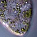
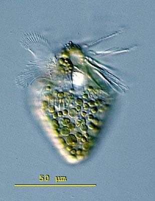
The body of Limnostrombidium viride is ob-conical (upside down egg) and living measure 50 - 70 X 40 - 45 microns. The euquatorial region is marked by a girdle of extrusomes. The posterior half of the cell has a pattern of hexagonal colourless platelets. The cytoplasm is greenish or yellow-green because of the symbiotic algae. No contractile vacuole. The macronucleus is ellipsoidal and located in the mid-body. This ciliate swims quickly using 16 buccal membranelles. This specimen was collected in the plankton from Lake Constance, Germany. This image shows the oral apparatus of Limnostrombidium viride with an adoral zone of membranelles that terminate above the mid-body. Measuring 70 microns. Differential interference contrast.
-
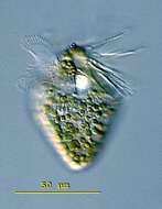
Limnostrombidium (lim-no-stom-bid-ee-um). The body of Limnostrombidium viride is ob-conical (upside down egg) and living measure 50 - 70 X 40 - 45 microns. The equatorial region is marked by a girdle of extrusomes. The posterior half of the cell has a pattern of hexagonal colourless platelets. The cytoplasm is greenish or yellow-green because of the symbiotic algae. No contractile vacuole. The macronucleus is ellipsoidal and located in the mid-body. This ciliate swims quickly using 16 buccal membranelles. This specimen was collected in the plankton from Lake Constance, Germany. This image shows outer adoral zone of membranelles and the symbiotic algae of Limnostrombidium viride. 70 microns. Differential interference contrast.
-
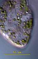
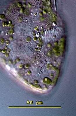
Limnostrombidium (lim-no-stom-bid-ee-um). The body of Limnostrombidium viride is ob-conical (upside down egg) and living measure 50 - 70 X 40 - 45 (m. The equatorial region is marked by a girdle of extrusomes. The posterior half of the cell has a pattern of hexagonal colourless platelets. The cytoplasm is greenish or yellow-green because of the symbiotic algae. No contractile vacuole. The macronucleus is ellipsoidal and located in the mid-body. This ciliate swims quickly using 16 buccal membranelles. This specimen was collected in the plankton from Lake Constance, Germany. This image shows the posterior end of a squashed specimen of Limnostrombidium viride with the focal plane on the hexagonal platelets. Each platelet measures 3.5 - 4 microns. Differential interference contrast.