-
Photo by Luxmmi Varathan & Craig Perl.
-
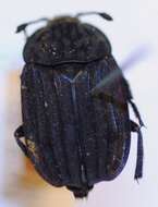
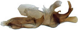
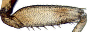
Protochiasmus mysticus, profemur (MZSP)
-
Michel P. Valim, Jason D. Weckstein
Zookeys
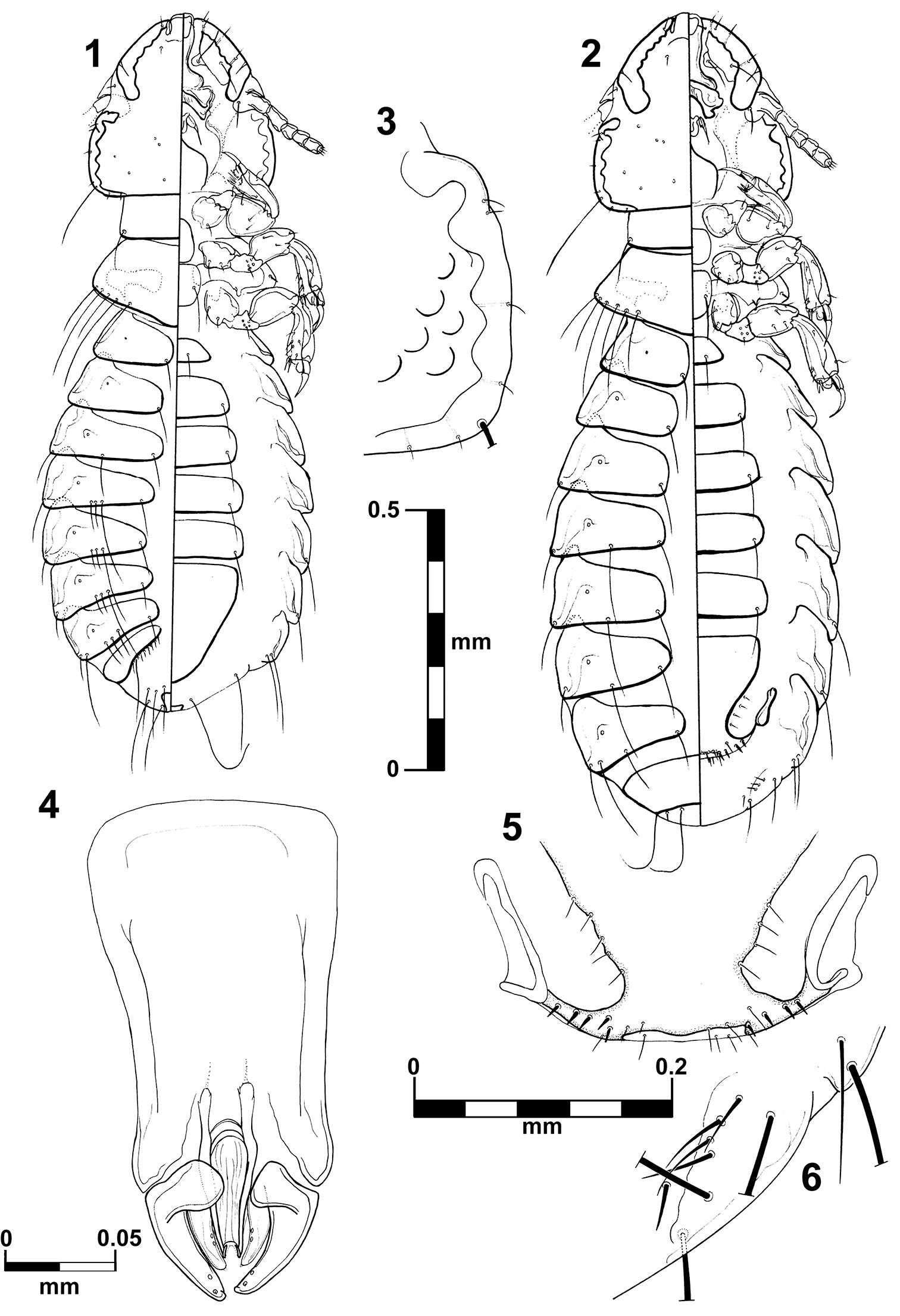
Figures 1–6.Brueelia sueta sp. n.: male, dorso-ventral views (1); female, dorso-ventral views (2); temporal carina (3); male genitalia (4); female vulvar margin (5); female gonapophysis (6).
-
Juli Pujade-Villar, Paul Hanson, Claudia A. Medina, Miguel Torres, George Melika
Zookeys
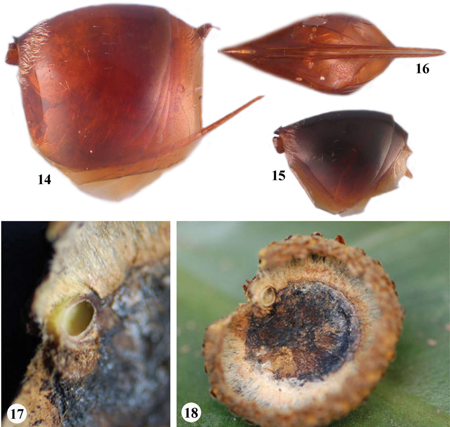
Figures 14–18.Zapatella grahami 14 metasoma, female (lateral view) 15 metasoma, male (lateral view) 16 ventral spine of hypopygium (ventral view) 17–18 gall.
-
Pierfilippo Cerretti, D. Monty Wood, James E. O’Hara
Zookeys
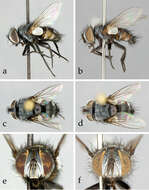
Figure 1.Neoethilla gen. n.ignobilis (New Mexico) a–d habitus a male in lateral view b female in lateral view c male in dorsal view d female in dorsal view d–e head in frontal view d male e female.
-
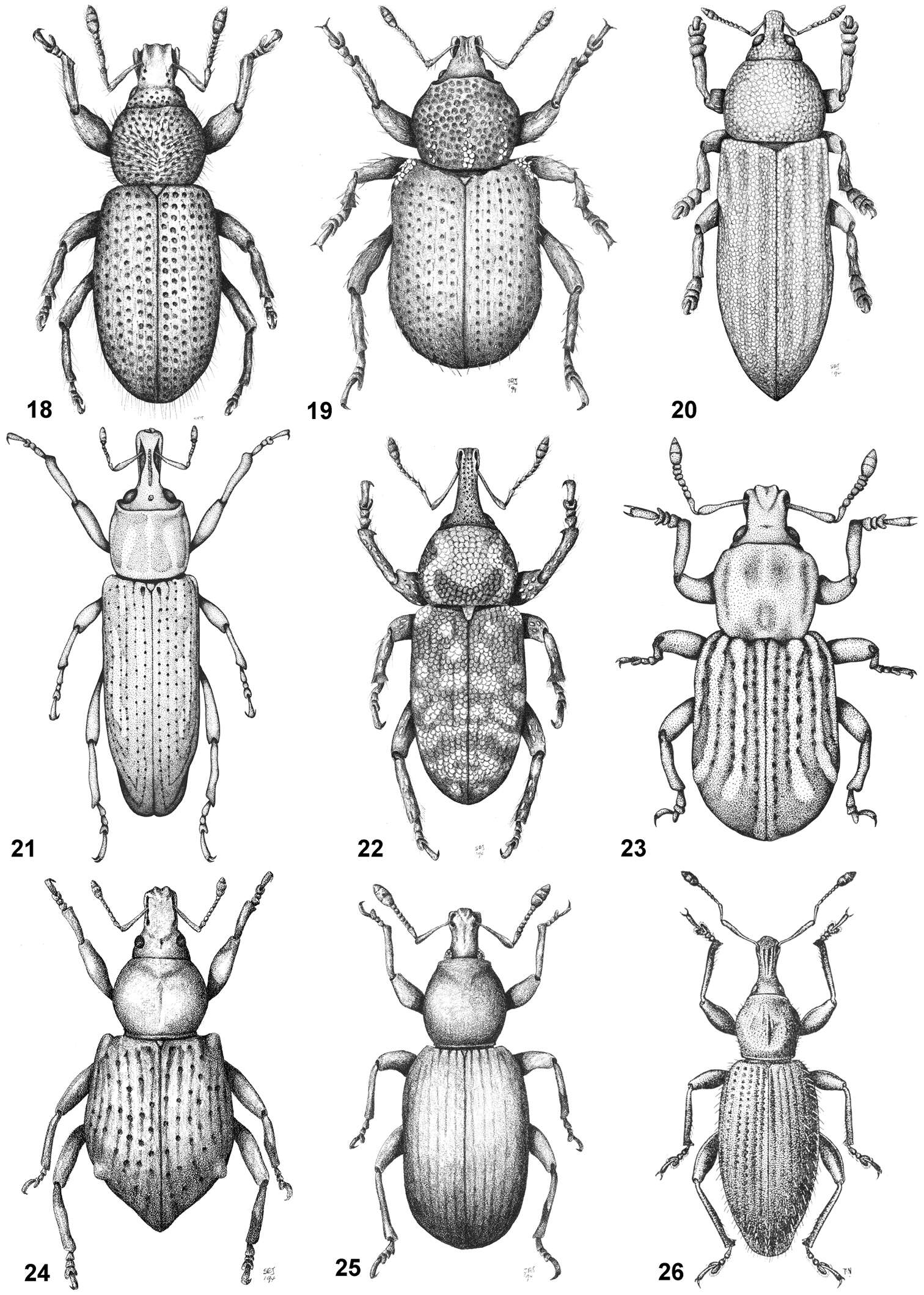
Figures 18–26.Habitus of representative Listroderini. 18 Adioristidius hirsutus 19 Puranius nigrinus 20 Haversiella albolimbata 21 Listronotus bosqi 22 Neopachytychius squamosus 23 Falklandiellus suffodens 24 Falklandiopsis magellanica 25 Falklandius antarcticus 26 Gromilus veneris.
-
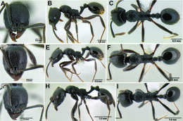
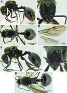
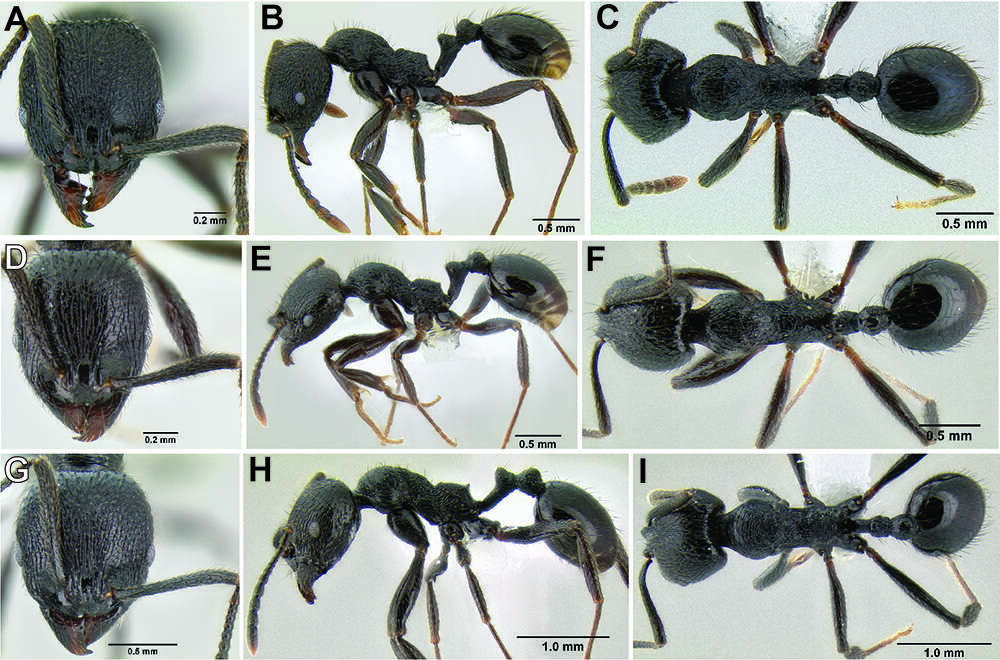
Figure 120.Stenamma megamanni worker variants. Face, profile, dorsal views A–C Variant 1 (CASENT0621836) D–F Variant 2 (CASENT0606734) G–I Variant 3 (CASENT0603924).
-
Felipe N. Soto-Adames, Steven J. Taylor
Zookeys
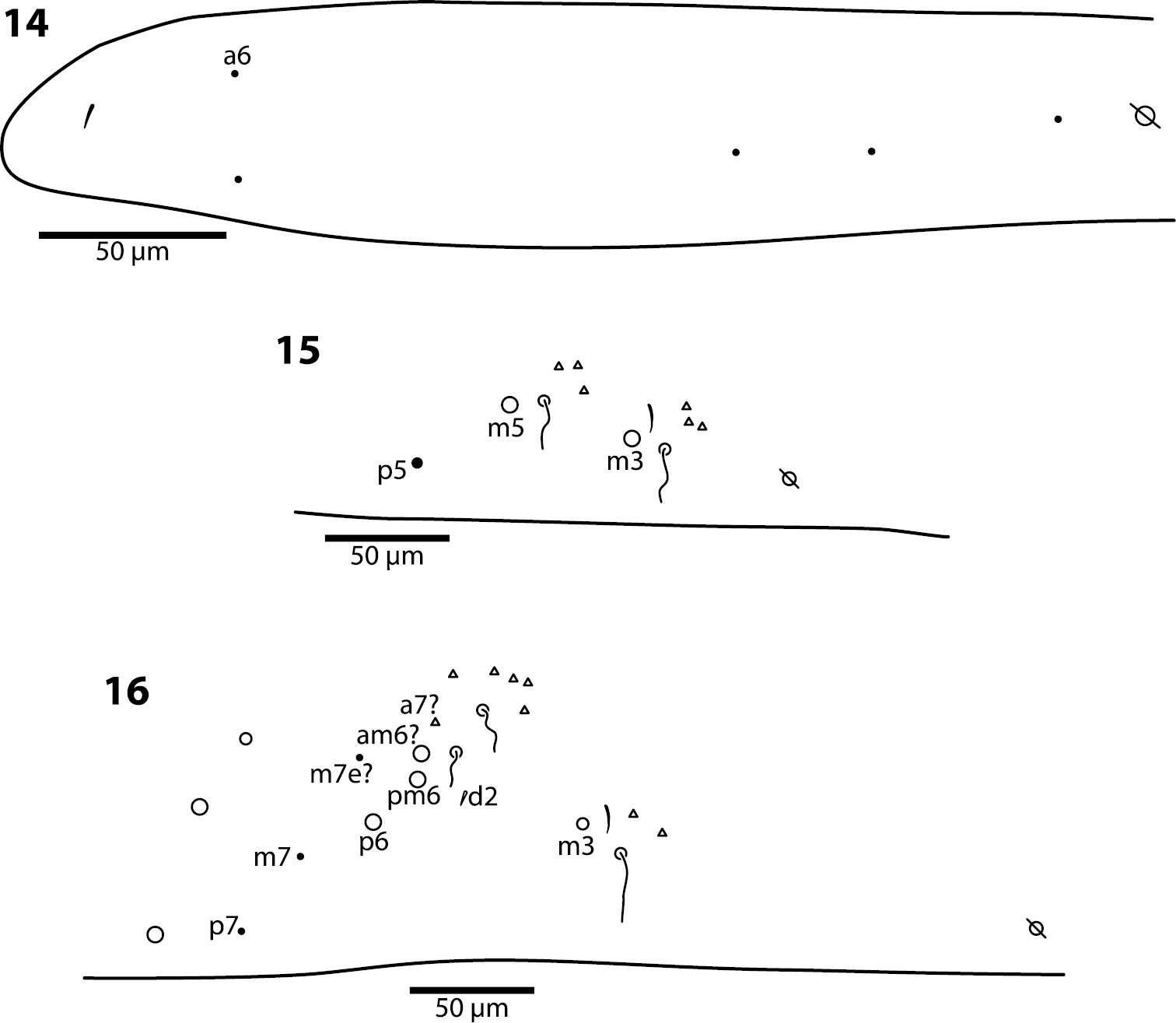
Figures 14–16.Trogolaphysa giordanoae sp. n. Dorsal chaetotaxy of abdominal segments 1–3, triangles are fan-shaped setae, circles are macrochaetae, filled are circles ciliate microchaeta14 First abdominal segment 15 Second abdominal segment 16 Third abdominal segment.
-
Figures 1–10.1–2. Symploce torchaceus Feng & Woo, male: 1 holotype, dorsal view 2 same, ventral view 3–4 Symploce bispot Feng and Woo, male: 3 holotype, dorsal view 4 same, ventral view 5–6 Symploce sphaerica sp. n., male: 5 holotype, dorsal view 6 same, ventral view 7–8 Symploce paramarginata sp. n., male: 7 holotype, dorsal view 8 same, ventral view 9–10 Symploce evidens sp. n., male: 9 holotype, dorsal view 10 same, ventral view. Scale bars = 1.0 cm.
-
Lizhi Huo, Xingmin Wang, Xiaosheng Chen, Shunxiang Ren
Zookeys
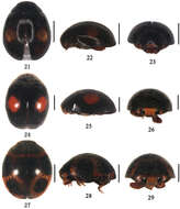
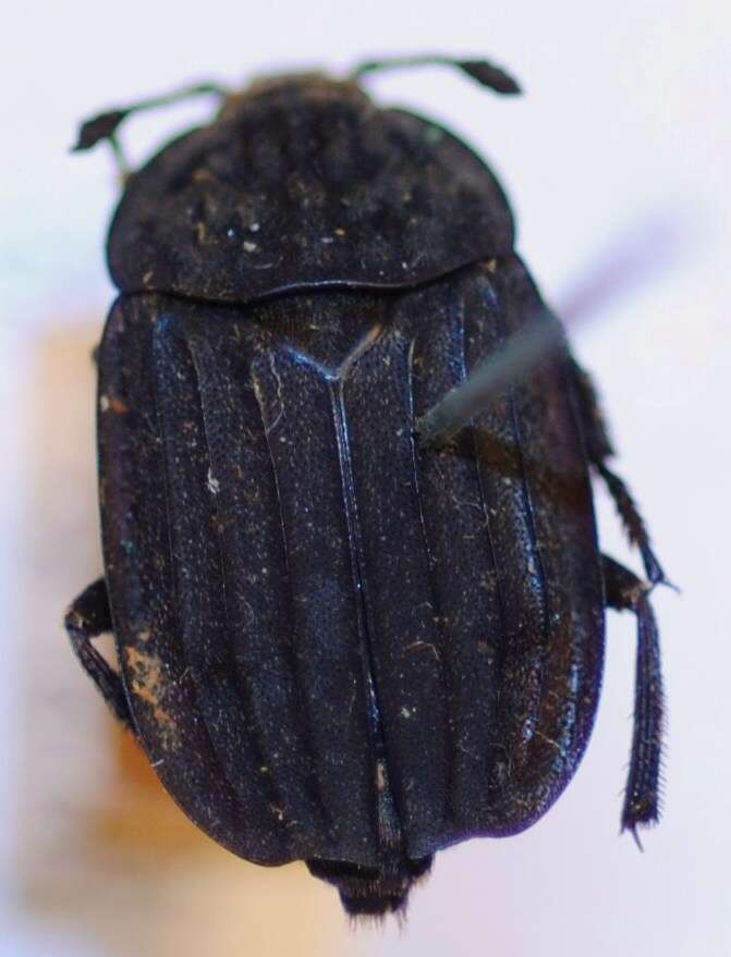
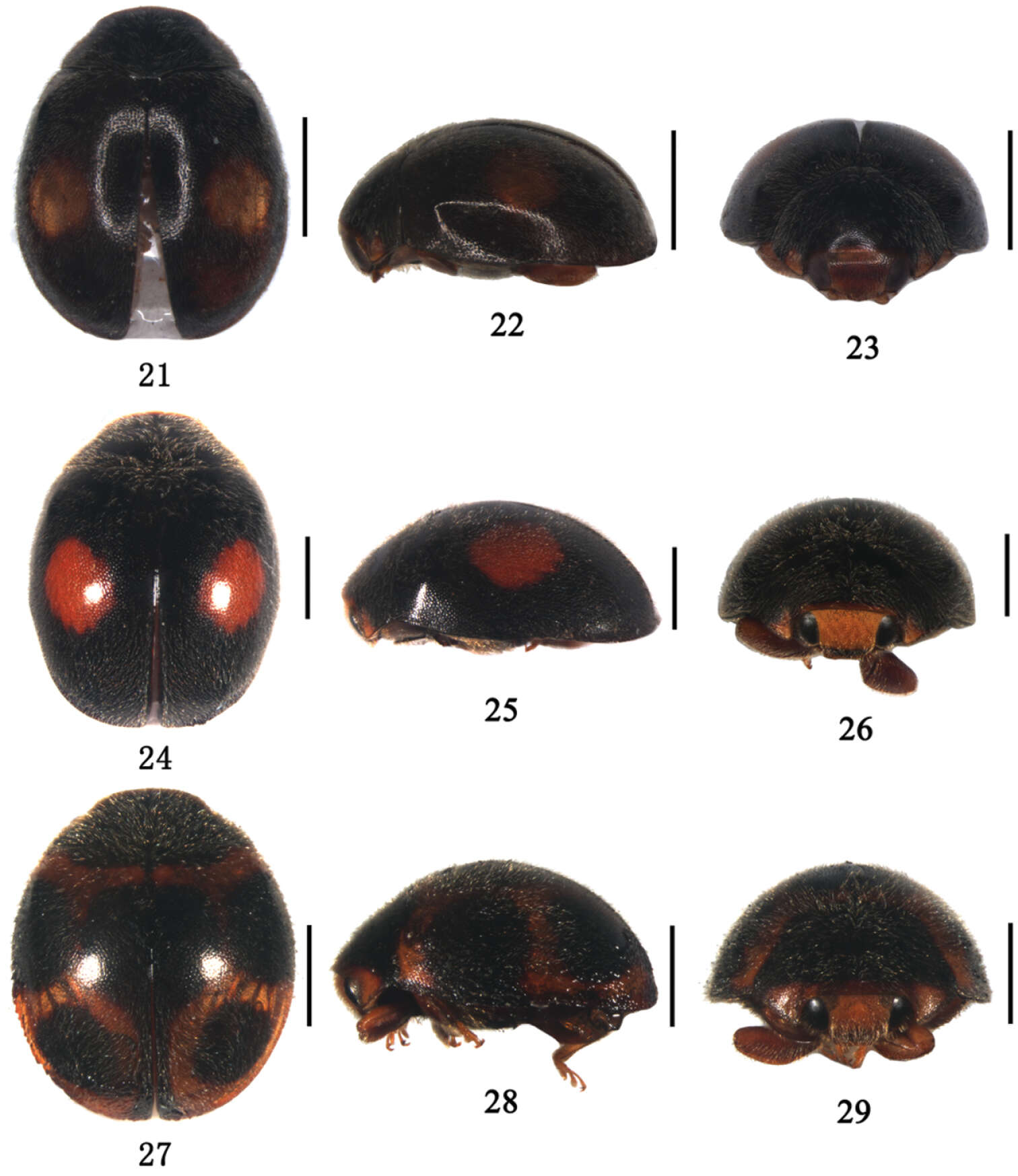
Figures 21–29.21–23 Aspidimerus guangxiensis Yu, 21 dorsal view 22 lateral view 23 frontal view 24–26 Aspidimerus matsumurai Sasaji, 24 dorsal view 25 lateral view 26 frontal view 27–29 Aspidimerus kabakovi Hoàng 27 dorsal view 28 lateral view 29 frontal view. Scale bars: 1.0mm.
-
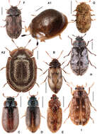
Figure 10.A Thymalus limbatus B Phloiophilus edwardsi C Eronyxa marginicollis D Decamerus haemorhoidalis E Diontolobus punctatipennis F Afrocyrona ciskeiensis G Afrocyrona dwesae H Grynoma sp., New Zealand I Grynoma diluta.
-
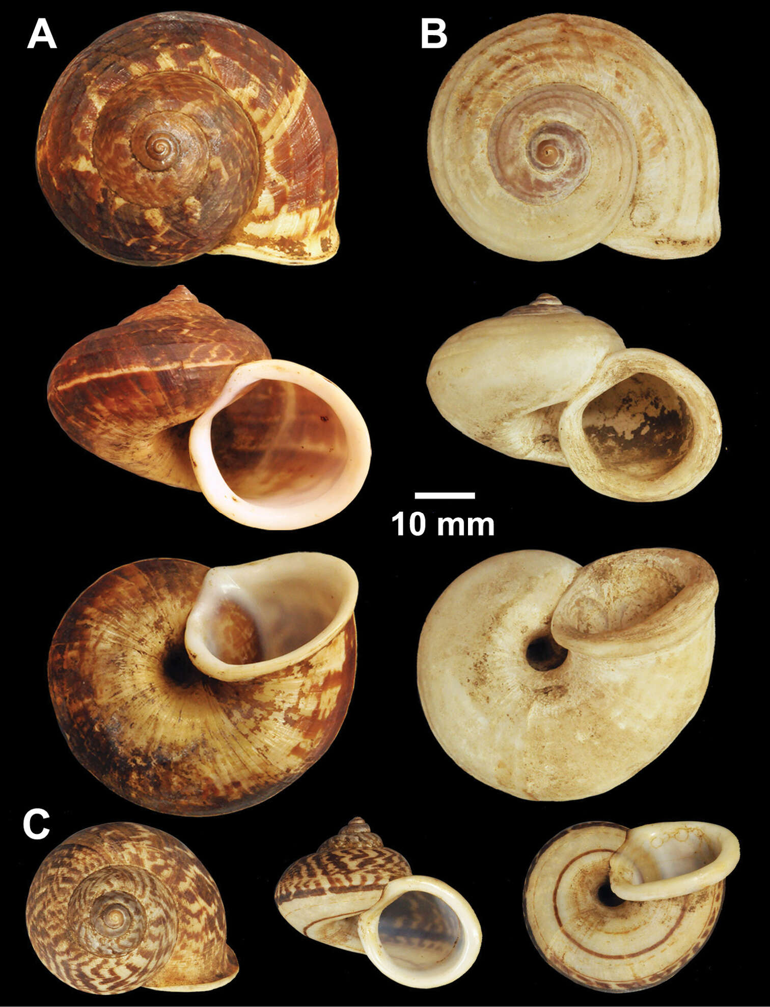
Nattawadee Nantarat, Chirasak Sutcharit, Piyoros Tongkerd, Jonathan Ablett, Fred Naggs, Somsak Panha
Zookeys
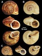
Figure 2.Types of Cyclophorus species. A, B Cyclophorus aborensis Godwin-Austen, 1915 A lectotype NHMUK 1903.7.1.3051 and B paralectotype NHMUK 1903.7.1.3048 C Cyclophorus affinis Theobald, 1858, lectotype NHMUK 1903.7.1.1454.
-
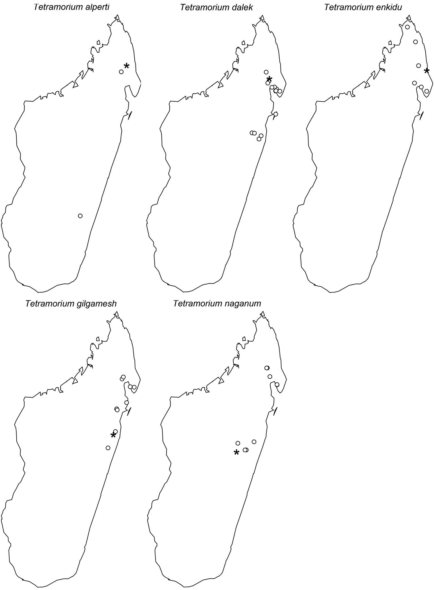
Francisco Hita Garcia, Brian L. Fisher
Zookeys
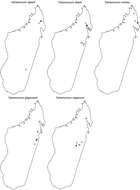
Figure 61.Geographic distribution maps for the species of the Tetramorium naganum species group. Star symbols represent type localities while circles represent non-type localities.
-
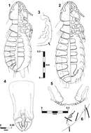
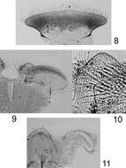
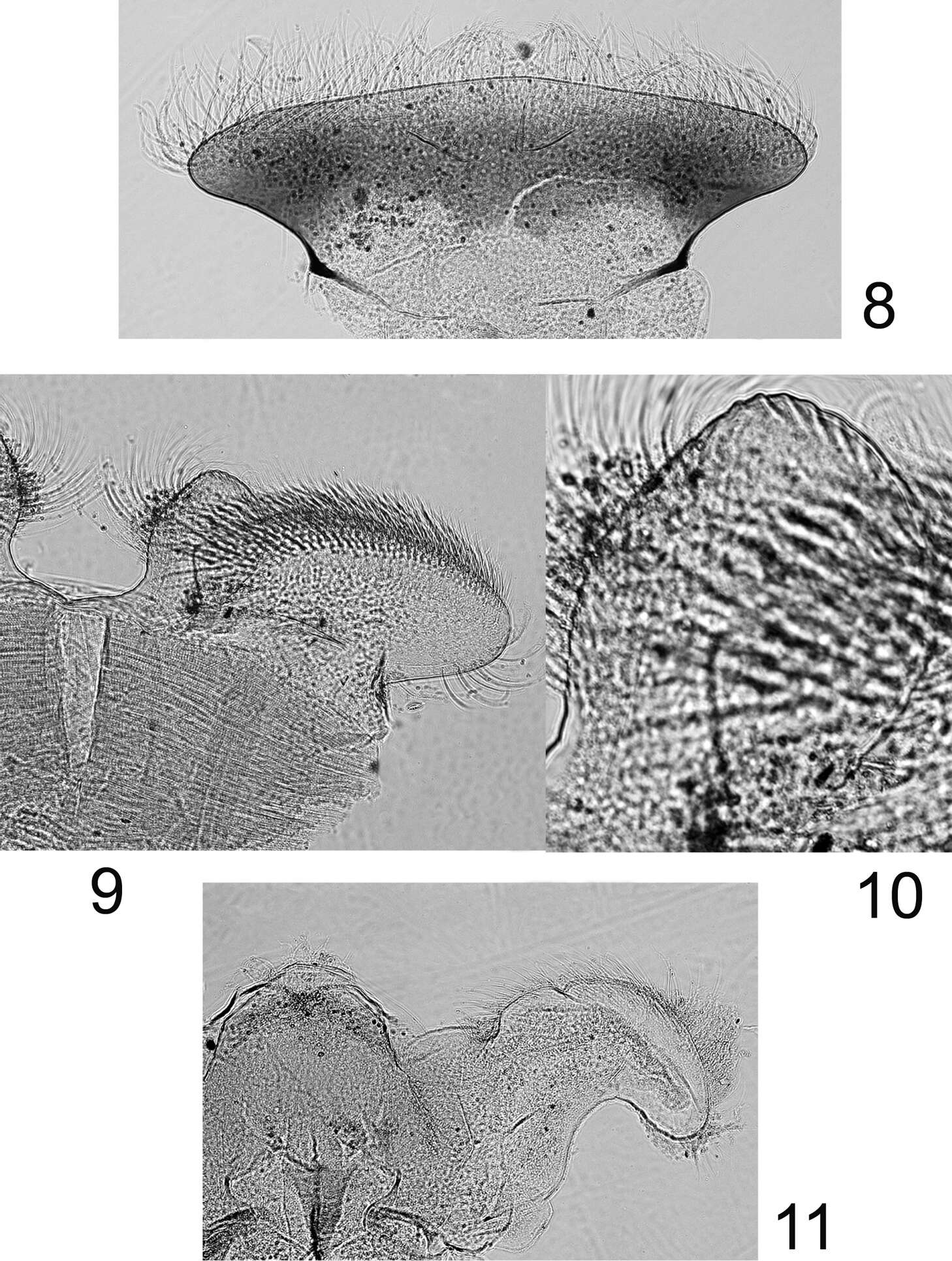
Figures 8–11.Rhithrogeniella ornata Ulmer, 1939, nymphal mouthparts. 8 Labrum in dorsal view 9 Left glossae and paraglossae of the labium 10 Detail of the glossae from 9 11 Hypopharynx, ventral view lingua and left superlingua.
-
Donald R. Davis, David L. Wagner
Zookeys
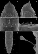
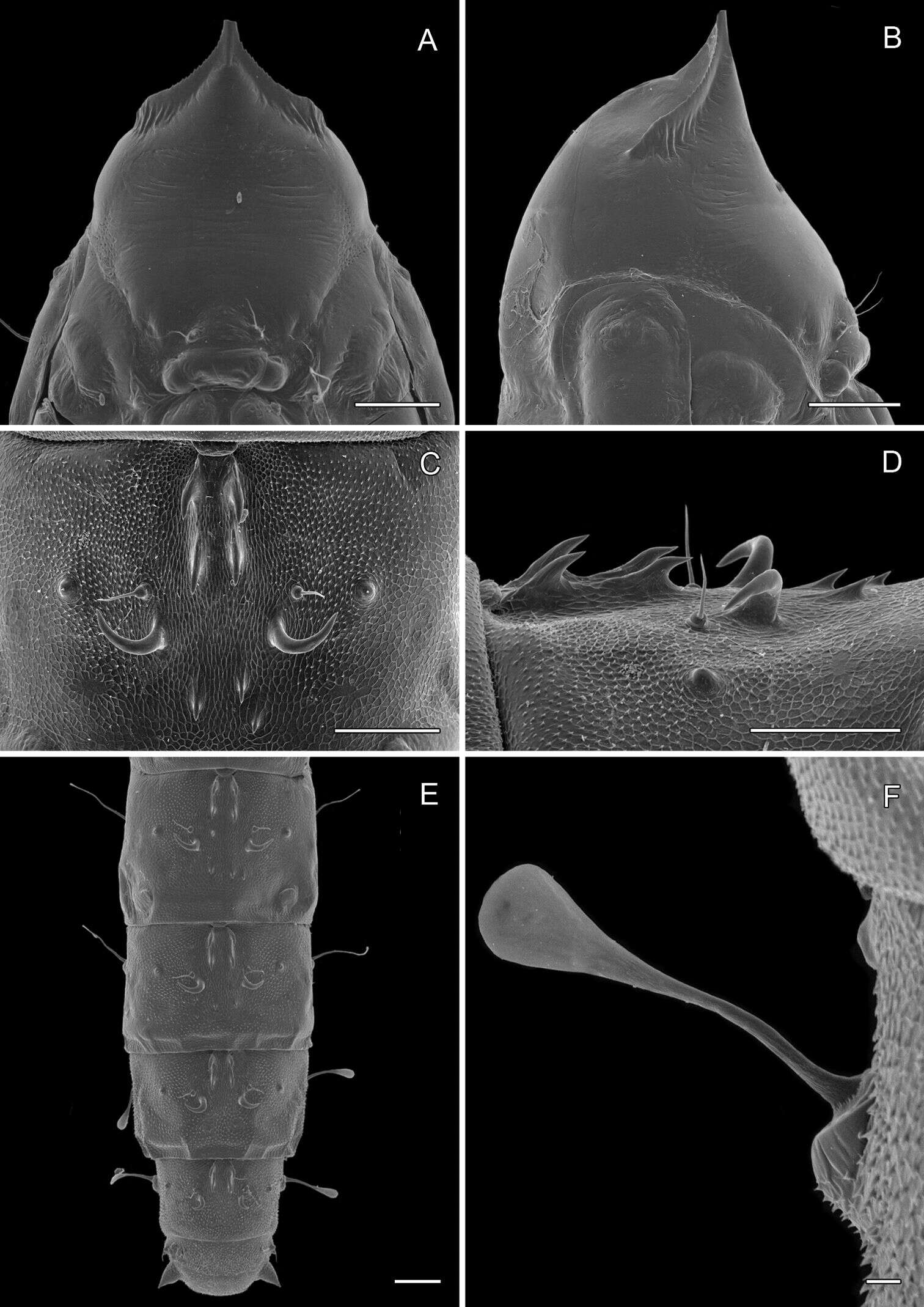
Figure 14.Phyllocnistis perseafolia sp. n. pupa. A Head, ventral view (176 µm) B Head, lateral view (200 µm) C Dorsal spines of abdominal tergum 5 (76 µm) D Lateral view of C (60 µm) E Abdominal terga 4-10 (100 µm) F Lateral seta of abdominal segment 6 (10 µm). (Length of bar scales shown in parentheses.)
-
-
Georgia, United States
-
Mukaria maculata, aedeagus, connective, and style, laterally (INHS)
-
Michel P. Valim, Jason D. Weckstein
Zookeys
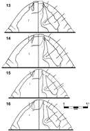
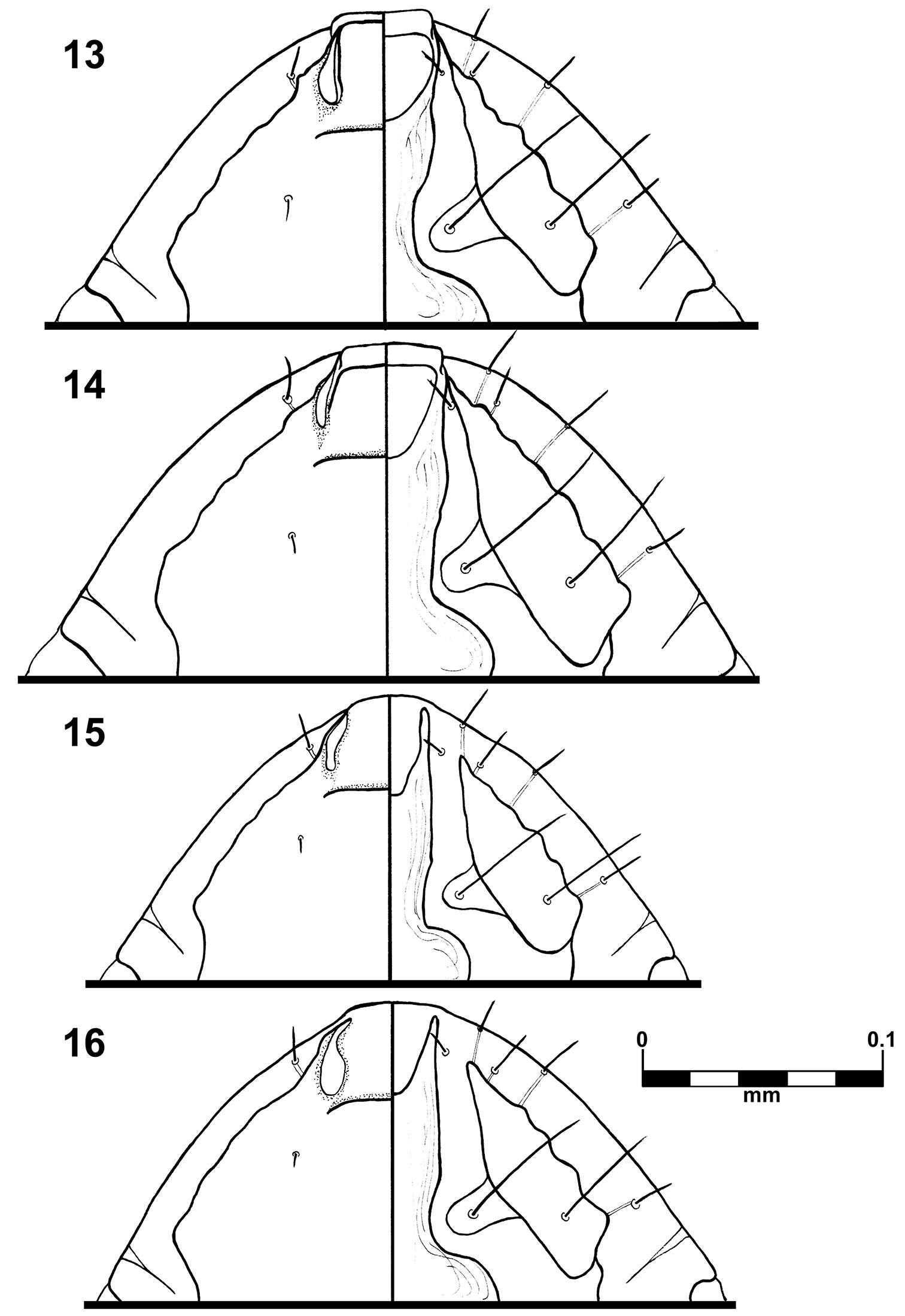
Figures 13–16.Brueelia sueta sp. n.: male preantenal region, dorso-ventral views (13); female preantenal region, dorso-ventral views (14); Brueelia cicchinoi sp. n.: male preantenal region, dorso-ventral views (15); female preantenal region, dorso-ventral views (16).
-
Juli Pujade-Villar, Paul Hanson, Claudia A. Medina, Miguel Torres, George Melika
Zookeys
Figures 19–24.Zapatella nievesaldreyi, female 19 head (anterior view) 20 head (dorsal view) 21 head (posterior view) 22 mesosoma (dorsal view) 23 mesosoma (lateral view) 24 tarsal claw.
-
Pierfilippo Cerretti, D. Monty Wood, James E. O’Hara
Zookeys
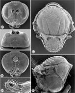
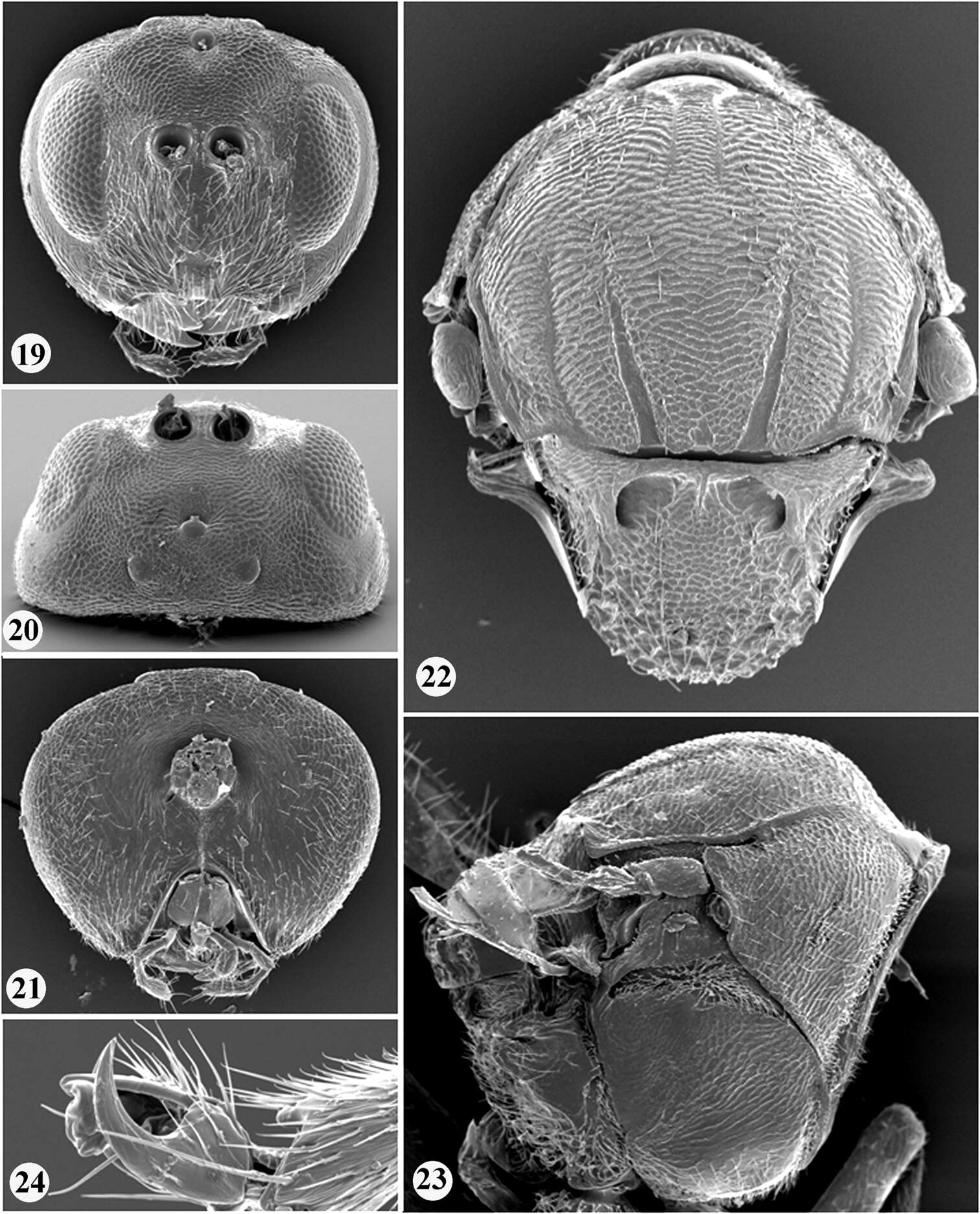
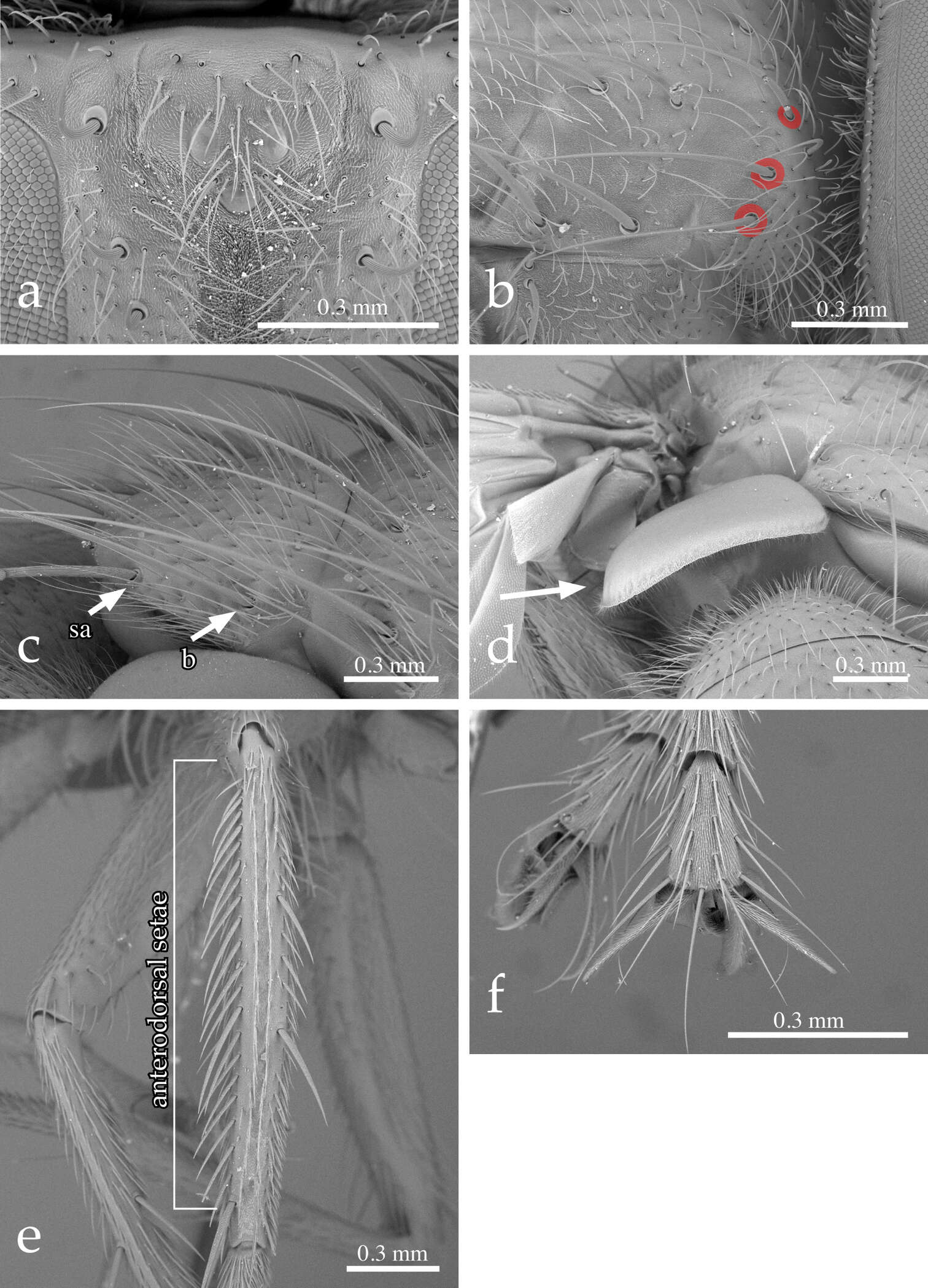
Figure 2.Neoethilla gen. n.ignobilis (male, New Mexico) a vertex in anterodorsal view b right postpronotum and part of presutural portion of scutum in laterodorsal view [circles indicate basal postpronotal saetae] c scutellum in laterodorsal view [b = basal scutellar seta; sa = subapical scutellar seta] d lower calypter in posterior view e left hind tibia in dorsal view f fore claws.
-
Figures 27–35.Habitus of representative Listroderini. 27 Lanteriella microphtalma 28 Telurus caudiculatus 29 Acrorius papallacta 30 Acrostomus bruchi 31 Antarctobius lacunosus 32 Germainiellus dentipennis 33 Lamiarhinus aelficus 34 Listroderes annulipes 35 Philippius superbus.
-
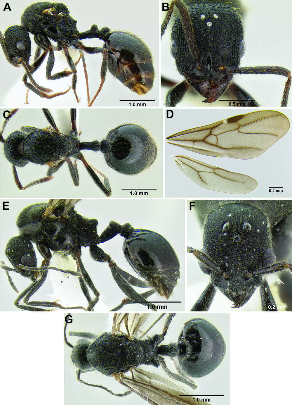
Figure 121.Stenamma megamanni A Paratype queen (CASENT0604840), profile B Same, face C Same, dorsum D Same, wings E Male (CASENT000007293), profile F Same, face G Same, dorsum.
-
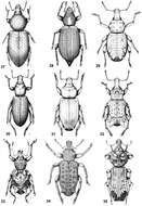
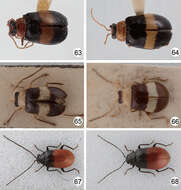
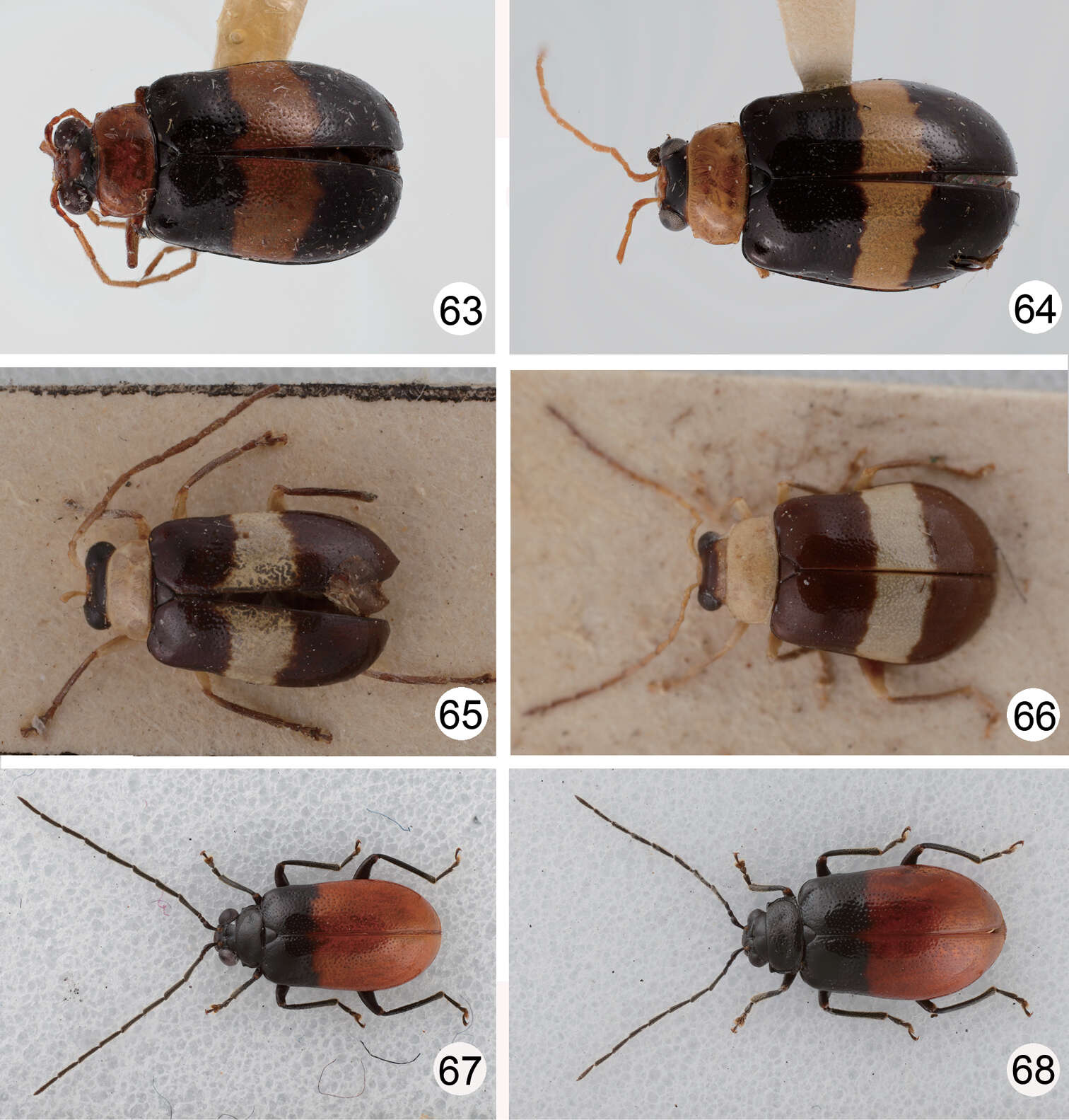
Figures 63–68.Dorsal habitus of Dercetina and Arthrotus species. 63 Dercetina unifasciata, male 64 Dercetina unifasciata, female 65 Arthrotus flavocincta, male 66 Arthrotus flavocincta, female 67 Arthrotus nakanei, male; 68. Arthrotus nakanei, female.