-
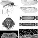
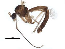
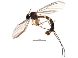
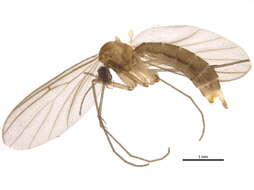
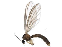
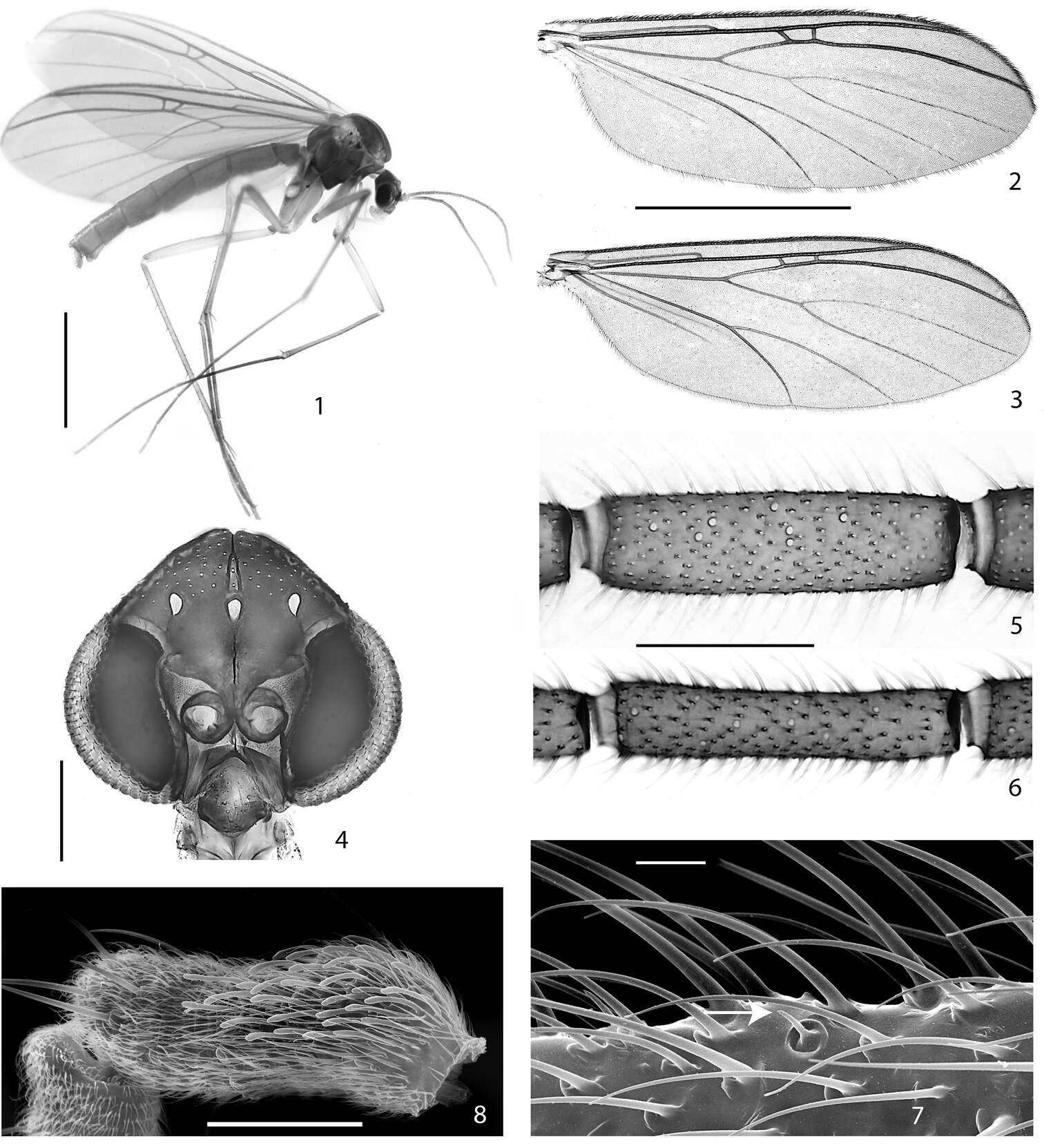
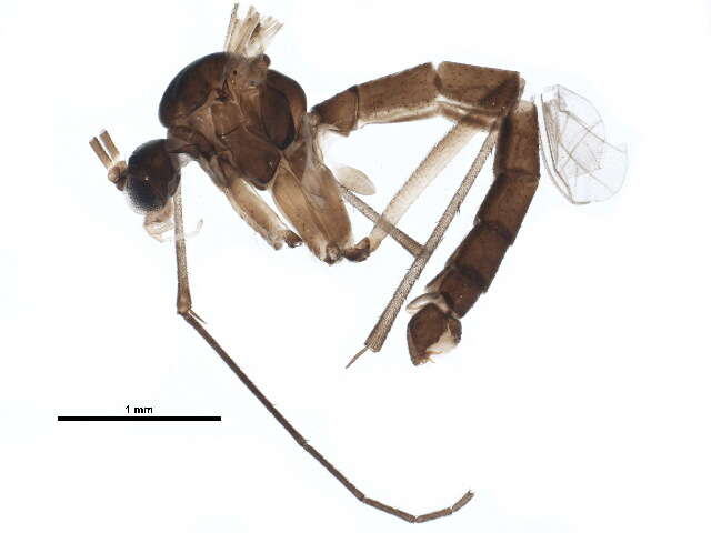
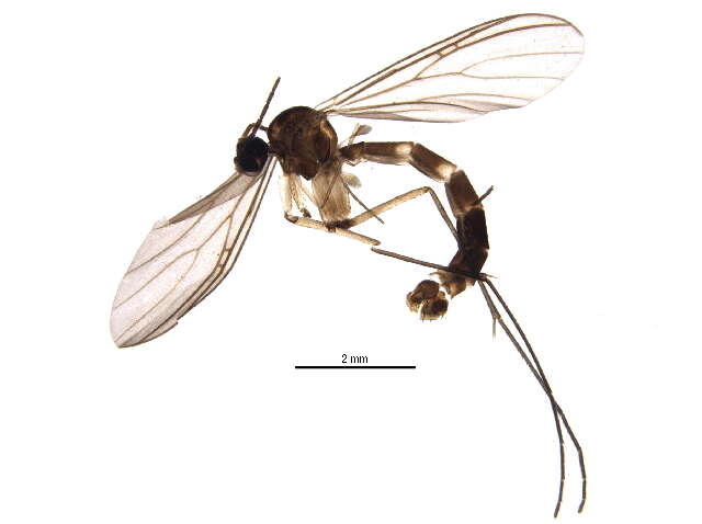
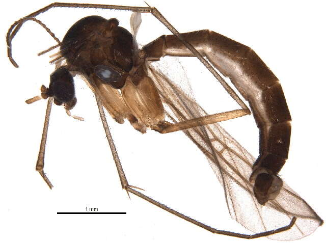
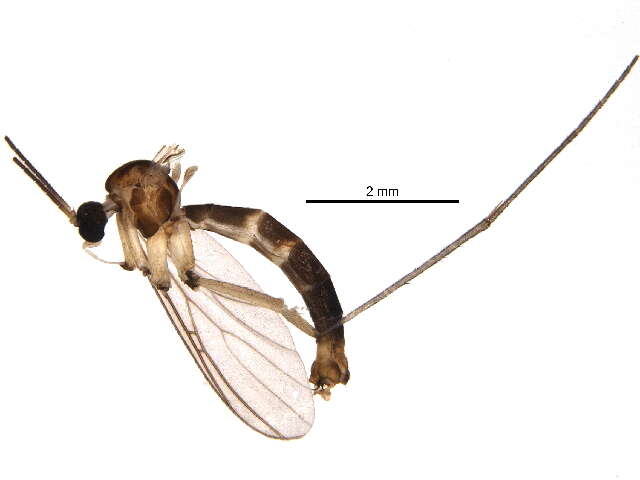
Figures 1–8.1–2, 4–5, 7–8. Acomopterella martinovskyi. 1 Female habitus 2 Male wing 4 Male head, frontal view 5 Flagellomere 4, male. Sensilla chaetica visible as pale spots. 7 Sensillum chaeticum (at arrowhead) on flagellomere 10, male 8 Sensilla on palpomere 3, male 3, 6 Acomopterella yoshiwaesp. n., male. 3 Wing 6 Flagellomere 4. Length of scale bar = 2 mm (for 1–3), 200 µm (for 4), 100 µm (for 5–6), 10 µm (for 7), 50 µm (for 8).
-
Figures 14–20.Terminalia of Acomopterella martinovskyi, male. 14 Terminalia in dorsal view, tergite IX removed 15 Gonocoxites in ventral view 16 Epiproct and hypoproct, dorsal view 17 Abdominal segments VII & VIII and terminalia in lateral view 18–19 Posterior view of terminalia 20 Cercal setae in detail. Length of scale bar = 100 µm (for 14–15, 18–19), 50 µm (for 16, 20), 0,5 mm (for 17).
-
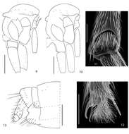
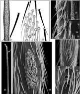
Figures 31–38.Mid tibial organ. Not on the same scale. 31–32 Acomopterella yoshiwae sp. n. 33–34 Acomopterella martinovskyi 33 General outer view of mid tibia, the arrowhead points to the tibial organ 35–36 Speolepta leptogaster 37–38 Ectrepesthoneura hirta.
-
Figures 1–8.1–2, 4–5, 7–8. Acomopterella martinovskyi. 1 Female habitus 2 Male wing 4 Male head, frontal view 5 Flagellomere 4, male. Sensilla chaetica visible as pale spots. 7 Sensillum chaeticum (at arrowhead) on flagellomere 10, male 8 Sensilla on palpomere 3, male 3, 6 Acomopterella yoshiwaesp. n., male. 3 Wing 6 Flagellomere 4. Length of scale bar = 2 mm (for 1–3), 200 µm (for 4), 100 µm (for 5–6), 10 µm (for 7), 50 µm (for 8).
-
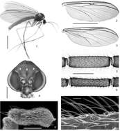
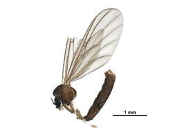
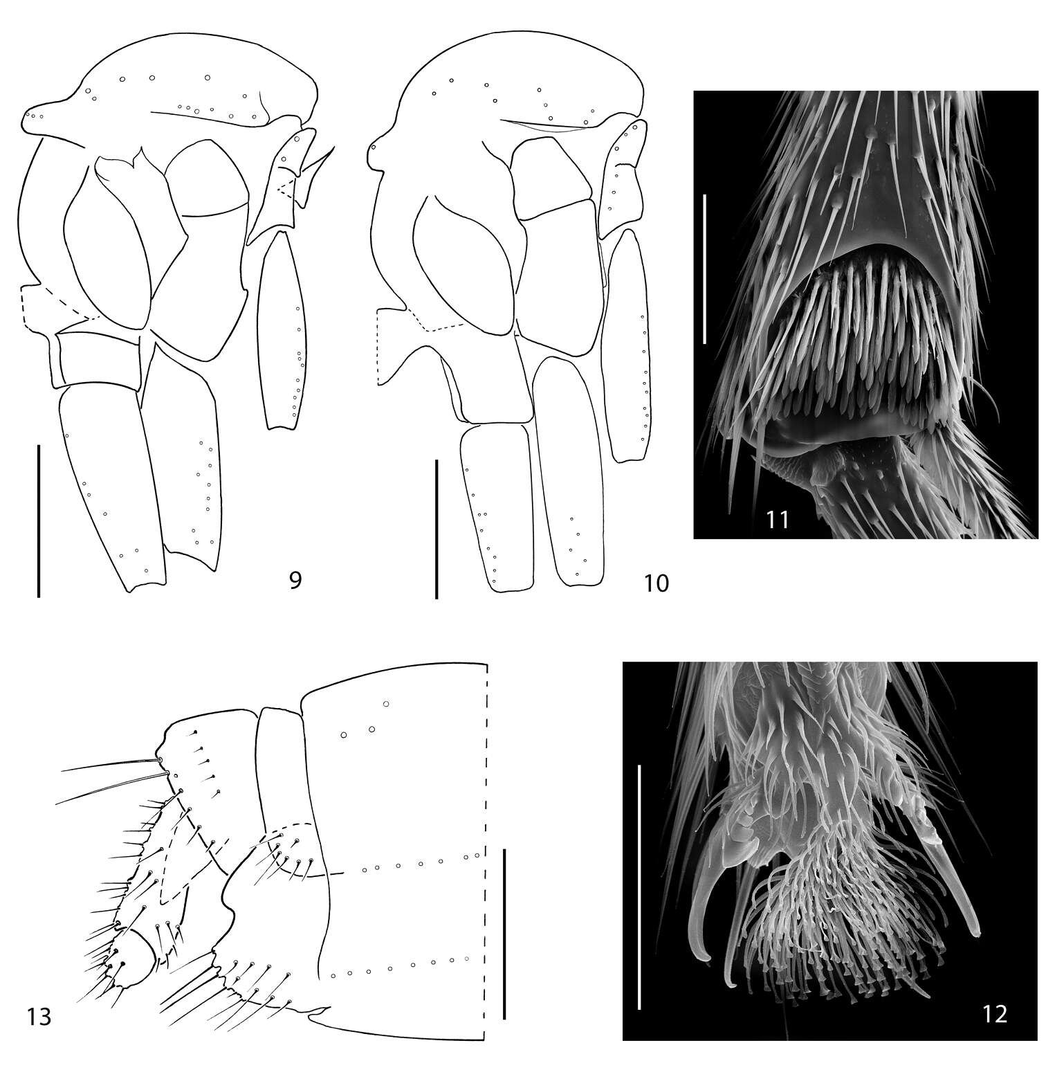
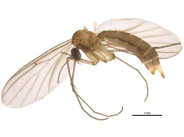
Figures 9–13.9 Acomopterella yoshiwaesp. n., thorax in lateral view. 10–13 Acomopterella martinovskyi. 10 Thorax in lateral view 11 Fore tibial organ, male 12 Fore claw, male 13 Female terminalia in lateral view. Length of scale bar = 0,5 mm (for 9–10), 50 µm (for 11–12), 250 µm (for 13).
-
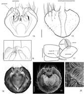
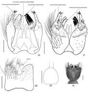
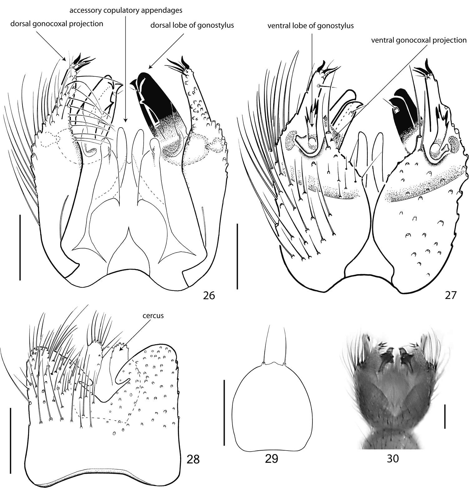
Figures 26–30.Terminalia of Acomopterella yoshiwae sp. n., male. 26 Terminalia in dorsal view, tergite IX removed 27 Terminalia in ventral view 28 Tergite IX, dorsal view 29 Hypoproct, ventral view 30 Terminalia incl. tergite IX in dorsal view. Length of scale bar = 100 µm.
-
Figures 31–38.Mid tibial organ. Not on the same scale. 31–32 Acomopterella yoshiwae sp. n. 33–34 Acomopterella martinovskyi 33 General outer view of mid tibia, the arrowhead points to the tibial organ 35–36 Speolepta leptogaster 37–38 Ectrepesthoneura hirta.
-
Lateral..
-
Lateral..
-
Lateral..
-
Lateral..
-
Lateral..
-
Lateral..
-
Lateral..