Liver flukes cause tremendous loss to farmers of cattle and sheep. They are responsible for such diseases as liver rot and black disease, which are detrimental to livestock. They are very hard to control in grazing animals. Though drugs will kill adults, they have no effect when the fluke is in a migratory stage. Vaccines given to livestock do not reduce infection. Grazing management reduces but does not eliminate infestation, probably because wild animals such as rabbits serve as reservoirs.
None.
Adult liver flukes feed on liver tissue while in the mammal host. The larvae stage known as redia feed on the digestive gland or liver while in the snail host. The free-living miracidium and metacercarium stages are non-feeding.
Liver flukes are found world-wide, especially in the U.S., Europe, Asia, and S. Africa. Basically they inhabit any region where mammals and snails are found.
Biogeographic Regions: nearctic (Native ); palearctic (Native ); oriental (Native ); ethiopian (Native ); neotropical (Native ); australian (Native )
The habitat of the liver fluke changes in relation to its current life stage.
Terrestrial Biomes: savanna or grassland ; forest ; rainforest ; scrub forest
Aquatic Biomes: lakes and ponds; rivers and streams
Other Physical Features: ectothermic ; bilateral symmetry
Liver flukes reproduce both sexually and asexually. Adults are hermaphroditic, capable of both cross- and self-fertilization. The larvae stage known as sporocyst reproduces asexually with its offspring developing into rediae, which also multiply asexually. Adults live in the bile ducts of their mammalian host. Their eggs enter the host gut and are passed on with feces. They hatch to form free-living egg larvae or miracidia, which can live only a few hours in water. If a suitable snail host is entered, the miracidium develop into a sporocyst, which produce, either more rediae or another type of larvae called cercaria. The cercaria exit the snail via the pulmonary cavity, free-swim until attaching to grass or some other object, and develop into cyst-encased metacercaria. The metacercaria remain secure in their cysts until eaten by a mammal. If eaten, a metacercarium bores through to the mammal's liver and remains until it matures into an egg producing adult, at which time it settles in the bile ducts.
Key Reproductive Features: sexual ; asexual
Parental Investment: no parental involvement
The trematodes Fasciola hepatica (the Sheep Liver Fluke) and Fasciola gigantica are parasites of herbivores that can infect humans accidentally, causing a condition known as fascioliasis. Fascioliasis occurs worldwide. Human infections with F. hepatica are found in areas where sheep and cattle are raised, and where humans consume raw watercress (see life cycle), including Europe, the Middle East, and Asia. Infections with F. gigantica have been reported, more rarely, in Asia, Africa, and Hawaii. Fascioliasis in Europe, the Americas, and Oceania involves only F. hepatica, but both F. hepatica and F. gigantica occur in many parts of Africa and Asia and there is evedince that hybridization occurs. (Centers for Disease Control Parasites and Health Website)
Immature eggs are discharged in the biliary ducts and in the stool. Eggs become embryonated in water and release miracidia, which invade a suitable snail intermediate host, including snails in the genera Galba, Fossaria, and Pseudosuccinea. In the snail, the parasites pass through several developmental stages: sporocyst, redia, and cercaria. The cercariae are released from the snail and encyst as metacercariae on aquatic vegetation or other surfaces. Mammals acquire the infection by eating vegetation containing metacercariae. Humans can become infected by ingesting metacercariae-containing freshwater plants, especially watercress. After ingestion, the metacercariae excyst in the duodenum and migrate through the intestinal wall, the peritoneal cavity, and the liver parenchyma into the biliary ducts, where they develop into adults. In humans, maturation from metacercariae into adult flukes takes approximately 3 to 4 months. The adult flukes (Fasciola hepatica: up to 30 mm by 13 mm; F. gigantica: up to 75 mm) reside in the large biliary ducts of the mammalian host. Fasciola hepatica infect a variety of mammals, but mostly herbivores. (Centers for Disease Control Parasites and Health Website)
Two hosts are needed for these parasites to complete their life cycle. The definitive host range is very broad and includes many herbivorous mammals, including humans. Intermediate hosts are freshwater snail species of the family Lymnaeidae (Gastropoda: Basommatophora). Fasciola hepatica has spread to other continents from Europe through the exportation of European livestock to other continents, where it has adapted to new hosts such as camelids in Africa and South America and Marsupials in Australia. This expansion is also related to the geographic expansion of its original European lymnaeid intermediate host species, G. truncatula, spread of the American intermediate host Pseudosuccinea columella, and the parasite's adaptation to lymnaeid species occurring in new areas. The more limited geographic distribution of F. gigantica seems to be related to the weaker diffusion capacity of its intermediate snail hosts, the African Radix natalensis and the European Radix auricularia. Mas-Coma et al. (2005) reviewed the biology, diagnosis, treatment, and epidemiology of fascioliasis. (Mas-Coma et al. 2005 and references therein)
Fasciola hepatica, also known as the common liver fluke or sheep liver fluke, is a parasitic trematode (fluke or flatworm, a type of helminth) of the class Trematoda, phylum Platyhelminthes. It infects the livers of various mammals, including humans, and is transmitted by sheep and cattle to humans the world over. The disease caused by the fluke is called fasciolosis or fascioliasis, which is a type of helminthiasis and has been classified as a neglected tropical disease.[2] Fasciolosis is currently classified as a plant/food-borne trematode infection, often acquired through eating the parasite's metacercariae encysted on plants.[3] F. hepatica, which is distributed worldwide, has been known as an important parasite of sheep and cattle for decades and causes significant economic losses in these livestock species, up to £23 million in the UK alone.[4] Because of its relatively large size and economic importance, it has been the subject of many scientific investigations and may be the best-known of any trematode species. F. hepatica's closest relative is Fasciola gigantica. These two flukes are sister species; they share many morphological features and can mate with each other.[5]

Fasciola hepatica occurs in the liver of a definitive host and its lifecycle is indirect. Definitive hosts of the fluke are cattle, sheep, and buffaloes. Wild ruminants and other mammals, including humans, can act as definitive hosts as well.[6] The life cycle of F. hepatica goes through the intermediate host and several environmental larval stages.[7] Intermediate hosts of F. hepatica are air-breathing freshwater snails from the family Lymnaeidae. Although several lymnaeid species susceptible to F. hepatica have been described, the parasite develops only in one or two major species on each continent. Galba truncatula is the main snail host in Europe, partly in Asia, Africa, and South America. Lymnaea viator, L. neotropica, Pseudosuccinea columella, and L. cubensis are most common intermediate hosts in Central and South America.[5][8][6] Several other lymnaeid snails may be naturally or experimentally infected with F. hepatica, but their role in transmission of the fluke is low.[5] The list of lymnaeid snails that may serve as natural or experimental intermediate hosts of F. hepatica include:[9]
The metacercariae are released from the freshwater snail as cercariae, and form cysts on various surfaces including aquatic vegetation. The mammalian host then eats this vegetation and can become infected. Humans can often acquire these infections through drinking contaminated water and eating freshwater plants such as watercress. Inside the duodenum of the mammalian host, the metacercariae are released from within their cysts. From the duodenum, they burrow through the lining of the intestine and into the peritoneal cavity. They then migrate through the intestines and liver, and into the bile ducts. Inside the bile ducts, they develop into an adult fluke.[10] In humans, the time taken for F. hepatica to mature from metacercariae into an adult fluke is roughly 3 to 4 months. The adult flukes can then produce up to 25,000 eggs per fluke per day.[11] These eggs are passed out via stools and into freshwater. Once in freshwater, the eggs become embryonated, allowing them to hatch as miracidia, which then find a suitable intermediate snail host of the Lymnaeidae family. Inside this snail, the miracidia develop into sporocysts, then to rediae, then to cercariae. The cercariae are released from the snail to form metacercariae and the life cycle begins again.[10]
Fasciola hepatica is one of the largest flukes of the world, reaching a length of 30 mm and a width of 13 mm (Fasciola gigantica, though, is even bigger and can reach up to 75 mm).[12] It is leaf-shaped, pointed at the back (posteriorly), and wide in the front (anteriorly). The oral sucker is small but powerful and is located at the end of a cone-shape projection at the anterior end. The acetabulum is a larger sucker than the oral sucker and is located at the anterior end.[10]
The outer surface of the fluke is called the tegument. This is composed of scleroprotein, and its primary function is to protect the fluke from the destructive digestive system of the host.[13] Its also used for renewal of the surface plasma membrane and the active uptake of nutrients, and the uptake of some compounds (e.g. taurine) make flukes even more resistant to be killed by the digestive system of host.[14][15] On the surface of the tegument are also small spines. Initially, these spines are single-pointed, then, just prior to the fluke entering the bile ducts, they become multipointed. At the anterior end of the fluke, the spines have between 10 and 15 points, whereas at the posterior end, they have up to 30 points.[16] The tegument is a syncytial epithelium. This means it is made from the fusion of many cells, each containing one nucleus, to produce a multinucleated cell membrane. In the case of F. hepatica, no nuclei are in the outer cytoplasm between the basal and apical membranes. Thus, this region is referred to as anucleate. Instead, the nuclei are found in the cell bodies, also known as tegumental cells, these connect to the outer cytoplasm via thin cytoplasmic strands. The tegumental cells contain the usual cytoplasmic organelles (mitochondria, Golgi bodies, and endoplasmic reticulum).[17] The tegument plays a key role in the fluke's infection of the host. Studies have shown that certain parts of the tegument (in this case, the antigen named Teg) can actually suppress the immune response of the mammalian host. This means that the fluke is able to weaken the immune response, and increase its chances of a successful infection. A successful infection is needed for the fluke to have enough time to develop into an adult and continue its lifecycle.[18]
The alimentary canal of F. hepatica has a single mouth which leads into the blind gut; it has no anus. The mouth is located within the anterior sucker on the ventral side of the fluke. This mouth leads to the pharynx, which is then followed by a narrow oesophagus. The oesophagus, which is lined with a thin layer of epithelial cells, then opens up into the large intestine. As no anus is present, the intestine branches, with each branch ending blindly near the posterior end of the body.[19] Flukes migrate into smaller capillaries and bile ducts when feeding within the host. They use their mouth suckers to pull off and suck up food, bile, lymph, and tissue pieces from the walls of the bile ducts.[19] F. hepatica relies on extracellular digestion which occurs within the intestine of the host. The waste materials are egested through the mouth. The nonwaste matter is adsorbed back in through the tegument and the general surface of the fluke. The tegument facilitates this adsorption by containing many small folds to increase the surface area.[19]
F. hepatica has no respiratory organs: the adult flukes respire anaerobically (without oxygen). Glycogen taken from within the host is broken down by glycolysis to produce carbon dioxide and fatty acids. This process provides the fluke with energy.[20] In contrast, the free-living miracidia stages of the parasite generally develop within oxygen-rich environments. The free-living stages of the parasite are thought to respire aerobically, to gain the most energy from their environment.[21]
F. hepatica's excretory system contains a network of tubules surrounding one main excretory canal. This canal leads to the excretory pore at the posterior end of the fluke. This main canal branches into four sections within the dorsal and ventral regions of the body. The role of F. hepatica's excretory system is excretion and osmoregulation.[20] Each tubule within the excretory system is connected to a flame cell, otherwise known as protonephridia. These cells are modified parenchyme cells. In F. hepatica, their role is to perform excretion, but more importantly, osmoregulatory functions. Flame cells are therefore primarily used to remove excess water.[20]
The nerve system of F. hepatica consists of a pair of nerve ganglia, each one is located on either side of the oesophagus. Around the oesophagus is a nerve ring, which connects the two nerve ganglia together. The nerves stem from this ring, reaching the posterior end of the body. At the posterior end, one pair of nerves becomes thicker than the others; these are known as the lateral nerve cords. From these lateral nerve cords, the other nerves branch. Sensory organs are absent from F. hepatica.[22][23]
F. hepatica adult flukes are hermaphrodite; each contains both male and female reproductive organs. The male and female reproductive organs open up into the same chamber within the body, which is called the genital atrium. The genital atrium is an ectodermal sac which opens up to the outside of the fluke via a genital pore.[22] The testes are formed of two branched tubules, these are located in the middle and posterior regions of the body. From the epithelium lining of the tubules, sperm is produced. The sperm then passes into the vas deferens and then into the seminal vesicle. From the seminal vesicle projects the ejaculatory duct, and this opens into the genital atrium, and many prostate glands surround this opening.[22] The right side of the anterior testis has a branched, tubular ovary. From here, a short oviduct passes to the vitelline duct. This duct connects, via a junction, the ovaries, the uterus, and the yolk reservoir. From this junction, the uterus opens into the genital atrium; this opening is surrounded by Mehlis glands. In some flukes, the terminal end of the uterus is strengthened with muscles and spines.[22]
F. hepatica reproduces both sexually, via the hermaphrodite adult flukes, and asexually. The miracidia can reproduce asexually within the intermediate snail host.[24]
With its draft genome sequence published in 2015, F. hepatica is known to have the largest nuclear genome size among trematodes so far sequenced. It is about 1.3 Gb,[25] which is two times that of Opisthorchis viverrini with 634.5 Mb, the second largest genome among trematodes.[26] The genome is contained in 10 pairs of chromosomes. The protein-coding sequence covers about 21.8 Mb and repetitive DNA sequence about 32% of the total genome.[25] The number of genes predicted is 14,642.[27] The mitochondrial genome consists of 14462 bp, containing 12 protein-encoding, 2 ribosomal and 22 transfer RNA genes.[28]
Currently, F. hepatica has one of the widest geographical spread of any parasitic and vector-borne disease. Originating in Europe, it has expanded to colonize over 50 countries, covering all continents except Antarctica.[31] In contrast, F. gigantica is generally considered more geographically restricted to the tropical regions of Africa, Asia, and the Middle East, with some overlap between the two species.[29]
Climate affects both F. hepatica and its intermediate host, the snail. For example, the development of F. hepatica miracidia and larvae, and the reproduction of Galba truncatula, require a temperature range of 10 to 25 °C. In addition, they both require high levels of moisture in the air, as both are at risk of desiccation. Due to this, the prevalence, along with the intensity of infection, of F. hepatica is primarily dependent on rainfall levels and temperature.[31]
F. hepatica's tegument protects it from the enzymes of the host's digestive system, whilst still allowing water to pass through.[15] Free-swimming larvae have cilia and the cercariae have a muscular tail to help them swim through the aquatic environment and also allow them to reach the plants on which they form a cyst.[30] To attach within the host, F. hepatica has oral suckers and body spines. Their pharynges also help them to suck onto the tissues within the body, particularly within the bile ducts.[32] The adult fluke's respiration is anaerobic; this is ideal, as no oxygen is available in the liver.[20] F. hepatica is adapted to produce a large number of eggs, which increases its chances of survival, as many eggs are destroyed on release into the environment. Also, F. hepatica is hermaphrodite, thus all flukes can produce eggs, increasing the number of offspring produced by the population.[22]
The genome for F. hepatica was published in 2015.[33] At 1.3 Gb, its genome is one of the largest known pathogen genomes. The genome contains many polymorphisms, and this represents the potential for the fluke to evolve and rapidly adapt to changes in the environment, such as host availability and drug or vaccine interventions.[25]
For more information on the epidemiology – see the disease page, fasciolosis
Infection begins when cyst-covered aquatic vegetation is eaten or when water containing metacercariae is drunk. In the United Kingdom, F. hepatica frequently causes disease in ruminants, most commonly between March and December.[34]
Humans become infected by eating watercress or by drinking 'Emoliente', a Peruvian drink that uses drops of watercress juice. Cattle and sheep are infected when they consume the infectious stage of the parasite from low-lying, marshy pasture.[34]
Human infections have been reported from more than 75 countries around the world. In Asia and Africa, people are infected both by F. hepatica and F. gigantica whereas human fasciolosis is caused only by F. hepatica in South and Central America and Europe.[35]
The presence of F. hepatica can interfere with the detection of bovine tuberculosis in cattle. Cattle co-infected with F. hepatica, compared to those infected with M. bovis alone, react weakly to the single intradermal comparative cervical tuberculin (SICCT) test.[36] Therefore, an infection from F. hepatica can make it difficult to detect bovine tuberculosis; this is, of course, a major problem in the farming industry.[37]
Both F. hepatica and F. gigantica can cause fasciolosis. Human symptoms vary depending on whether the disease is chronic or acute. During the acute phase, the immature worms begin penetrating the gut, causing symptoms of fever, nausea, swollen liver (caused by Fh8), skin rashes, and extreme abdominal pain.[38] The chronic phase occurs when the worms mature in the bile duct, and can cause symptoms of intermittent pain, jaundice, and anemia.[38] In cattle and sheep, classic signs of fasciolosis include persistent diarrhea, chronic weight loss, anemia, and reduced milk production.[39] Some remain asymptomatic. F. hepatica can cause sudden death in both sheep and cattle, due to internal hemorrhaging and liver damage.[4]
Fasciolosis is an important cause of both production and economic losses in the dairy and meat industries. Over the years, the prevalence has increased and it is likely to continue increasing in the future.[40] Livestock are often treated with flukicides, chemicals toxic to flukes, including bromofenofos,[41][42] triclabendazole, and bithionol. Ivermectin, which is widely used for many helminthic parasites, has low effectivity against F. hepatica, as does praziquantel.[43][44] For humans, the type of control depends on the setting. One important method is through the strict control over the growth and sales of edible water plants such as watercress. This is particularly important in highly endemic areas. Some farms are irrigated with polluted water, hence, vegetables farmed from such land should be thoroughly washed and cooked before being eaten.[10]
The best way to prevent fasciolosis is by reducing the lymnaeid snail population or separating livestock from areas with these snails.[39] These two methods are not always the most practical, so control by treating the herd before they are potentially infected is commonly practiced.
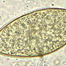
A diagnosis may be made by finding yellow-brown eggs in the stool. They are indistinguishable from the eggs of Fascioloides magna, although the eggs of F. magna are very rarely passed in sheep, goats, or cattle. If a patient has eaten infected liver, and the eggs pass through the body and out via the faeces, a false positive result to the test can occur. Daily examination during a liver-free diet will unmask this false diagnosis.[45]
An enzyme-linked immunosorbent assay (ELISA) test is the diagnostic test of choice. ELISA is available commercially and can detect antihepatica antibodies in serum and milk; new tests intended for use on faecal samples are being developed.[46] Using ELISA is more specific than using a Western blot or Arc2 immunodiffusion.[34] Proteases secreted by F. hepatica have been used experimentally in immunizing antigens.[47]
Fasciola hepatica, also known as the common liver fluke or sheep liver fluke, is a parasitic trematode (fluke or flatworm, a type of helminth) of the class Trematoda, phylum Platyhelminthes. It infects the livers of various mammals, including humans, and is transmitted by sheep and cattle to humans the world over. The disease caused by the fluke is called fasciolosis or fascioliasis, which is a type of helminthiasis and has been classified as a neglected tropical disease. Fasciolosis is currently classified as a plant/food-borne trematode infection, often acquired through eating the parasite's metacercariae encysted on plants. F. hepatica, which is distributed worldwide, has been known as an important parasite of sheep and cattle for decades and causes significant economic losses in these livestock species, up to £23 million in the UK alone. Because of its relatively large size and economic importance, it has been the subject of many scientific investigations and may be the best-known of any trematode species. F. hepatica's closest relative is Fasciola gigantica. These two flukes are sister species; they share many morphological features and can mate with each other.


_(14598306110).jpg)
_(17576890133).jpg)


