Distribution
provided by Echinoderms of Panama
In Panama this species has been collected in the Caribbean from Galeta Island (USNM E 36448, USNM E 36447; Centroid Latitude: 9.4050, Centroid Longitude: -79.8633, depth 10.7 m); Isla Grande (USNM E 14573; Centroid Latitude: 9.6283, Centroid Longitude: -79.5700, depth 6 m); and from Corgetupo Island (USNM E 25681) and Pico Feo (USNM E 18765), San Blas.
References and links
provided by Echinoderms of Panama
Mortensen, T. (1951): A Monograph of the Echinoidea. V, 2. Spatangoida II. Amphisternata II. Spatangidæ, Loveniidæ, Pericosmidæ, Schizasteridæ, Brissidæ. - 593 pp., Copenhagen (C. A. Reitzel); pages: 496-498.
The Echinoid Directory
World Echinoidea Database
LSID urn:lsid:marinespecies.org:taxname:422511
Comprehensive Description
provided by Smithsonian Contributions to Zoology
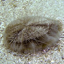
Plagiobrissus grandis (Gmelin)
Echinus grandis Gmelin, 1791:3200. [For synonymy, see Mortensen, 1951:496.]
Immature specimens of Plagiobrissus grandis (Gmelin) and Meoma ventricosa (Lamarck) were found living buried in the sand at a depth of 50 to 100 mm, in sand fields in the grooves of the Spur and Groove Zone on the transect off Carrie Bow Cay at a depth of 10 m.
The immature of the two species resemble each other in general appearance (compare Plate 1: figure 1 with Plate 6: figure 1); but the young of Plagiobrissus grandis can be distinguished by their whiter spines and lack of large, black pedicellariae on their dorsal surface. If all specimens found had been less than 6 mm, it would have been difficult to identify these species; but specimens nearly 20 mm long are enough like the adults to make identification possible. These identifications were confirmed by the comparison of specific features in the tests of both immature and adult that are not affected by growth. Chesher (1968a) has shown that the fascioles remain on the same plates throughout the growth of the echinoid, that the number of plates beyond the petals are constant, and that the periproct remains enclosed by the same plates. These features were compared between the immature specimens and specimens of adults believed to be the same species and no differences were found.
No adult specimens of either species were living with these immature. Considering the great differences between the immature and the adult, it is not surprising that they live in different environments.
Three immature specimens were found of P. grandis varying in length from 5.8 to 20 mm. The smallest specimen is far smaller than any of this species previously described (Kier and Grant, 1965: 36, described a specimen 35 mm long) and shows features never seen before. These features and changes that occur in the larger specimens are described in detail below.
SHAPE.—The smaller specimens have a higher, more smoothly rounded test with a far larger periproct and peristome relative to the test’s total length. The specimen 5.8 mm long has a very high test—a height equal to 67 percent of the length. In a specimen 35 mm long, the height is 45 percent of the length; and in an adult 150 mm long, only 33 percent (Figure 2). The test is smoothly rounded in the smallest specimen; but in an adult the margin is sharp, the adoral surface is more flattened, and the dorsal interambulacra are slightly ridged.
The periproct in the smallest specimen is very large, with its height equal to 38 percent of the length of the test and its width equal to 24 percent. In contrast, the periproct of a specimen 35 mm long has a height of only 18 percent and a width of only 10 percent. Finally, in an adult 150 mm long, the height of the periproct is only 10 percent; and the width is only 7 percent of the length of the test.
The periproct remains surrounded by the same interambulacral plates throughout its growth. The surrounding plates just become fewer in the adult. The smallest specimen’s periproct occurs within interambulacral plates 5 through 10. An adult’s falls within plates 5 through 8. The anus is situated so far dorsally in the smaller specimens (Figure 4A; Plate 1: figure 1) that it is almost entirely visible when viewed from the top. In an adult (Plate 1: figure 2) it has shifted down to a posterior truncation where most of it is now visible from a ventral view.
The peristome, too, is much larger (Figure 3G–I; Plate 1: figures 3, 4) relative to the length of the test in the very young. The width of the peristome in the smallest specimen is 34 percent of the length of the test; in a specimen 20 mm long, only 13 percent. A decrease also occurs in the height of the peristome. The smallest specimen, 5.8 mm long, has a height equal to 22 percent of the length of the test; whereas, the peristome in a specimen 20 mm long is only 11 percent of the total length, and is only 4 percent in an adult 150 mm long. Part of this reduction in the height of the peristome is due to the development of a protruding labrum in the adult.
PLASTRON.—The plastron on the smaller specimens is far wider than that in the adult. A specimen 6.9 mm long has a plastron with a width equal to 43 percent of the length of the test (Figure 3G); whereas, a specimen 20 mm long has a plastron width of only 30 percent (Figure 3H). In an adult the width is only 20 percent (Figure 3I).
The plastron in the immature specimens bears many large tubercles, but in the adult the tubercles are smaller than those on the broad interambulacral areas. Presumably, the spines attached to the plastron in the immature (Plate 3: figure 2) are the primary means of locomotion and burrowing. During growth a change takes place, and these locomotive spines in the adult are attached to the interambulacra.
APICAL SYSTEM.—The apical system of an immature specimen differs from an adult in being much larger relative to the size of the test, lacking genital pores, having only one madreporic pore, and lacking an extension of the madreporite (genital 2) into the posterior interambulacrum.
The apical system in the smallest specimen (5.8 mm long) is 0.99 mm wide (measured at its greatest width across oculars II and IV). This is 17 percent of the length of the test. The next largest specimen (6.9 mm long) has an apical system 0.98 mm wide or 14 percent of its length. A specimen 20 mm long has a system 1.2 mm wide or 6 percent of its length. An adult 150 mm long has a system 4.7 mm wide, which is only 3.1 percent of its length. Although the apical system does increase in size during the growth of the animal, its rate of growth is much less than the growth of the test.
No genital pores are present in the two smallest specimens, 5.8 and 6.9 mm long (Figure 3C,D); but small pores occur in a specimen 20 mm long (Figure 3E). They are quite large in a specimen 35 mm long; in an adult 150 mm long, they are extremely large, occupying most of the genital plates excluding the madreporite (Figure 3F).
Only one madreporic pore is present in each of the two smaller specimens (Figure 3C,D), and this occurs in the right anterior portion of genital 2. Sixteen pores are present in a specimen 20 mm long (Figure 3E); 35 in a specimen 35 mm long, and hundreds in an adult 150 mm long (Figure 3F). Although the single pore in the two smaller specimens is large relative to the size of the test, the madreporic pores in all the specimens are approximately the same size, 0.03 to 0.04 mm in diameter.
The madreporite (genital 2) extends very far into the posterior interambulacrum in the adult specimen (Figure 3F), but in the smaller specimens (Figure 3D,E) this posterior extension does not occur.
PETALS.—The ambulacral plates of the petals are just beginning to be developed in the smallest specimen 5.8 mm long. These petaloid plates can be distinguished from the ambulacral plates beyond the petals by their smaller size and by their slightly larger pores (Plate 1: figure 1). The pores in the ambulacral plates beyond the petals are so small that they cannot be distinguished from the holes in the meshwork of the calcite plates. Each of the petaloid ambulacra have six to eight plates. The pore in each plate is single, not double as in an adult. Each of these pores was studied under high magnification (Plate 4: figures 1, 2) with a scanning electron microscope, and in none of them is there any indication of a partition developing to divide the pore in two. However, in the nonpetaloid ambulacrum III, each pore is constricted medially (Figure 3B; Plate 4: figure 1), suggesting that the ambulacral pores are originally single and then are divided in two by this constriction.
In the next larger specimen, 6.9 mm long, most of the petaloid pores are double (Figure 3C). The pores, though, are still very close together; and the ambulacra are not petal-like in appearance. Only eight plates are present in each petaloid ambulacra. A specimen 20 mm long has well-developed petals with the pores elongated transversely.
The petals are slightly depressed, not flush to the general surface of the test as in the smaller specimens. Each anterior petal has 26 plates, and each posterior has 32. The petals in an adult 150 mm long (Plate 1: figure 2) are flexuous, not straight as in the smaller specimens. There are 70 plates in each anterior paired petal and 90 in each posterior.
FASCIOLES.—The peripetalous fasciole is only slightly developed in the immature specimens, but the subanal fasciole is distinct and clearly functional. The fascioles increase in size, not by an increase in the size of the fasciole tubercles but by an increase in the number of these tubercles.
The peripetalous fasciole on a specimen 5.8 mm long is composed of a single row of tubercles. A second row of tubercles is present for part of the length of this fasciole in a specimen 6.9 mm long (Plate 1: figure 1). Most of this fasciole has 2 rows of tubercles in a specimen 20 mm long; 5 rows in a specimen 35 mm long; and 17 rows in an adult 150 mm long.
The subanal fasciole is very distinct in the smallest specimen (5.8 mm long) and consists of 2 to 3 rows of tubercles. There are 3 to 5 rows in a specimen 20 mm long; 9 to 13 in a specimen 35 mm long; and 30 to 35 in an adult 150 mm long.
The tubercles in both fascioles do not increase in size during the growth of the echinoid. They are between 0.05 and 0.06 mm in diameter in all the specimens regardless of the size of the echinoid.
DORSAL TUBERCLES.—The large dorsal tubercles that typically occur within the peripetalous fasciole of an adult are absent or just forming in the smallest specimen. The plates that bear them in the adult are just beginning to be developed in the smaller specimens. A specimen 20 mm long has approximately 20 of these larger tubercles. A specimen 35 mm long has less than 50, and an adult has nearly 200.
VENTRAL RODS.—Rod-like structures (Figure 5) are very apparent on the ventral side of the smaller specimens, particularly when the specimens are wettened with xylene (Plate 2: figure 2). Under high magnification it can be seen that the rods are formed of the calcite matrix of the test and are produced by thickening of the calcite with a corresponding reduction in the size of the holes in the calcite meshwork (Plate 2: figures 3, 6). Where a rod meets the suture of an adjacent plate, it is overlapped by a rod extending from that plate (Plate 2: figures 4, 5). The rods are very conspicuous on the smaller specimens because of their thin tests, but much less apparent on an adult.
I suspect that these rods serve to strengthen the test, which is very thin in this species, especially in the immature. The rods are most developed where large tubercles occur, suggesting that they are especially needed here because of the strain exerted on the test by the strenuous movement of the ventral spines. The rods are not present in the immature of Meoma ventricosa (described below), but the test in that species is considerably thicker and perhaps needs no such reinforcement.
ADULTS.—Although I found no adults in the region, Richard Chesher (pers. comm., 1974) points out that, in his experience, they are one of the most difficult echinoids to find because they bury very deep, have no surface burrow, and do not come up at night.
- bibliographic citation
- Kier, Porter M. 1975. "The echinoids of Carrie Bow Cay, Belize." Smithsonian Contributions to Zoology. 1-45. https://doi.org/10.5479/si.00810282.206