Description
provided by Zookeys
MBL = 1.67-1.96 mm; MBH = 1.60-1.80 mm; FBL = 2.01-2.27 mm; MBH = 1.90-2.09 mm; AL/BL = 0.60±0.05; MBW = 1.02–1.13 mm; EL/EW = 2.42–2.49; PW/PL = 1.67–1.68; EWB/PWB = 1.10±0.05; EWM/PWM = 1.40–1.41.
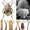
Color of elytra, pronotum and head consistently copperish. Antennomere 1 partly dark brown. Antennomeres 2–3 yellow. Antennomere 4 yellow or partly brown. Antennomere 5 partly brown. Remaining antennomeres black. Pro- and mesofemora brown with yellow on the apex. Metafemora brown. Tarsi brown with yellow on base of each tarsomere.
Base of pronotum with two short, obscure longitudinal impressions without punctures near basal margin. Deep row of large punctures at base of pronotum present on sides, lacking in middle. Pronotal base evenly convex. Lateral sides of pronotum slightly convex with maximum width near base. Anterolateral prothoracic callosity protruding laterally forming round angle. Posterolateral prothoracic callosity projects up to lateral margin of pronotum. Diameter of pronotal punctures 2 to 4 times smaller than distance between them.
Elytra with convex sides. Scutellar row of punctures on elytron regular and single. Remaining rows of punctures regular. Elytral humeral calli well developed. Interspaces between rows of punctures smooth and glabrous. Two lines of minute punctures on each interspace.
Head hypognathous. Frontal ridge between antennal sockets narrow and convex. Frontolateral sulcus present. Suprafrontal sulcus shallow and faint or deep laterally, shallow in middle. Suprafrontal sulcus slightly concave. Orbital sulcus (above the antennal socket) deep, but rather narrow. Width of frontal ridge to width of antennal socket: 0.900–1.005. Width of orbital sulcus to width of frontolateral sulcus: 0.611–0.614. Surface of vertex sparsely and unevenly covered with 6–7 punctures near each eye. Numbers of punctures on each orbit: 2–3. Numbers of setae along frontolateral sulcus on each side: 8–10. Numbers of setae on frons (triangular area surrounded by frontolateral sulci and clypeus): 0. Numbers of setae on clypeus: 7. Numbers of setae on labrum: 6. Anterior margin of labrum slightly concave in middle.
First male protarsomere distinctly larger than second one. First male protarsomere, length to width ratio: 1.63–1.67. First and second male protarsomeres, length to length ratio: 2.00–2.03; width to width ratio: 1.55–1.59. First male protarsomere, width at apex to width at base: 2.58–2.64. Length of metatibia to distance between denticle and metatibial apex: 2.50–2.55. Large lateral denticle on metatibia sharp. Metatibial serration proximal to large lateral denticle present, obtuse. Metatibia proximal to denticle in dorsal view concave. First male metatarsomere, length to width ratio: 3.01–3.05. First and second male metatarsomeres, length to length ratio: 1.87–1.89. First and second male metatarsomeres, width to width ratio about 0.98. Third and fourth male metatarsomeres, length to length ratio: 1.64–1.68. Metatibia length to metafemora length: 0.81±0.05. Length of hind leg to length of body: 0.92±0.05.
Median lobe of aedeagus parallel-sided with apical third slightly widening. Apical part of median lobe in ventral view narrowing abruptly. Ventral longitudinal groove of median lobe absent in apical part and poorly developed in middle and basal part. Apical denticle of aedeagus in ventral view poorly differentiated, straight in lateral view. Minute transverse wrinkles on ventral side of median lobe absent. Median lobe in lateral view narrow and evenly curved. Width (in middle) to length of median lobe (in ventral view) about 0.15.
Spermathecal receptacle pear-shaped. Spermathecal pump much shorter than receptacle. Apex of spermathecal pump cylindrical. Spermathecal pump attached to middle of receptacle top. Maximum width of receptacle situated basally. Basal part of receptacle wider than apical. Posterior sclerotization of tignum spoon-shaped, wider than mid section. Anterior sclerotization of tignum wider than mid section. Apex of vaginal palpus subdeltoid, with lateral side slightly arching. Sides of middle part of vaginal palpus (before apex) narrowing from base, slightly widening towards apex. Anterior sclerotization of vaginal palpus slightly widening anteriorly. Anterior sclerotization of vaginal palpus slightly and evenly curved along length. Anterior end of anterior sclerotization broadly rounded. Length of posterior sclerotization greater than width. Posterior sclerotization about as wide as anterior sclerotization.
- license
- cc-by-3.0
- copyright
- Yongying Ruan, Alexander S. Konstantinov, Siqin Ge, Xingke Yang
- bibliographic citation
- Ruan Y, Konstantinov A, Ge S, Yang X (2014) Revision of the Chaetocnema picipes species-group (Coleoptera, Chrysomelidae, Galerucinae, Alticini) in China, with descriptions of three new species ZooKeys 387: 11–32
- author
- Yongying Ruan
- author
- Alexander S. Konstantinov
- author
- Siqin Ge
- author
- Xingke Yang
Distribution
provided by Zookeys
Heilongjiang, Liaoning, Inner Mongolia, Beijing, Hebei, Tianjin, Shanxi, Shandong, Gansu, Qinghai, Shaanxi; Europe, North Asia (Konstantinov et al. 2011); Madgascar (alien) (Biondi 2001).
- license
- cc-by-3.0
- copyright
- Yongying Ruan, Alexander S. Konstantinov, Siqin Ge, Xingke Yang
- bibliographic citation
- Ruan Y, Konstantinov A, Ge S, Yang X (2014) Revision of the Chaetocnema picipes species-group (Coleoptera, Chrysomelidae, Galerucinae, Alticini) in China, with descriptions of three new species ZooKeys 387: 11–32
- author
- Yongying Ruan
- author
- Alexander S. Konstantinov
- author
- Siqin Ge
- author
- Xingke Yang