About
Education
Discuss
TraitBank
Sign In
Sign Up
Language
Deutsch
English
Español
français
italiano
Nederlands
Piemontèis
Português do Brasil
suomi
Türkçe
Čeština
Ελληνικά
македонски
Українська
العربية
简体中文
繁體中文
names in breadcrumbs
vernacular
scientific
About
Education
Discuss
TraitBank
Sign In
Sign Up
en
Deutsch
English
Español
français
italiano
Nederlands
Piemontèis
Português do Brasil
suomi
Türkçe
Čeština
Ελληνικά
македонски
Українська
العربية
简体中文
繁體中文
names in breadcrumbs
vernacular
scientific
Life
»
…
»
Mushrooms, Lichens, Molds, Yeasts And Relatives
»
…
»
Sac Fungi
»
…
Life
»
Cellular
»
Eukaryotes
»
Opisthokonts
»
Nucletmycea
»
Mushrooms, Lichens, Molds, Yeasts And Relatives
»
Dikarya
»
Sac Fungi
»
Sordariomycetes
«
Ophiostomatales
collect
overview
data
media
articles
maps
names
CC-BY
any license
CC-BY
CC-BY-NC
CC-BY-NC-SA
CC-BY-SA
No copyright
provider
any provider
iNaturalist
Wikimedia Commons
Barcode of Life Data Systems
Flickr Group
Public Health Image Library
1
2
Last »
cc-publicdomain
trusted
cc-publicdomain
trusted
cc-publicdomain
trusted
cc-publicdomain
trusted
cc-publicdomain
trusted
cc-publicdomain
trusted
cc-publicdomain
trusted
cc-publicdomain
trusted
cc-by-4.0
trusted
cc-by-4.0
trusted
cc-by-4.0
trusted
cc-publicdomain
trusted
cc-by-4.0
trusted
cc-by-4.0
trusted
cc-by-4.0
trusted
cc-by-4.0
trusted
cc-publicdomain
trusted
cc-by-4.0
trusted
cc-publicdomain
trusted
cc-publicdomain
trusted
cc-publicdomain
trusted
cc-publicdomain
trusted
cc-publicdomain
trusted
cc-publicdomain
trusted
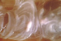
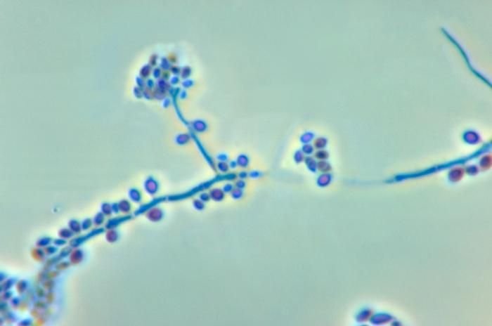
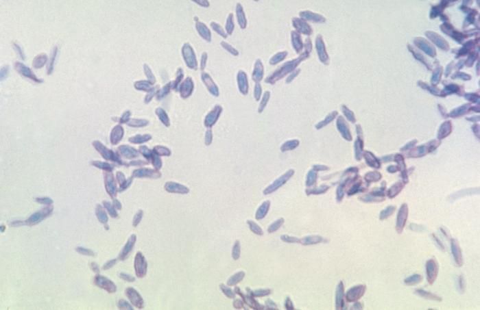
Image of Sporothrix
cc-publicdomain
Public Health Image Library
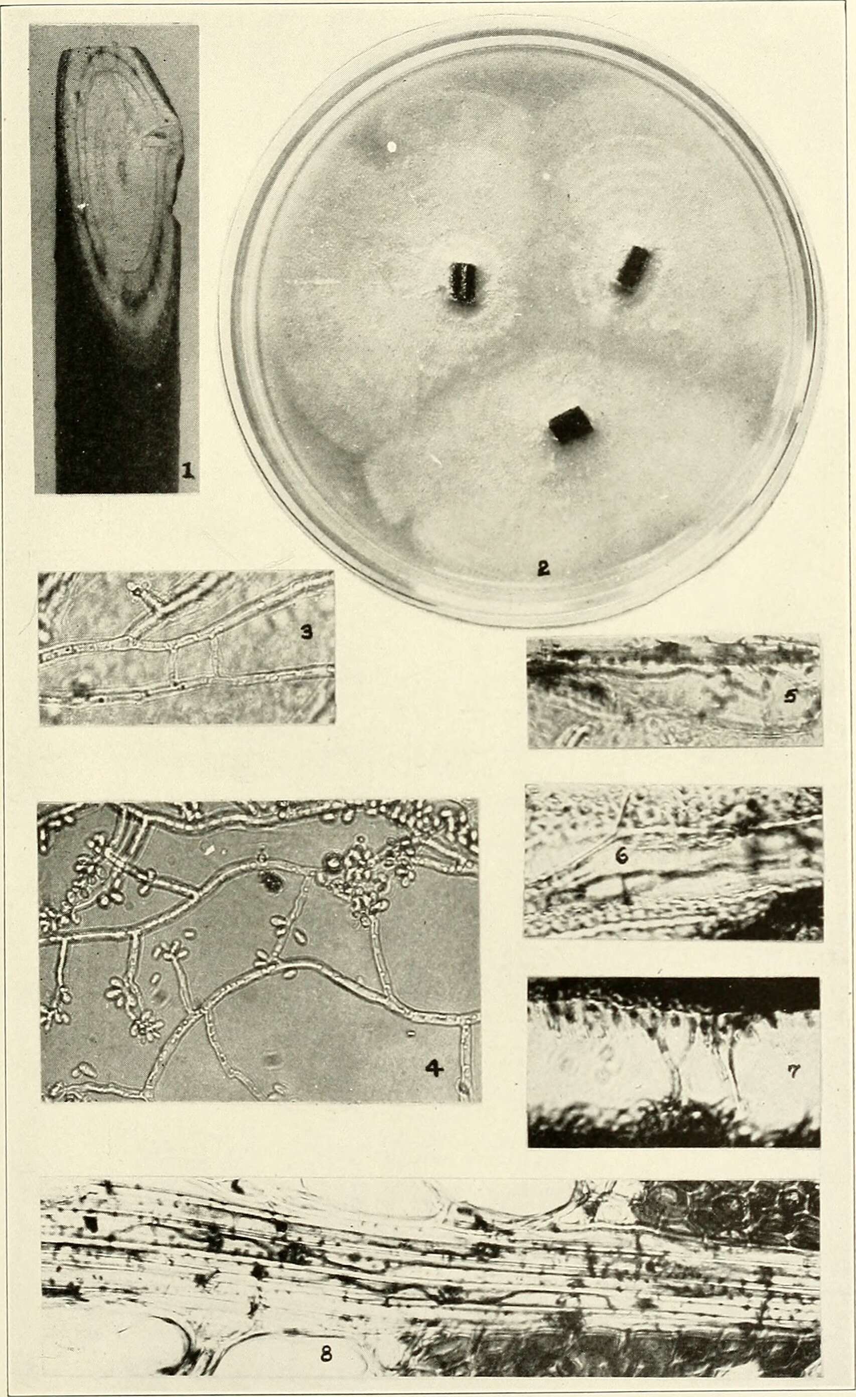
Magnified 500X, this photomicrograph revealed the presence of
Sporothrix sp.
fungal organisms that were isolated from a peat moss specimen.Created: 1971
Image of Sporothrix schenckii Hektoen & C. F. Perkins 1900
cc-publicdomain
Public Health Image Library
This photomicrograph reveals the conidiophores and conidia of the fungus
Sporothrix schenckii
.Created: 1972
Image of Sporothrix schenckii Hektoen & C. F. Perkins 1900
cc-publicdomain
Public Health Image Library
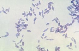
Shown here is a photomicrograph of the fungus
Sporothrix schenckii
during yeast phase.Created: 1964
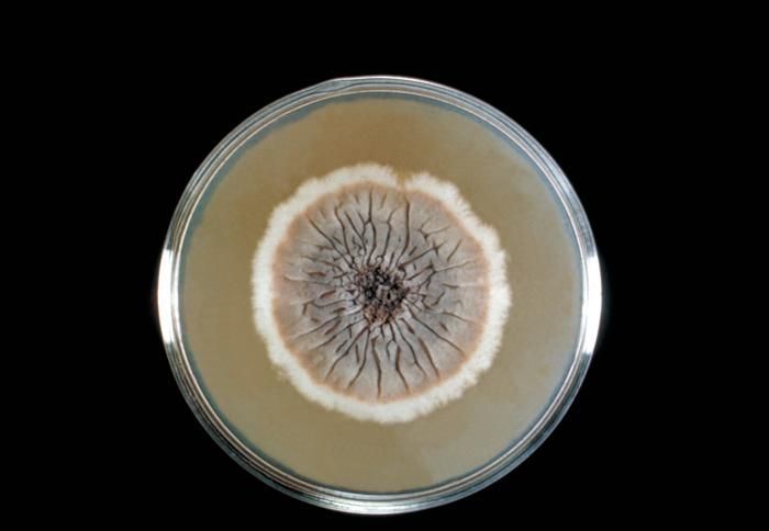
Image of Sporothrix schenckii Hektoen & C. F. Perkins 1900
cc-publicdomain
Public Health Image Library
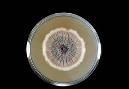
This Sabouraud's dextrose agar plate culture is growing the fungus
Sporothrix schenckii
.Created: 1964
Image of Sporothrix schenckii Hektoen & C. F. Perkins 1900
cc-publicdomain
Public Health Image Library
Shown here is a close-up of a
Sporothrix schenckii
culture during yeast phase.Created: 1964
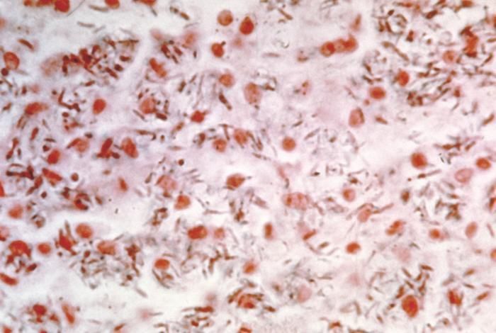
Image of Sporothrix schenckii Hektoen & C. F. Perkins 1900
cc-publicdomain
Public Health Image Library
This photomicrograph shows the presence of
Sporothrix schenckii
in a smear obtained from a rat.Created: 1964
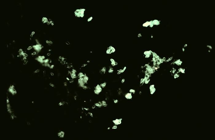
Image of Sporothrix schenckii Hektoen & C. F. Perkins 1900
cc-publicdomain
Public Health Image Library
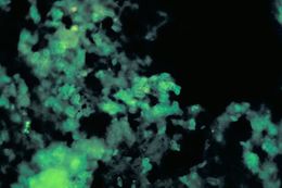
Using a direct FA stain, this slide demonstrates the histopathology of sporotrichosis due to
Sporothrix schenckii
.Created: 1977
Image of Sporothrix
cc-publicdomain
Public Health Image Library
Using a direct FA stain, this slide demonstrates the histopathology of sporotrichosis due to
Sporothrix schenckii
.Created: 1977
"
cc-by-4.0
Michael Ellis
iNaturalist
"
cc-by-4.0
Michael Ellis
iNaturalist
"
cc-by-4.0
Michael Ellis
iNaturalist
"
cc-publicdomain
Garrett Taylor
iNaturalist
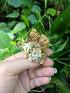
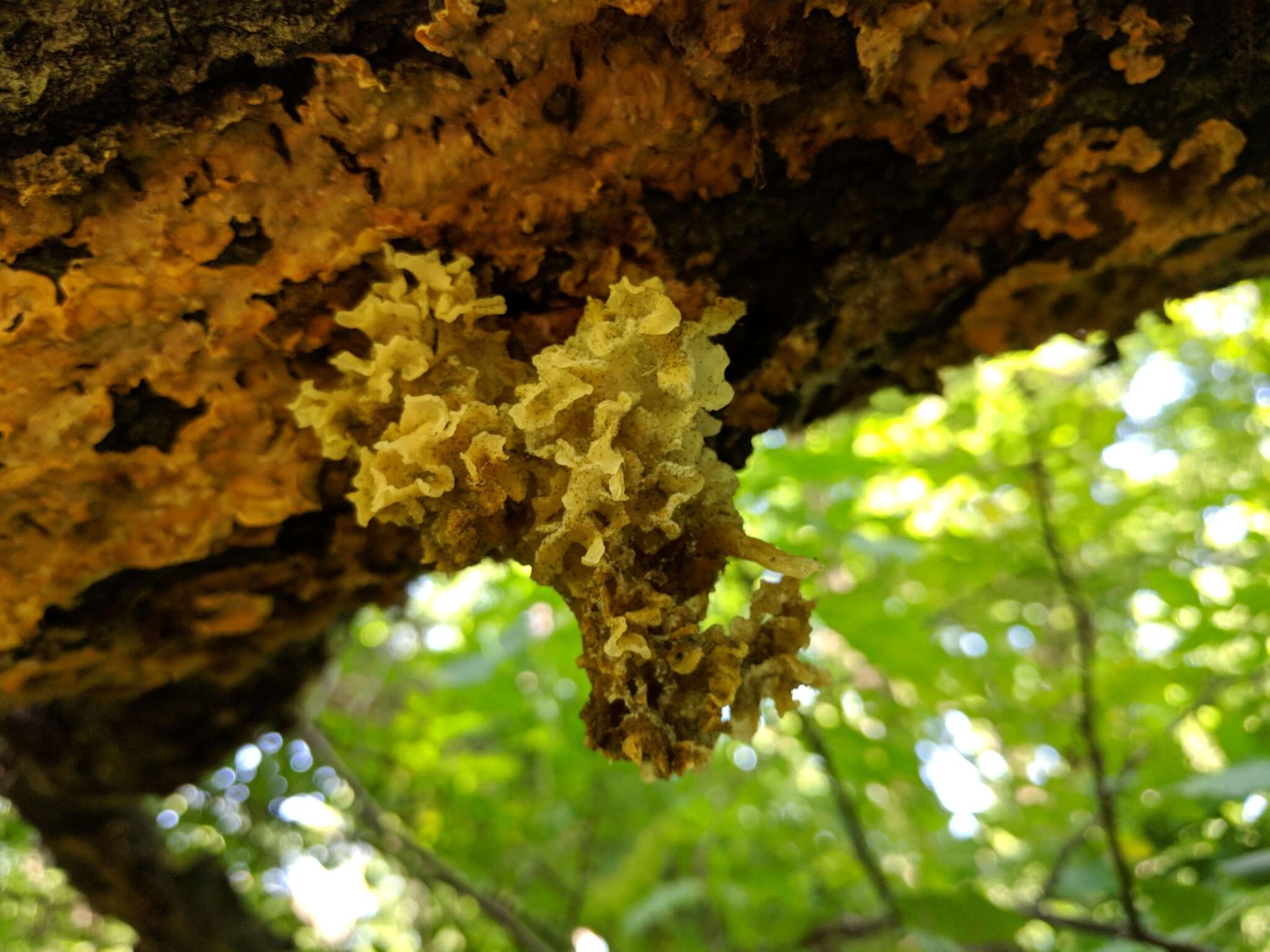
"
cc-by-4.0
Sarah DeLong-Duhon
iNaturalist
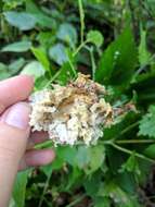
"
cc-by-4.0
Sarah DeLong-Duhon
iNaturalist
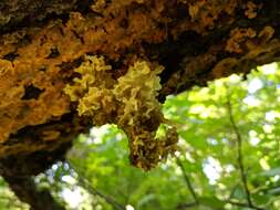
"
cc-by-4.0
Sarah DeLong-Duhon
iNaturalist
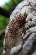
"
cc-by-4.0
Sarah DeLong-Duhon
iNaturalist
"
cc-publicdomain
Garrett Taylor
iNaturalist
"
cc-by-4.0
Caleb Catto
iNaturalist
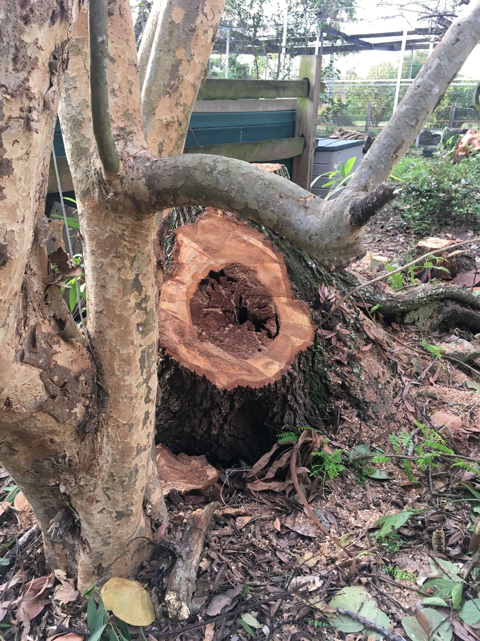
"
cc-publicdomain
Jade Fortnash
iNaturalist
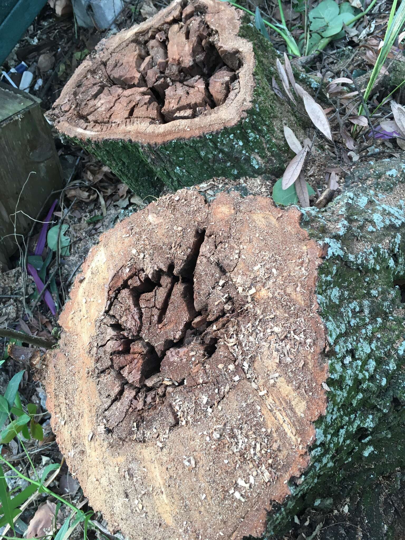
"
cc-publicdomain
Jade Fortnash
iNaturalist
"
cc-publicdomain
Jade Fortnash
iNaturalist
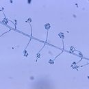
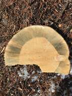
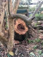
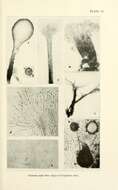
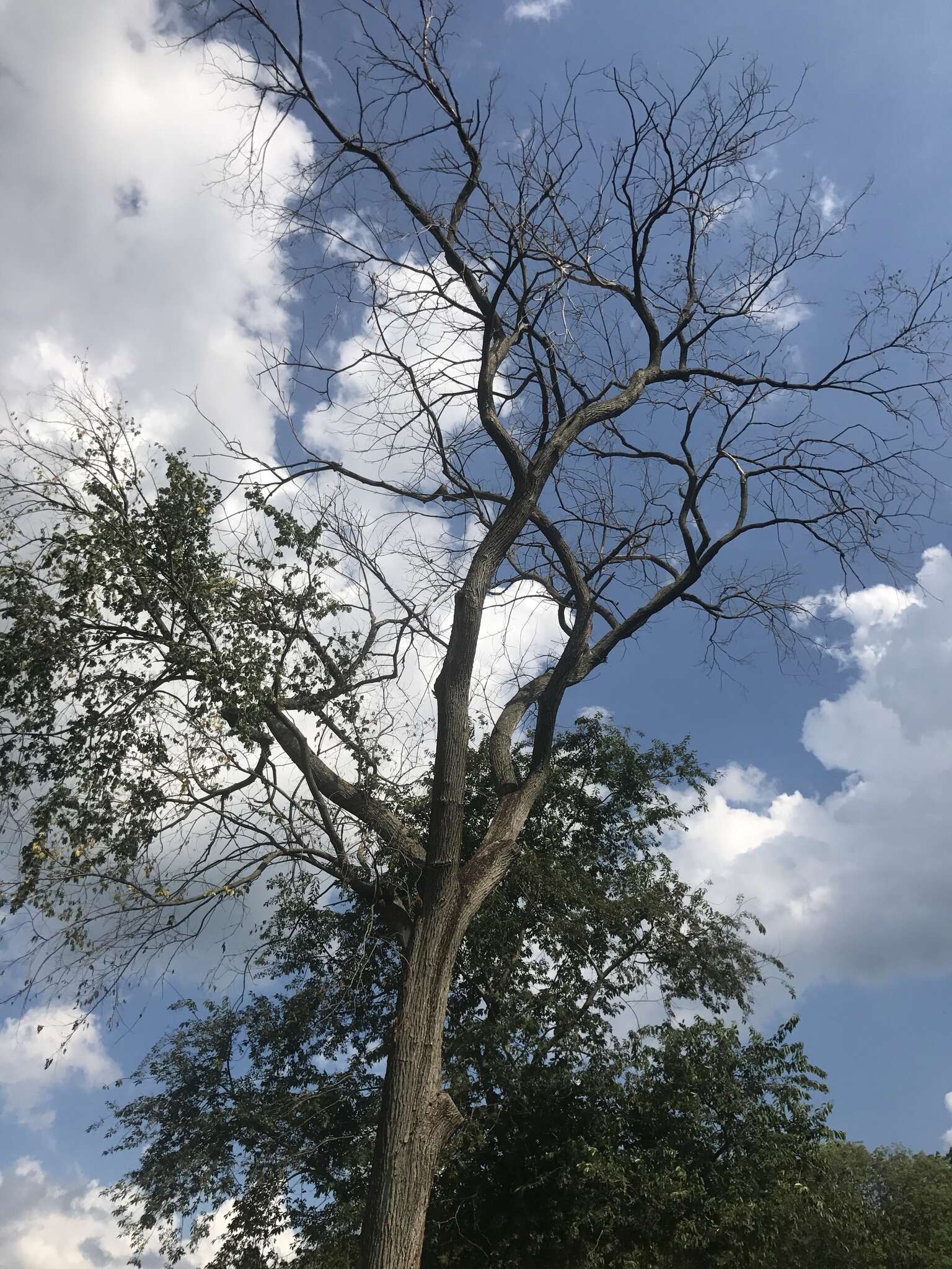
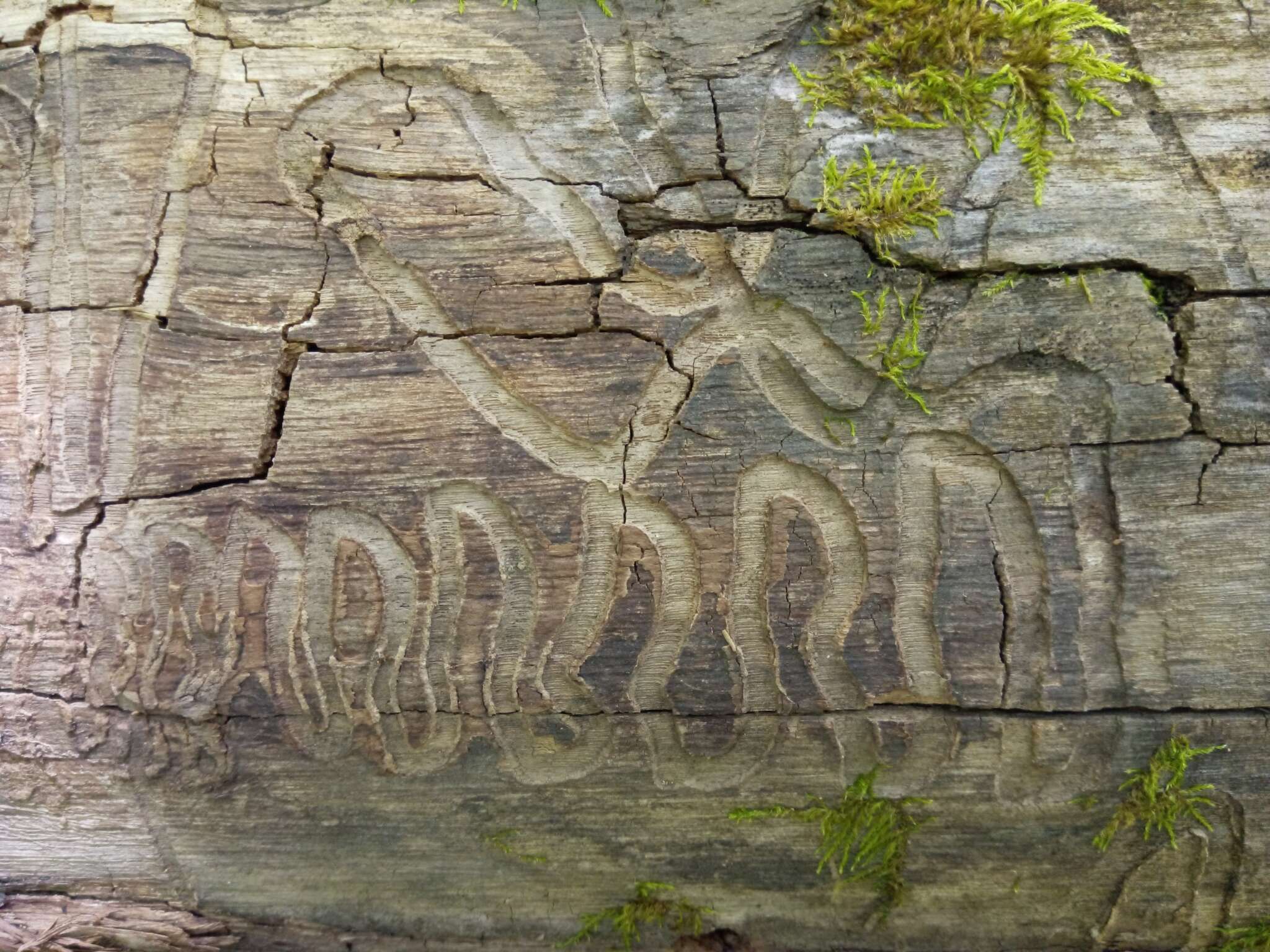
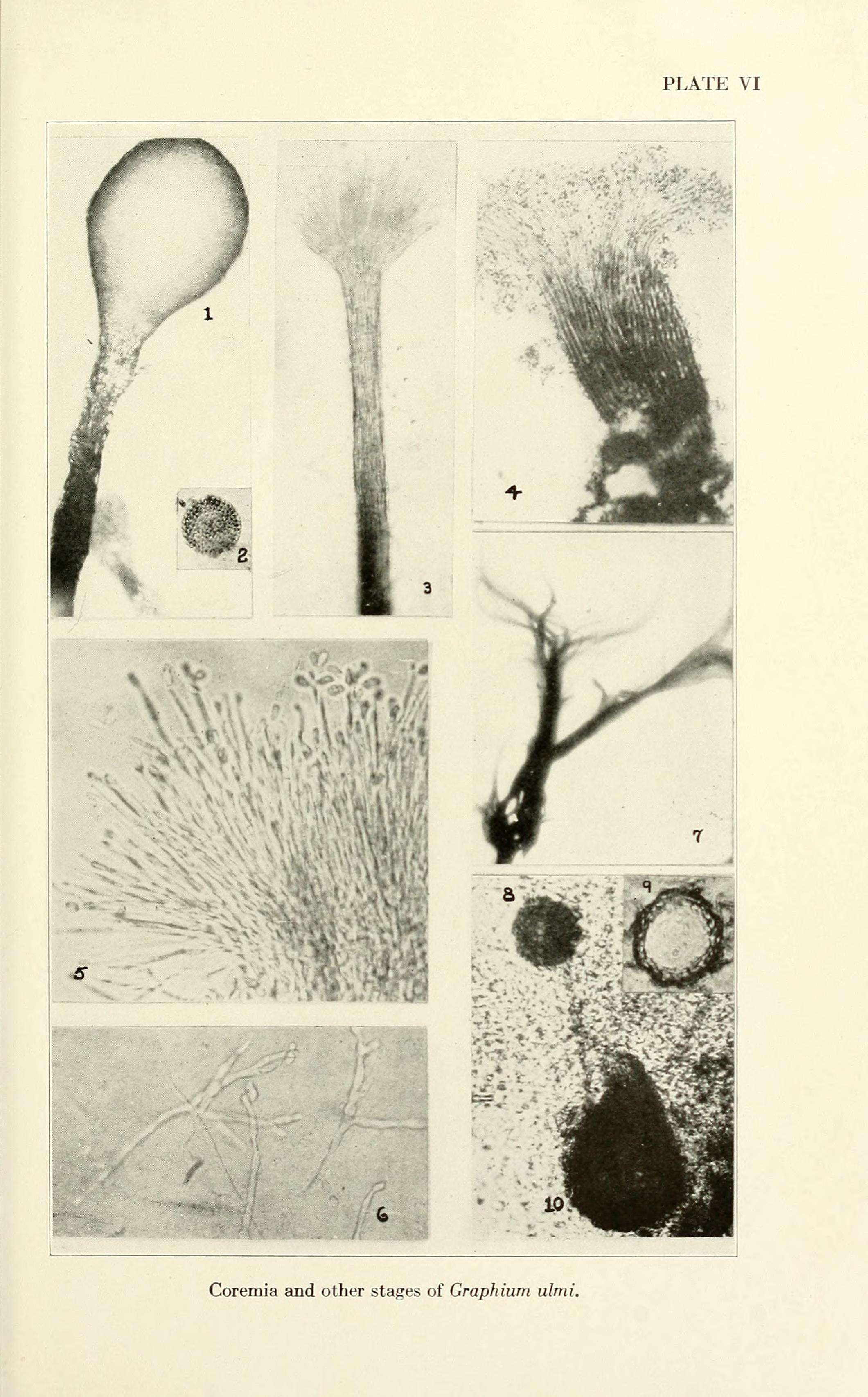
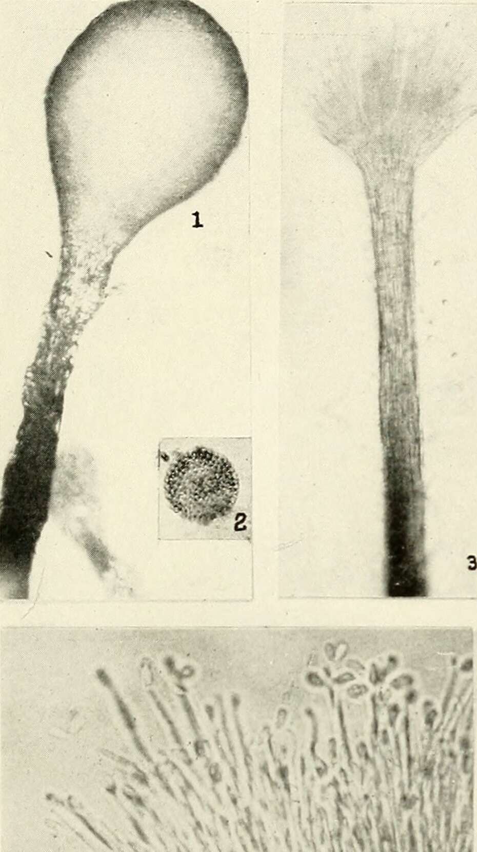
Ophiostoma ulmi coremium (1)
cc-publicdomain
Wikimedia Commons
Description: Plate VI. Ophiostoma ulmi: coremium (synnema). Date: 1936. Source:
https://archive.org/stream/dutchelmdiseaseg00clin/#page/n70/mode/1up
. Author: Clinton, G. P. (George Perkins), McCormick, Florence A. (Florence Anna).
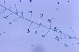
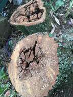
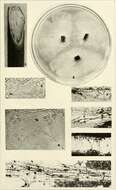
Ophiostoma ulmi
cc-publicdomain
Wikimedia Commons
Summary =[
edit
] Description: Plate V. Ophiostoma ulmi. Date: 1936. Source:
https://www.flickr.com/photos/internetarchivebookimages/21114916805/in/photolist-yaRt8K-y8xhwo-xTm3yn-xTmjW2-xTfcXo-y9Xyus-xTedh5-xTmwAK-y9Xz8G-wBq4FT-xdYDii-yaRvWa-xTmi3c-xdXZzx-xTf6hm-y9Y3Do-ybx6Wc-tEqDir-wXmH5G-x4RhcG-ovTtb2-tiDwfo-t4wy2a-wajeRR-tCL3qX-wPGdXa-tj6wgs-wPGiyz-xxMUjF-xi1YgL-xfG3qH-tm3BKZ-wmitcK-w55FaA-soXEWw-x7bPsg-w56AxC-x28KgX-tkZh9p-w5gkge-xfQQ8S-x2725V-xzGpYN-x5JhH8-sGdkNP-x1xWn7-tkYb76-wPxf4s-xvYgmy-wPwHyE
. Author: Clinton, G. P. (George Perkins), McCormick, Florence A. (Florence Anna). Permission (
Reusing this file
): 1936.
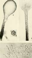
Ophiostoma ulmi coremium
cc-publicdomain
Wikimedia Commons
Summary =[
edit
] Description: Plate VI. Ophiostoma ulmi: coremium (synnema). Date: 1936. Source:
https://www.flickr.com/photos/internetarchivebookimages/20493848153/in/photolist-xdYk4x-y9YiLh-y9Yu1W-xdQu17-y8xTnL-xdYEfP-w9AhL3-xdYMJM-yaS1Mg-xdYK6F-xTf6VW-xTeeK5-xdYT4p-xTg7L9-xTepVq-ybwFF8-xTeXhJ-xTek5U-y8xRfj-xTfK5d-xTf3uY-xTmHPr-xTg87E-y8wVy1-yaRddV-xTehEd-xTfVMq-yaRt8K-y8xhwo-xTm3yn-xTmjW2-xTfcXo-y9Xyus-xTedh5-xTmwAK-y9Xz8G-wBq4FT-xdYDii-yaRvWa-xTmi3c-xdXZzx-xTf6hm-y9Y3Do-ybx6Wc-tEqDir-wXmH5G-x4RhcG-ovTtb2-tiDwfo-wajeRR
. Author: Clinton, G. P. (George Perkins), McCormick, Florence A. (Florence Anna).
1
2
Last »