-
Epalxella (ee-palk-sell-a) is an odontostome ciliate. This is a small group of small flattened and sparsely ciliated ciliates which are most usually found in anoxic habitats. They have a small group of ciliary organelles associated with the mouth - the clear area at about 4 o clock. Differential interference contrast.
-

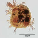
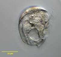
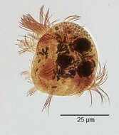
Portrait (right side) of the sapropelic odontostome ciliate, Epalxella antiquorum (Penard, 1922) Corliss, 1960 (synonymous with the preoccupied Epalxis). The cell has a clear rigid pellicle with a broad lenticular outline. The body is strongly laterally compressed. The dorsum is broadly rounded. The right side is broadly truncate posteriorly with a blunt spine on the ventral margin. The posterior side on the left terminates in 5-7 blunt spines. The spines do not terminate in needle-like processes seen in the similar genus, Saprodinium. The right and left surfaces of the pellicle bear longitudinal folds and ridges. A perizonal ciliary complex with 5 kineties and two shorter kineties runs across the ventral surface for a short distance onto the left and a longer distance onto the right surface anterior to the cytostome. The cytostome and adoral zone of membranelles is in the middle of the ventral surface. There is a prominent tooth-like spine at the anterior edge of the cytostome. The longitudinal somatic kineties are located on the pellicular ridges of the left surface and the dorsal and ventral edge of the right side. They do not extend more than 1/3 body length. The single contractile vacuole is located posteriorly. There are either two or four macronuclei. The cytoplasm contains methanogenic bacteria. Refractile brown cytoplasmic granules accumulate anteriorly. Collected from hydrogen sulfide- rich bottom sediments of slow-flowing freshwater stream near Boise, Idaho March 2004. DIC.
-
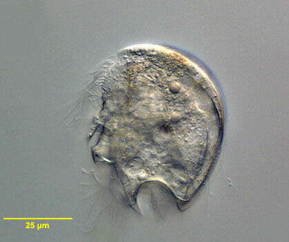
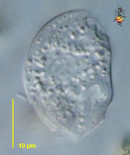
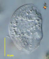
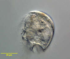
Portrait (left side) of the sapropelic odontostome ciliate, Epalxella antiquorum (Penard, 1922) Corliss, 1960 (synonymous with the preoccupied Epalxis). The cell has a clear rigid pellicle with a broad lenticular outline. The body is strongly laterally compressed. The dorsum is broadly rounded. The right side is broadly truncate posteriorly with a blunt spine on the ventral margin. The posterior side on the left terminates in 5-7 blunt spines. The spines do not terminate in needle-like processes seen in the similar genus, Saprodinium. The right and left surfaces of the pellicle bear longitudinal folds and ridges. A perizonal ciliary complex with 5 kineties and two shorter kineties runs across the ventral surface for a short distance onto the left and a longer distance onto the right surface anterior to the cytostome. The cytostome and adoral zone of membranelles is in the middle of the ventral surface. There is a prominent tooth-like spine at the anterior edge of the cytostome. The longitudinal somatic kineties are located on the pellicular ridges of the left surface and the dorsal and ventral edge of the right side. They do not extend more than 1/3 body length. The single contractile vacuole is located posteriorly. There are either two or four macronuclei. The cytoplasm contains methanogenic bacteria. Refractile brown cytoplasmic granules accumulate anteriorly. Collected from hydrogen sulfide- rich bottom sediments of slow-flowing freshwater stream near Boise, Idaho March 2004. DIC.
-
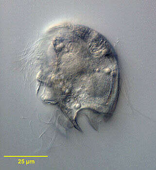
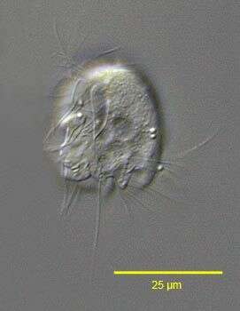
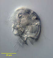
Portrait (right side) of the sapropelic odontostome ciliate, Epalxella antiquorum (Penard, 1922) Corliss, 1960 (synonymous with the preoccupied Epalxis). The cell has a clear rigid pellicle with a broad lenticular outline. The body is strongly laterally compressed. The dorsum is broadly rounded. The right side is broadly truncate posteriorly with a blunt spine on the ventral margin. The posterior side on the left terminates in 5-7 blunt spines. The spines do not terminate in needle-like processes seen in the similar genus, Saprodinium. The right and left surfaces of the pellicle bear longitudinal folds and ridges. A perizonal ciliary complex with 5 kineties and two shorter kineties runs across the ventral surface for a short distance onto the left and a longer distance onto the right surface anterior to the cytostome. The cytostome and adoral zone of membranelles is in the middle of the ventral surface. There is a prominent tooth-like spine at the anterior edge of the cytostome. The longitudinal somatic kineties are located on the pellicular ridges of the left surface and the dorsal and ventral edge of the right side. They do not extend more than 1/3 body length. The single contractile vacuole is located posteriorly. There are either two (as seen in this image) or four macronuclei. The cytoplasm contains methanogenic bacteria. Refractile brown cytoplasmic granules accumulate anteriorly. Collected from hydrogen sulfide- rich bottom sediments of slow-flowing freshwater stream near Boise, Idaho March 2004. DIC.
-
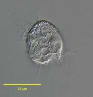
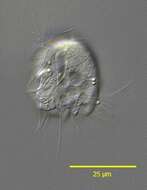
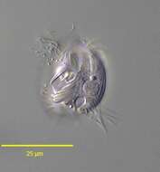
Portrait (left side) of the sapropelic odontostome ciliate, Epalxella exigua (Penard, 1922) Corliss, 1960. Synonymous with the preoccupied Epalxis. Collected from bottom sediments of a rain barrel; Boise, Idaho. DIC. This image was taken by William Bourland. He now uses a Zeiss Axioskop 2 with a Spot Insight CCD camera (Diagnostic Instruments).
-
Portrait (right side) of the sapropelic odontostome ciliate, Epalxella exigua (Penard, 1922) Corliss, 1960. Synonymous with the preoccupied Epalxis. Collected from bottom sediments of a rain barrel; Boise, Idaho. DIC. This image was taken by William Bourland. He now uses a Zeiss Axioskop 2 with a Spot Insight CCD camera (Diagnostic Instruments).
-
Infraciliature (left side) of the sapropelic odontostome ciliate, Epalxella exigua (Penard, 1922) Corliss, 1960. Synonymous with the preoccupied Epalxis. Collected from bottom sediments of a rain barrel; Boise, Idaho.Stained by the silver carbonate technique (see Foissner, W. Europ. J. Protistol., 27:313-330;1991).Brightfield. This image was taken by William Bourland. He now uses a Zeiss Axioskop 2 with a Spot Insight CCD camera (Diagnostic Instruments).